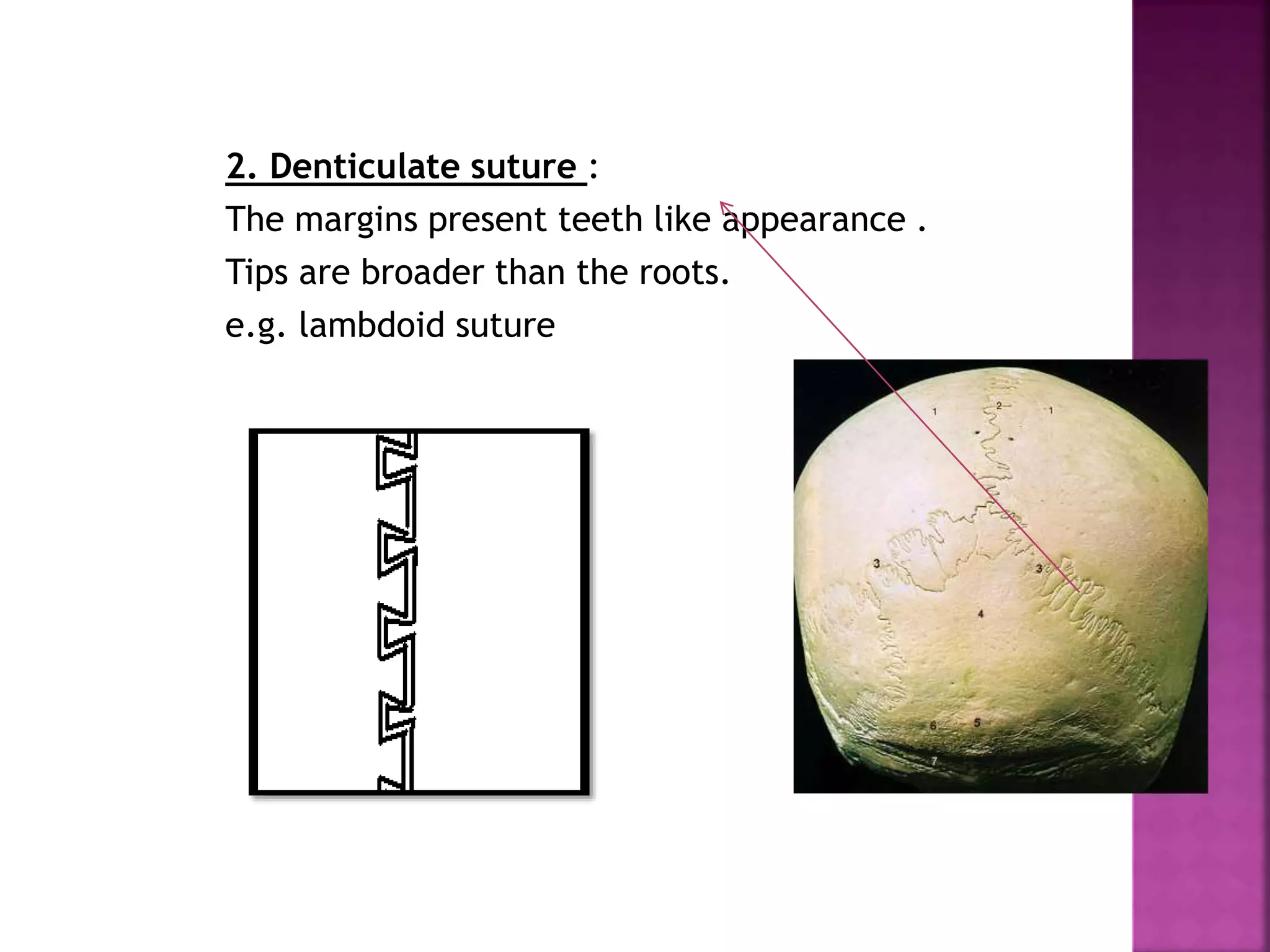
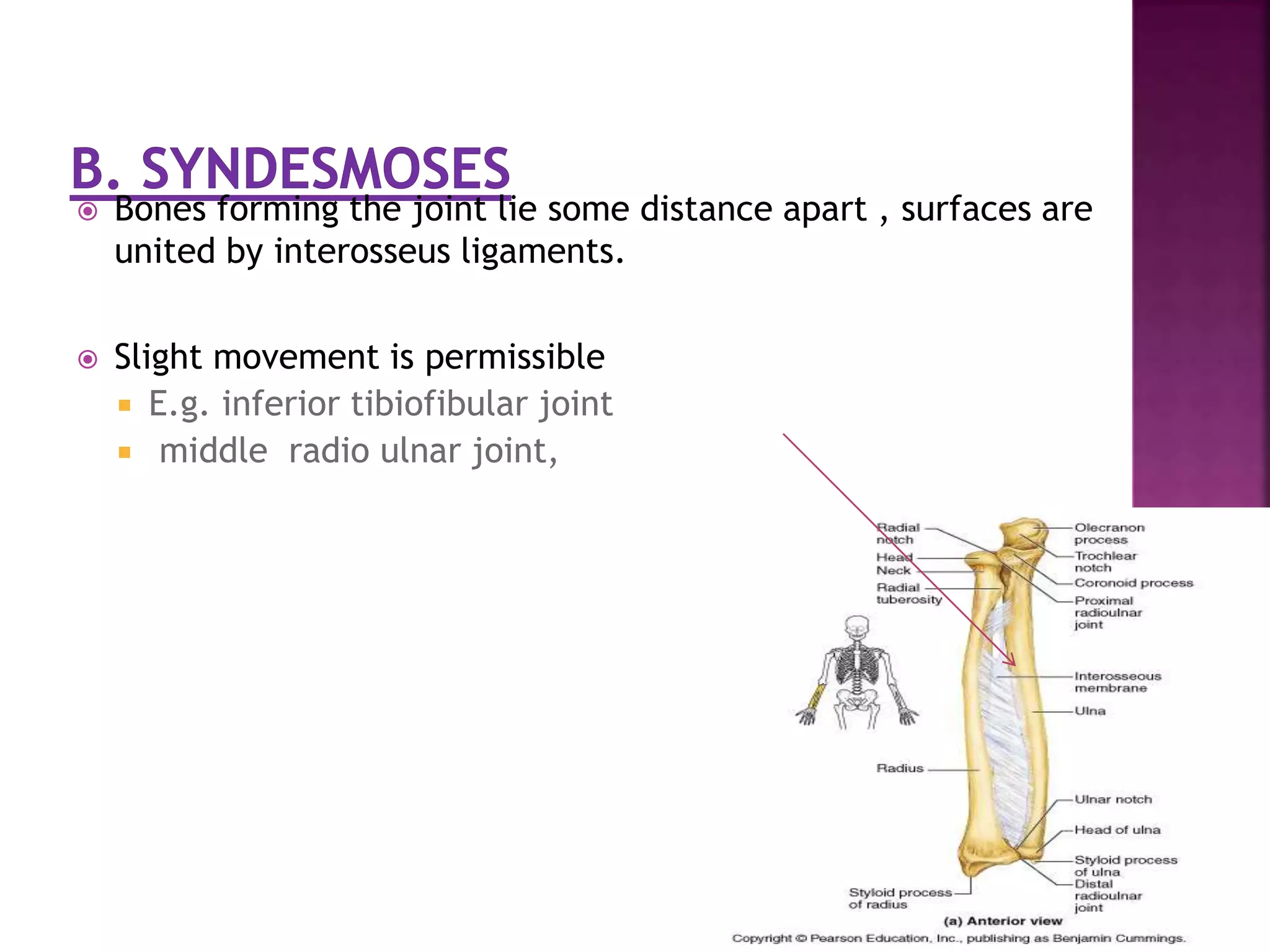
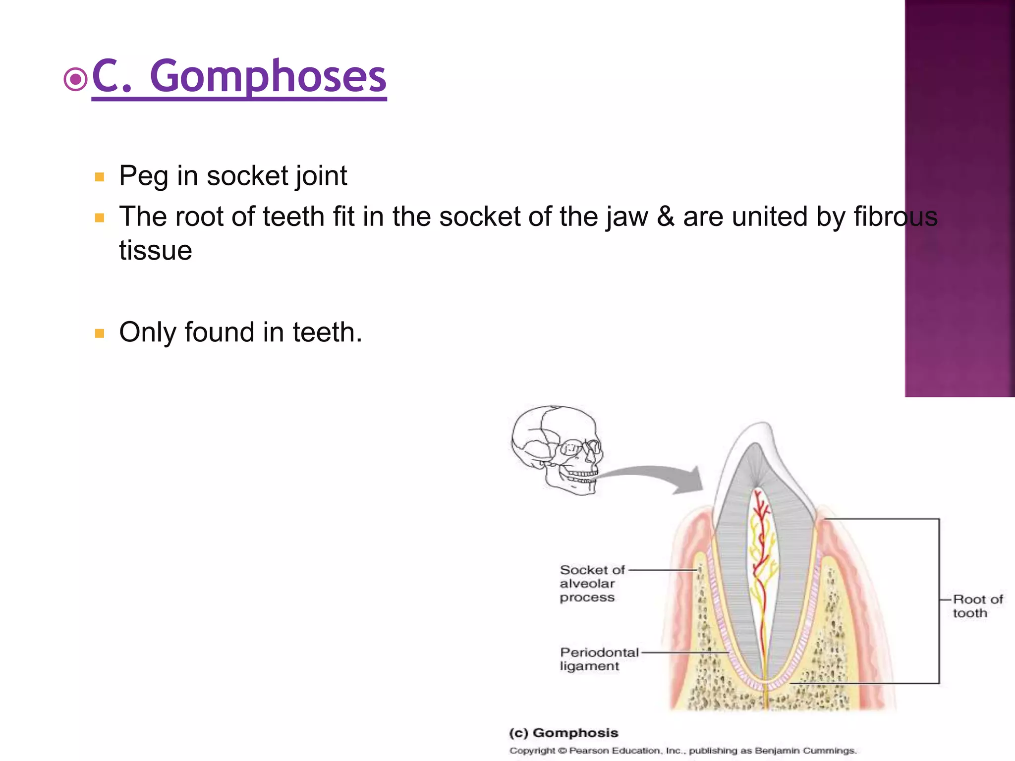
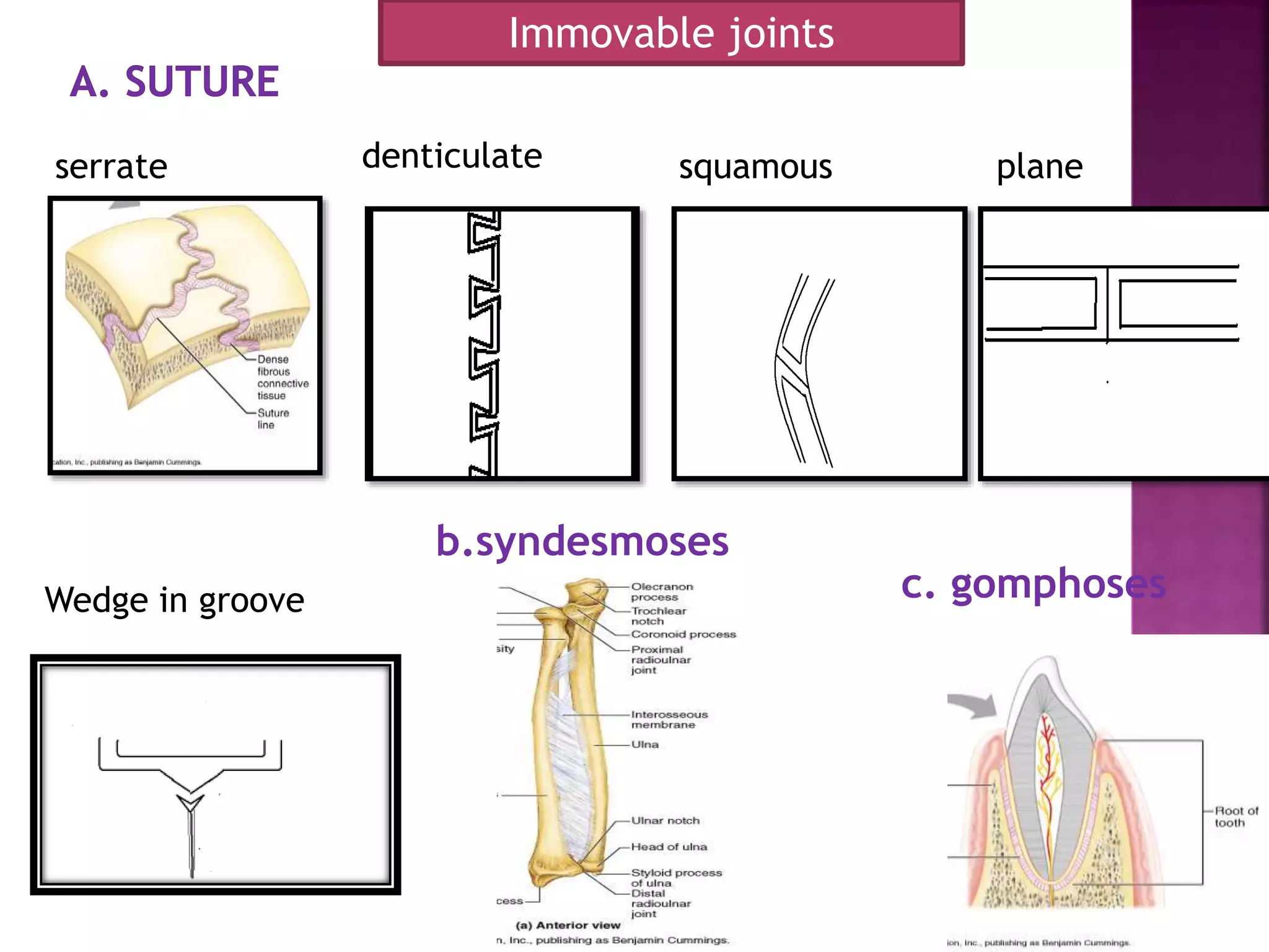
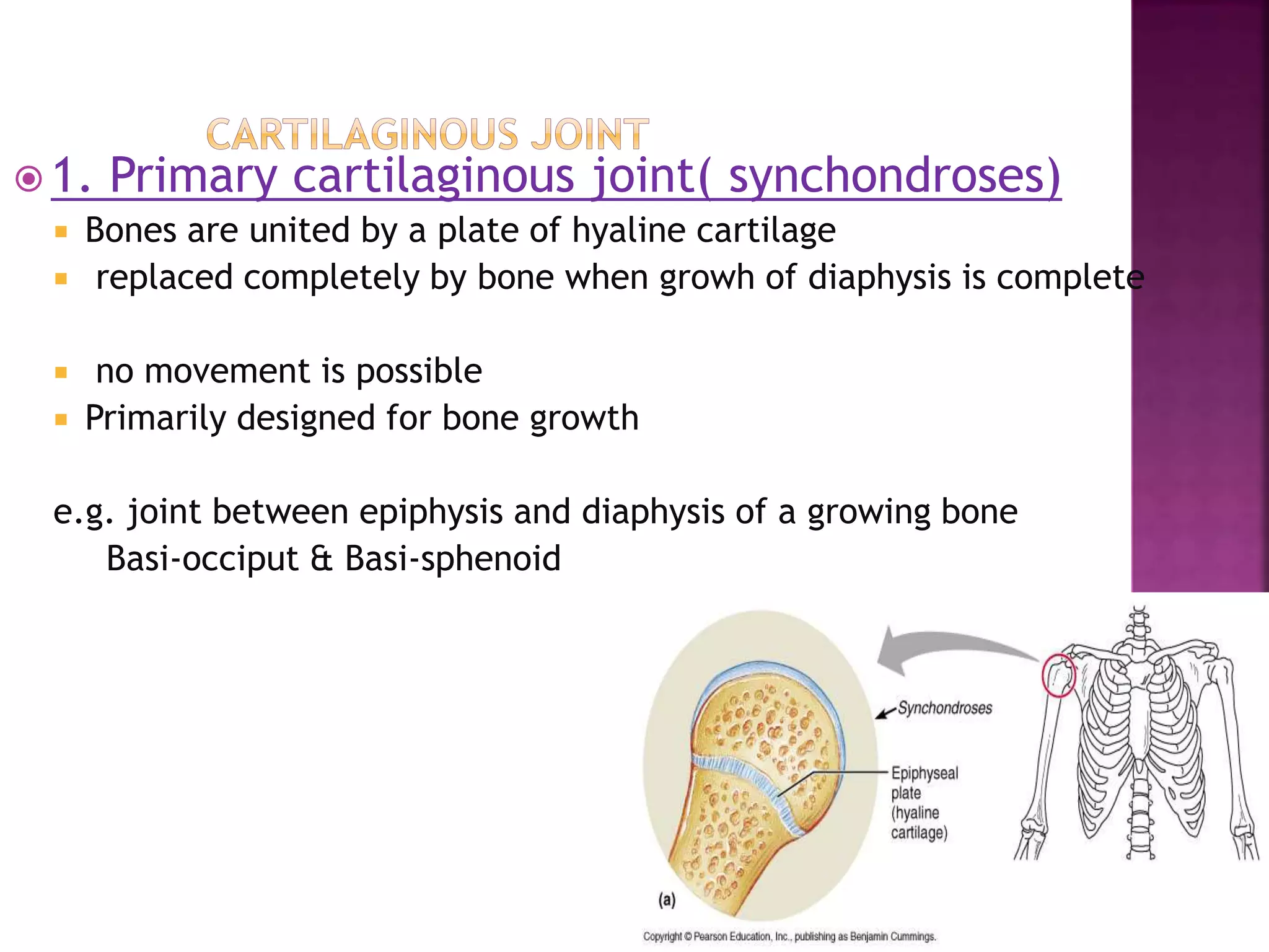
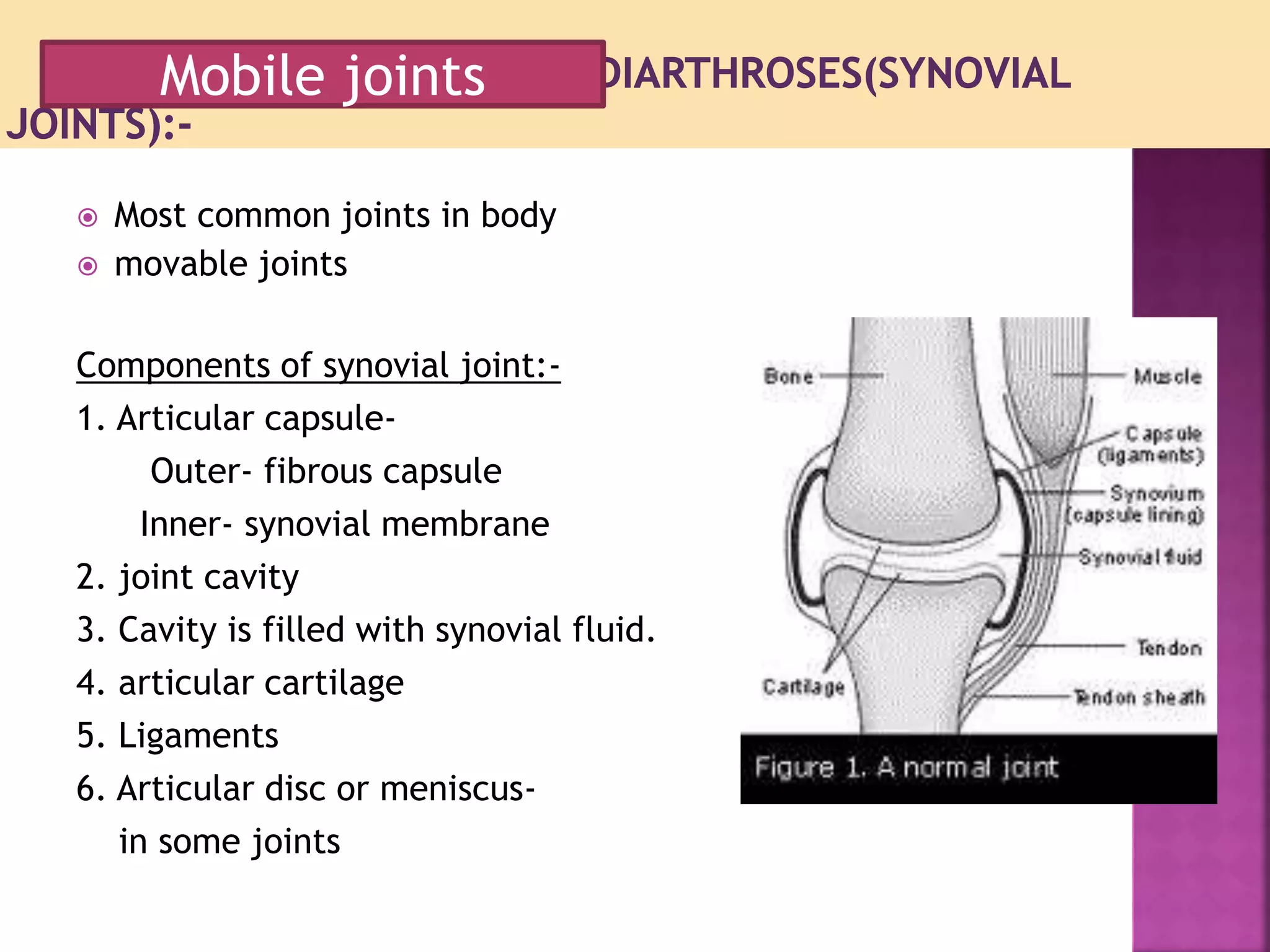
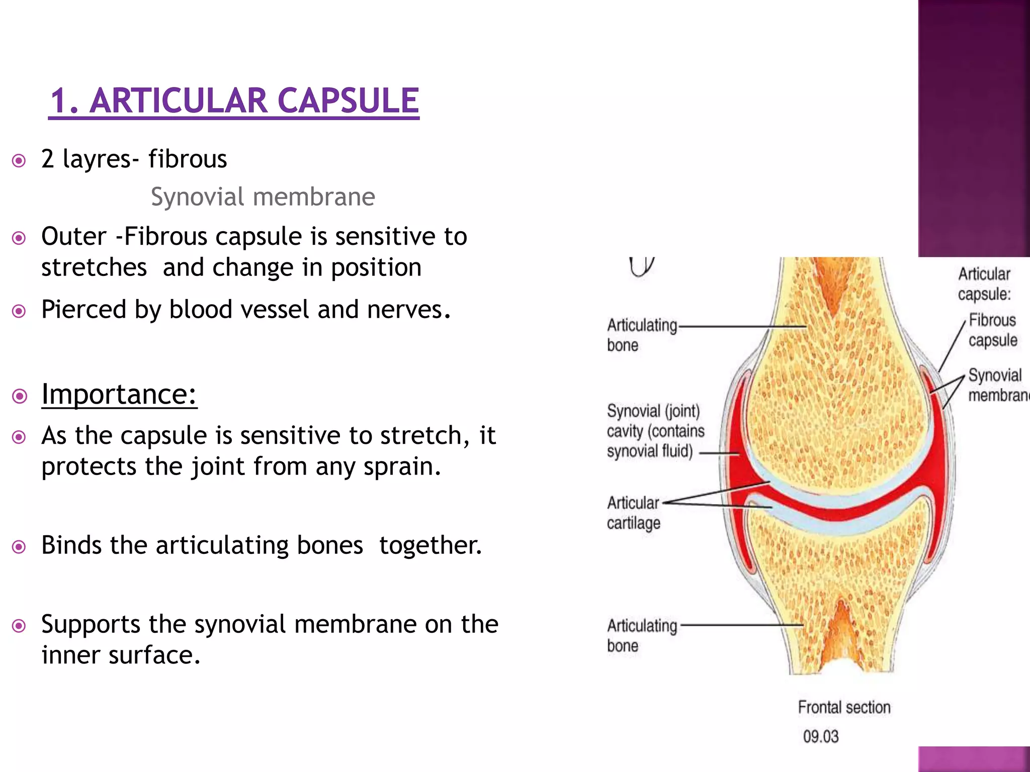
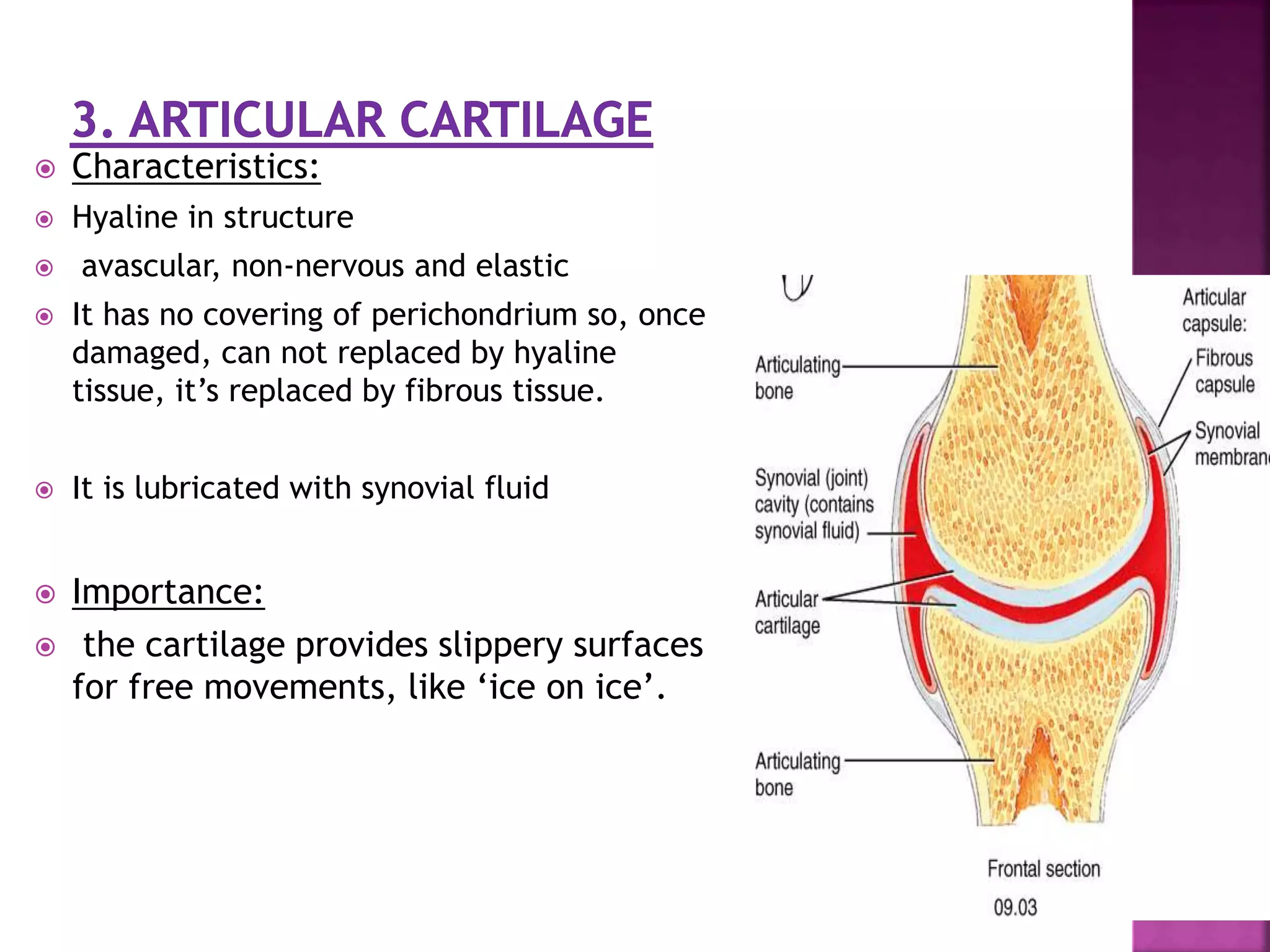
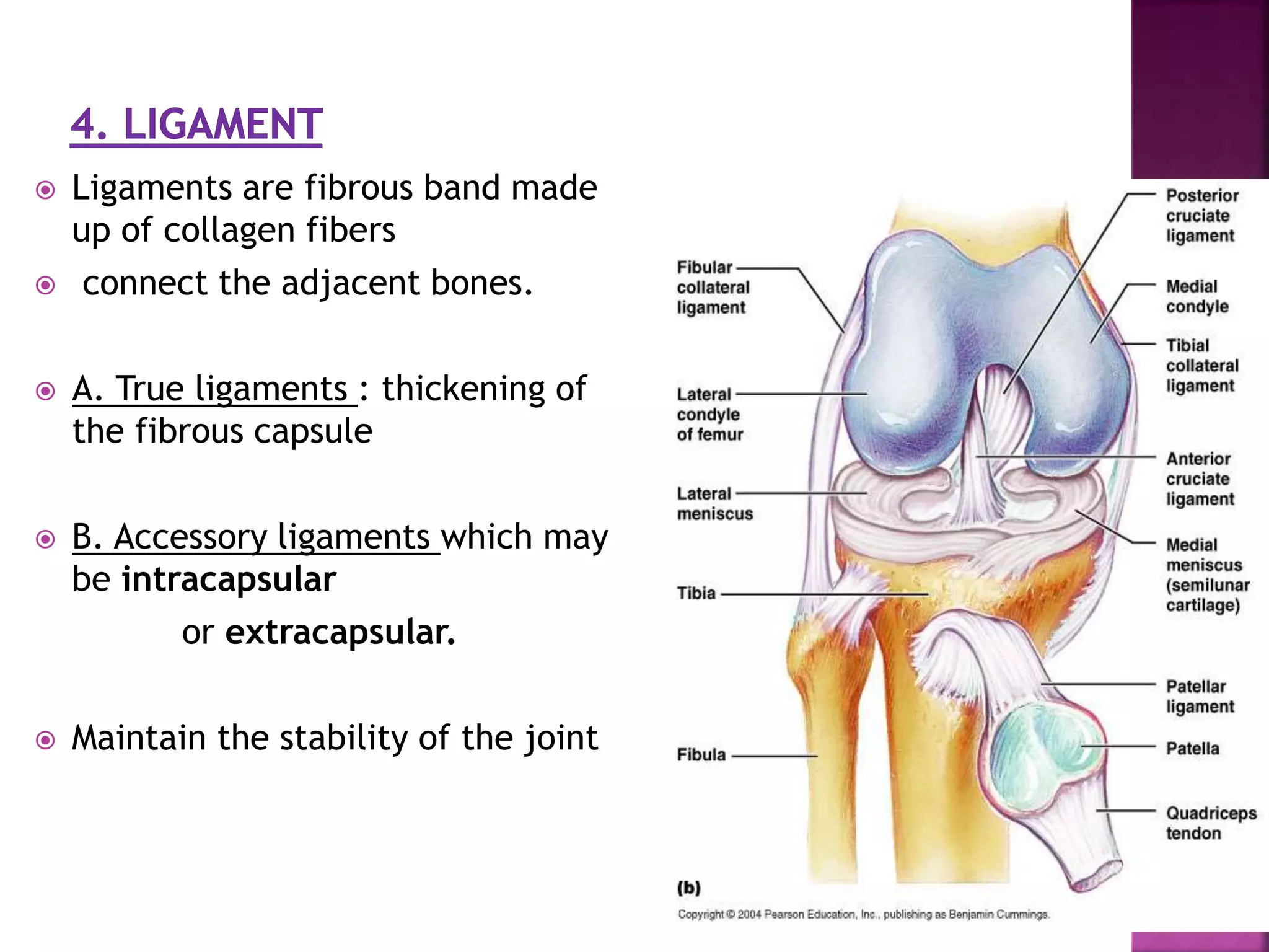
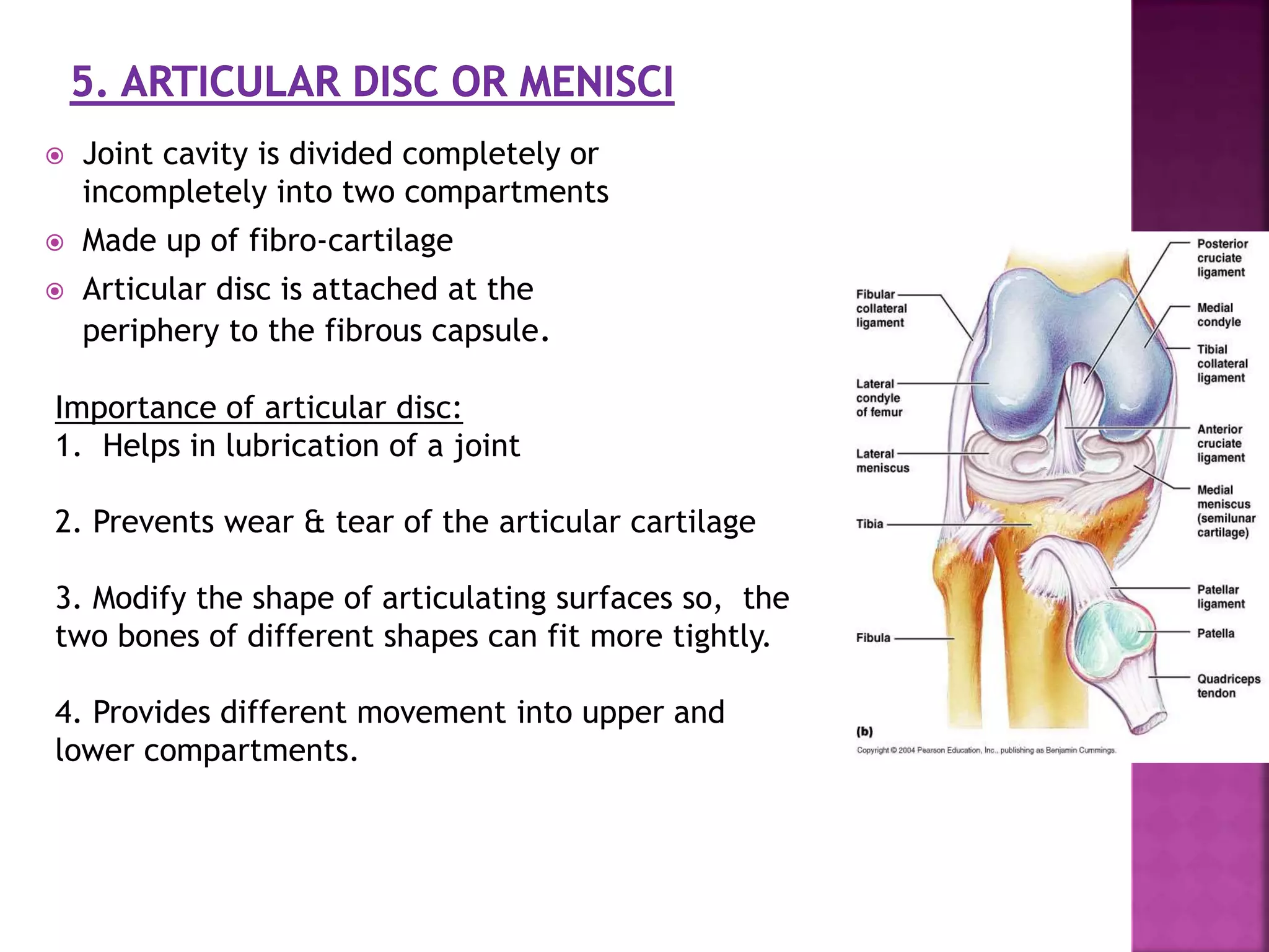
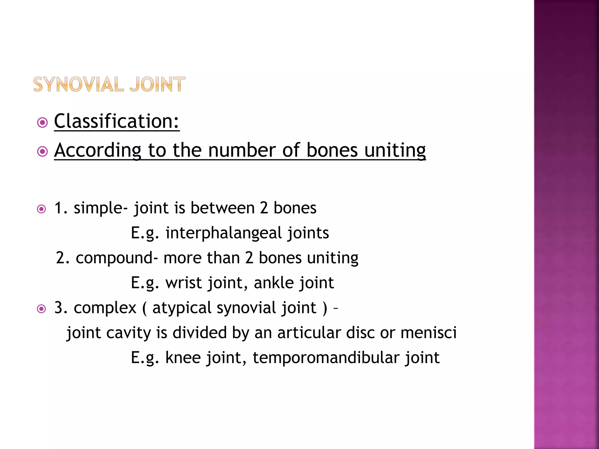
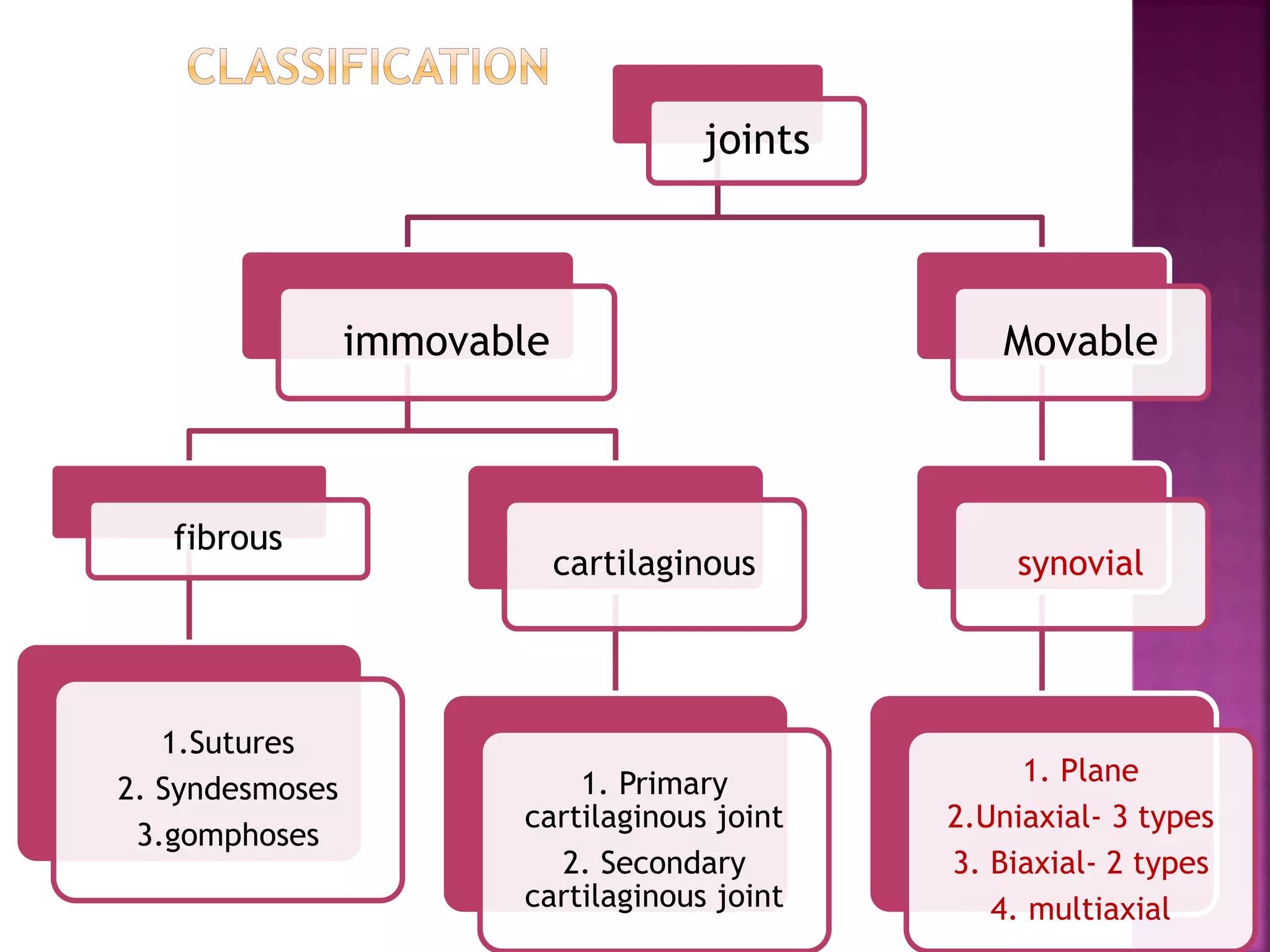
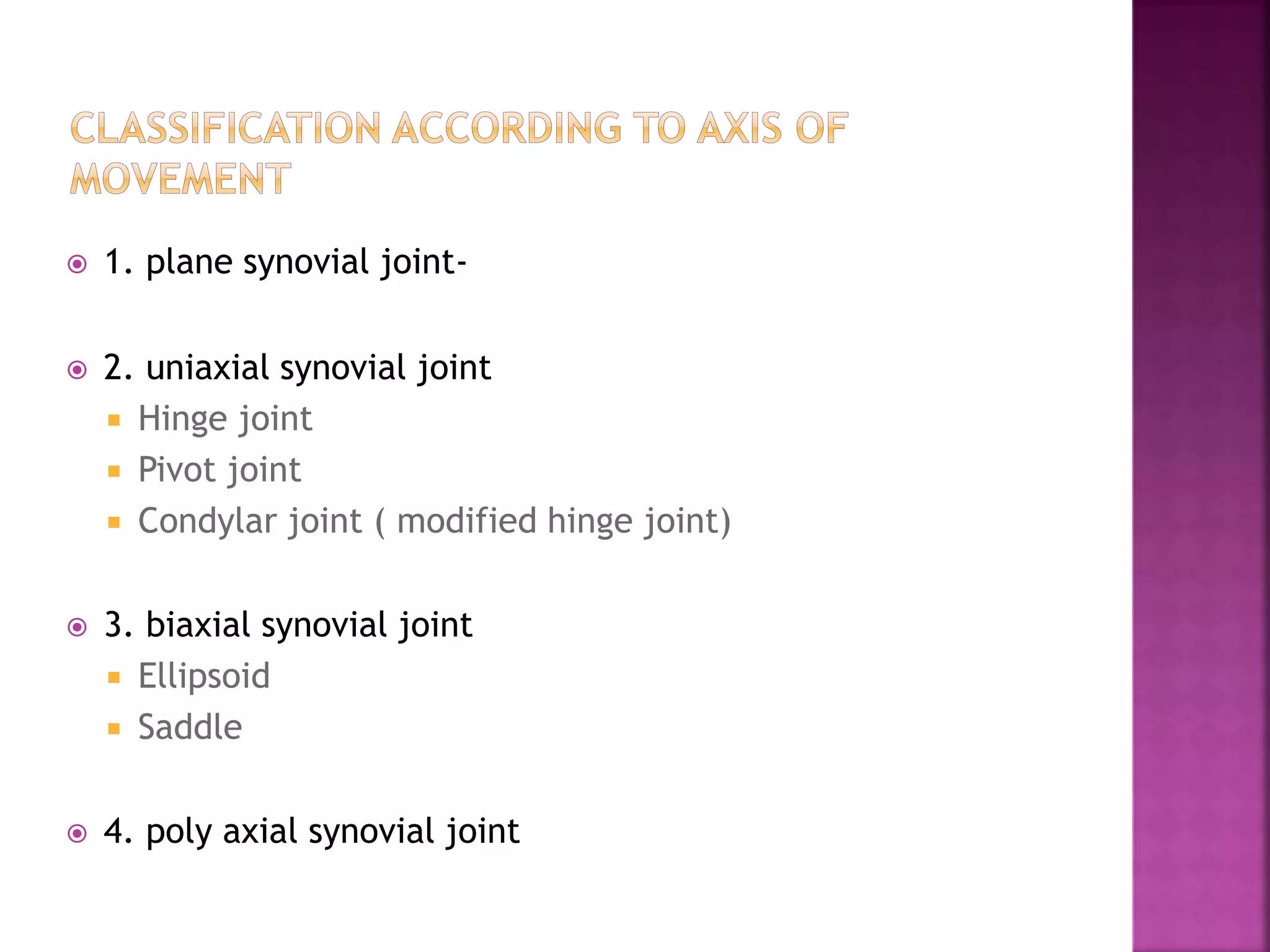
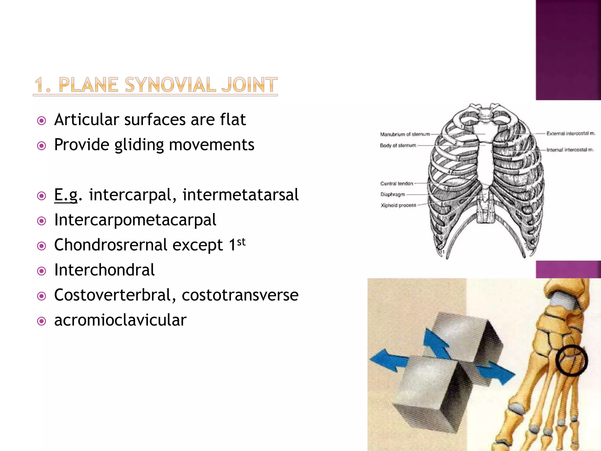
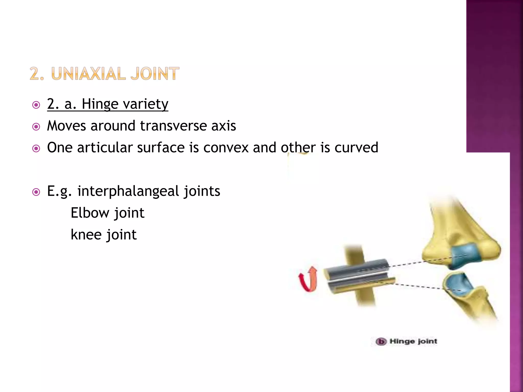
Presented by:
Mr.Manthan Kapadiya,
Assistant Professor
NCN,Visnagar
 Definition
 Introduction
 Classification-
 Immovable
 Movable
 Blood supply
 Nerve supply
 Lymphatic drainage
 Clinical notes
 Joint is a junction between two or more bones or
cartilages
 Related term: arthron
Definition
Importance:
Joints are responsible for movements
weight bearing
& growth of bones.
Clinical importance:
1. Age related degenerative changes occur e.g. arthritis
2. injury
3. Medicolegal importance
 Note:
Long bones articulate by their ends
Flat bones by their margins
Short and irregular bones by surfaces
SHOULDER JOINT
ELBOW JOINT
WRIST JOINT
HIP JOINT
KNEE JOINT
ANKLE JOINT
TEMPORO-MANDIBULAR JOINT
STERNO-CLAVICULAR JOINT
1ST CHONDRO-STERNAL JOINT
METACARPAL-
PHALANGEAL JOINT
INTERCARPAL JOINT
INFERIOR TIBIO-FIBULAR JOINT
PUBIC
SYMPHYSIS
INTERVERTEBRAL JOINT
SAGITTAL
SUTURE
joints
immovable
fibrous
1.Sutures- 5types
2. Syndesmoses
3.gomphosis
cartilaginous
1. Primary
cartilaginous joint
2. Secondary
cartilaginous joint
Movable
synovial
1. Plane
2.Uniaxial- 3 types
3. Biaxial- 2 types
4. multiaxial
 A. Fibrous joints
 In these joints bones are united by fibrous tissue.
 3 types:
a. suture-
1.serrate suture
2.denticulate suture
3.squamous suture
4.plane suture
5.wedge in groove suture
b.Syndesmoses
c. gomphosis
Immovable joints
 Most of the joints of the skull are suture joints.
 Here, ligament or suture membrane connect the
periosteum
 Types: 5
1. Serrate suture : The edge of bones present saw tooth
appearance.
e.g.- sagittal suture of the skull
2. Denticulate suture :
The margins present teeth like appearance .
Tips are broader than the roots.
e.g. lambdoid suture
BONE
SUTURAL
MEMBRANE
PERIOSTEUM
BONEBONE
SUTURAL
MEMBRANE
PERIOSTEUM
ROSTRUM OF
SPHENOID
VOMER
 Bones forming the joint lie some distance apart , surfaces are
united by interosseus ligaments.
 Slight movement is permissible
 E.g. inferior tibiofibular joint
 middle radio ulnar joint,
C. Gomphoses
 Peg in socket joint
 The root of teeth fit in the socket of the jaw & are united by fibrous
tissue
 Only found in teeth.
serrate denticulate squamous plane
Wedge in groove
b.syndesmoses
c. gomphoses
Immovable joints
joints
immovable
fibrous
1.Sutures
2. Syndesmoses
3.gomphoses
cartilaginous
1. Primary
cartilaginous joint
2. Secondary
cartilaginous joint
Movable
synovial
1. Plane
2.Uniaxial
3. Biaxial
4. multiaxial
 1. Primary cartilaginous joint( synchondroses)
 Bones are united by a plate of hyaline cartilage
 replaced completely by bone when growh of diaphysis is complete
 no movement is possible
 Primarily designed for bone growth
e.g. joint between epiphysis and diaphysis of a growing bone
Basi-occiput & Basi-sphenoid
 These joints are permanent and persist throughout the life.
 Here, joint surface is covered by hyaline cartilage and united by fibro-
cartilage.
 Occupy median plane of body.
 E.g. pubic symphysis
 Intervertebral joint
Secondary cartilaginous joint
Immovable joints
Cartilaginous joints:
Synovial joints
 Most common joints in body
 movable joints
Components of synovial joint:-
1. Articular capsule-
Outer- fibrous capsule
Inner- synovial membrane
2. joint cavity
3. Cavity is filled with synovial fluid.
4. articular cartilage
5. Ligaments
6. Articular disc or meniscus-
in some joints
Mobile joints
 2 layres- fibrous
Synovial membrane
 Outer -Fibrous capsule is sensitive to
stretches and change in position
 Pierced by blood vessel and nerves.
 Importance:
 As the capsule is sensitive to stretch, it
protects the joint from any sprain.
 Binds the articulating bones together.
 Supports the synovial membrane on the
inner surface.
 2. Inner layer – synovial membrane
 lines whole of the interior the joint except the
articular surfaces .
 Highly vascular and cellular
 Connective tissue membrane
 Importance:
 The membrane secretes a synovial fluid.
 Liberates hyaluronic acid which maintains
viscosity of fluid
 Removes particulate matters
 Synovial fluid:
 It nourieses the articular cartilage
 Lubricates the joint surfaces
 Characteristics:
 Hyaline in structure
 avascular, non-nervous and elastic
 It has no covering of perichondrium so, once
damaged, can not replaced by hyaline
tissue, it’s replaced by fibrous tissue.
 It is lubricated with synovial fluid
 Importance:
 the cartilage provides slippery surfaces
for free movements, like ‘ice on ice’.
 Ligaments are fibrous band made
up of collagen fibers
 connect the adjacent bones.
 A. True ligaments : thickening of
the fibrous capsule
 B. Accessory ligaments which may
be intracapsular
or extracapsular.
 Maintain the stability of the joint
 Joint cavity is divided completely or
incompletely into two compartments
 Made up of fibro-cartilage
 Articular disc is attached at the
periphery to the fibrous capsule.
Importance of articular disc:
1. Helps in lubrication of a joint
2. Prevents wear & tear of the articular cartilage
3. Modify the shape of articulating surfaces so, the
two bones of different shapes can fit more tightly.
4. Provides different movement into upper and
lower compartments.
 1. The movable joint is-
 A. serrate suture
 B. denticulate suture
 C. synovial joint
 D. cartilaginous joint
 A. serrate suture
 B. denticulate suture
 C. plane suture
 D. primary cartilaginous joint
 A. primary cartilagionus joint
 B. secondary cartilaginous joint
 c. synovial joint
 D. none of the above
 An articulat cartilage is avascular,
nonnervous and elastic structure.
 A. immobile
 B. possess a joint cavity
 C. covered by an articular capsule
 D. synovial fluid fills the joint cavity
 1. The movable joint is-
 A. serrate suture
 B. denticulate suture
 C. synovial joint
 D. cartilaginous joint
 A. serrate suture
 B. denticulate suture
 C. plane suture
 D. primary cartilaginous joint
 A. primary cartilagionus joint
 B. secondary cartilaginous joint
 c. synovial joint
 D. none of the above
 An articulat cartilage is avascular,
nonnervous and elastic structure.
 Ans: true
 A. immobile
 B. possess a joint cavity
 C. covered by an articular capsule
 D. synovial fluid fills the joint cavity
Dr. sanjay vikani
 Classification:
 According to the number of bones uniting
 1. simple- joint is between 2 bones
E.g. interphalangeal joints
2. compound- more than 2 bones uniting
E.g. wrist joint, ankle joint
 3. complex ( atypical synovial joint ) –
joint cavity is divided by an articular disc or menisci
E.g. knee joint, temporomandibular joint
joints
immovable
fibrous
1.Sutures
2. Syndesmoses
3.gomphoses
cartilaginous
1. Primary
cartilaginous joint
2. Secondary
cartilaginous joint
Movable
synovial
1. Plane
2.Uniaxial- 3 types
3. Biaxial- 2 types
4. multiaxial
 1. plane synovial joint-
 2. uniaxial synovial joint
 Hinge joint
 Pivot joint
 Condylar joint ( modified hinge joint)
 3. biaxial synovial joint
 Ellipsoid
 Saddle
 4. poly axial synovial joint
 Articular surfaces are flat
 Provide gliding movements
 E.g. intercarpal, intermetatarsal
 Intercarpometacarpal
 Chondrosrernal except 1st
 Interchondral
 Costoverterbral, costotransverse
 acromioclavicular
 2. a. Hinge variety
 Moves around transverse axis
 One articular surface is convex and other is curved
 E.g. interphalangeal joints
Elbow joint
knee joint
 Articular surfaces comprise a central bony pivot (peg)
surrounded by an osteroligamentous ring
 Movements are permitted in one plane around vertical axis
 E.g. superior and inferior radioulnar joint
Median atlanto axial joint
 Moves mainly around a transverse axis and partly around a
vertical axis
 Each bone consist of two distinct articular surfaces known
as condyles.
 E.g. knee joint
 temporo mandibular joint
 These joints have 2 degree freedom of movements.
 3.a. ellipsoid variety
 3.b. saddle variety
 3.a. ellipsoid variety:
 one articular surface is convex and elliptical ( oval ) in
outline. The other articular surface is concave and
reciprocally curved.
 Movement takes place around transverse and anterio-
posterior axes.
 Flexion, extension, adduction, abduction, circumduction
 E.g. wrist joint, atlanto-occipital joint ,
metatarsophalangeal jojint etc.
 Articular surfaces are saddle shaped and the other bone
has the reciprocally concavo- convex surface.
 E.g. sternoclavicular, calcaneo-cuboid, between femur and
patella, 1st carpo-metacarpal
 Known as ball and socket joint
 Possess 3 degree freedom of movements
 Articular surfaces include a globular head fitting into cup
shaped socket
-movement take place around transverse, antero-
posterior, vertical axis.
e.g.- shoulder joint,
& hip joint,
talo-calcaneo-navicular joint.
1. Plane synovial joint
hinge variety
Pivot variety
Condyler variety
Ellipsoid variety
Saddle variety
Ball & socket variety
Factors maintaining stability of joint:-
1. Muscles:
tone of different muscle groups maintain the joint stability,
important factor e.g. shoulder joint
2. Shape of articulating surfaces of bones:
e.g. hip joint, head of femur is completely received by acetabular
socket
3. Ligaments:
permits desirable movements and prevents undesirable movements.
1. shape of articulating bones:
disproportionate shape of glenoid cavity and head of humerus
provides wider range of moment.
2. Tension of ligament:
e.g. over-extension of hip joint is pervented by tension of
iliofemoral ligament
3. Tension of antagonist muscle
4. The approximation of soft part:
flexion at elbow joint
 At a particular phase of movement, the convex articular
surface is perfectly congruent with the reciprocal concave
surface of the other bone. This condition is known as close
packed condition.
 Here, no joint space is available for flushing of the
synovial fluid and articular capsule is maximum stretched.
 Arterial branches from several different arteries merge
around a joint, & form a rich plexus before penetrating
the articular capsule
 Articular cartilage is avascular & gets nutrition by
diffusion.
 Capsule and ligaments possess a rich nerve supply.
 3 different types-
 1. sensory nerves- convey pain sensation
Persive as a diffuse pain
 2. sensory nerve conveying proprioseptive sensations.
 3. autonomic nerves- vasomotor, vasosensory
∗ therefore, the joint pain is associated with, nausea,
vomiting, slowing of pulse and blood pressure
 The law states that the nerves supplying a joint, also suppy muscles
regulating the movements of the joint & the skin over the joint
 This mechanism of innervation helps to protect the joint.
 Lymphatic drainage:
 Form a plexus in the synovial membrane
 1. synovial fluid
 2. articular cartilage
 3. bursae
 4. sesamoid bone
 This is a condition in which the
articular surfaces of the joint are
abnormally displaced, so one surface
loses its contact completely from the
other.
 If partial contact is still retained, it is
called subluxation.
2. Osteoarthritis
• Degenerative joint disease
- aging, wear & tear
- Affects large weight bearing joints
- Patient feels lots of pain due to rubbing of bones during
movements.
 1. tearing of – menisci
 E.g. football players
 2. Intraarticular fractures- can lead to collection of blood
and broken particles in the joint cavity
• 4. Arthroscopy- examination
of joint
- By an instrument size of a
pencil
- Requires small incisions only
- Diagnostic and theraputic
purpose
- remove torn knee cartilage
 Related to weather with reduction in the temperature (
cold weather), viscosity of synovial fluid increses – leads to
joint stiffness.
a. Hip joint
b. Elbow joint
c. Lateral vertebral joint
d. Knee joint
 A. condyler
 B. plane
 C. concavo- convex
 D. none of the above
 A. arthritis
 B. joint dislocation
 C. paralysis
 D. stiffness of a joint
a. Hip joint - ball and socket variety of synovial
joint
b. Elbow joint – hinge variety of synovial joint
c. Lateral vertebral joint- plane variety of
synovial joint
d. Knee joint- condyler variety of synovial joint
 A. condyler
 B. plane
 C. concavo- convex
 D. none of the above
 A. arthritis
 B. joint dislocation
 C. paralysis
 D. stiffness of a joint
 Total knee replacement surgery
GOOD DAY

Joints anatomy and physiology

  • 1.
  • 2.
     Definition  Introduction Classification-  Immovable  Movable  Blood supply  Nerve supply  Lymphatic drainage  Clinical notes
  • 3.
     Joint isa junction between two or more bones or cartilages  Related term: arthron Definition
  • 4.
    Importance: Joints are responsiblefor movements weight bearing & growth of bones. Clinical importance: 1. Age related degenerative changes occur e.g. arthritis 2. injury 3. Medicolegal importance
  • 5.
     Note: Long bonesarticulate by their ends Flat bones by their margins Short and irregular bones by surfaces
  • 6.
    SHOULDER JOINT ELBOW JOINT WRISTJOINT HIP JOINT KNEE JOINT ANKLE JOINT TEMPORO-MANDIBULAR JOINT STERNO-CLAVICULAR JOINT 1ST CHONDRO-STERNAL JOINT METACARPAL- PHALANGEAL JOINT INTERCARPAL JOINT INFERIOR TIBIO-FIBULAR JOINT PUBIC SYMPHYSIS INTERVERTEBRAL JOINT SAGITTAL SUTURE
  • 7.
    joints immovable fibrous 1.Sutures- 5types 2. Syndesmoses 3.gomphosis cartilaginous 1.Primary cartilaginous joint 2. Secondary cartilaginous joint Movable synovial 1. Plane 2.Uniaxial- 3 types 3. Biaxial- 2 types 4. multiaxial
  • 8.
     A. Fibrousjoints  In these joints bones are united by fibrous tissue.  3 types: a. suture- 1.serrate suture 2.denticulate suture 3.squamous suture 4.plane suture 5.wedge in groove suture b.Syndesmoses c. gomphosis Immovable joints
  • 9.
     Most ofthe joints of the skull are suture joints.  Here, ligament or suture membrane connect the periosteum  Types: 5 1. Serrate suture : The edge of bones present saw tooth appearance. e.g.- sagittal suture of the skull
  • 10.
    2. Denticulate suture: The margins present teeth like appearance . Tips are broader than the roots. e.g. lambdoid suture
  • 11.
  • 12.
  • 13.
  • 14.
     Bones formingthe joint lie some distance apart , surfaces are united by interosseus ligaments.  Slight movement is permissible  E.g. inferior tibiofibular joint  middle radio ulnar joint,
  • 15.
    C. Gomphoses  Pegin socket joint  The root of teeth fit in the socket of the jaw & are united by fibrous tissue  Only found in teeth.
  • 16.
    serrate denticulate squamousplane Wedge in groove b.syndesmoses c. gomphoses Immovable joints
  • 17.
    joints immovable fibrous 1.Sutures 2. Syndesmoses 3.gomphoses cartilaginous 1. Primary cartilaginousjoint 2. Secondary cartilaginous joint Movable synovial 1. Plane 2.Uniaxial 3. Biaxial 4. multiaxial
  • 18.
     1. Primarycartilaginous joint( synchondroses)  Bones are united by a plate of hyaline cartilage  replaced completely by bone when growh of diaphysis is complete  no movement is possible  Primarily designed for bone growth e.g. joint between epiphysis and diaphysis of a growing bone Basi-occiput & Basi-sphenoid
  • 19.
     These jointsare permanent and persist throughout the life.  Here, joint surface is covered by hyaline cartilage and united by fibro- cartilage.  Occupy median plane of body.  E.g. pubic symphysis  Intervertebral joint
  • 20.
    Secondary cartilaginous joint Immovablejoints Cartilaginous joints:
  • 21.
  • 22.
     Most commonjoints in body  movable joints Components of synovial joint:- 1. Articular capsule- Outer- fibrous capsule Inner- synovial membrane 2. joint cavity 3. Cavity is filled with synovial fluid. 4. articular cartilage 5. Ligaments 6. Articular disc or meniscus- in some joints Mobile joints
  • 23.
     2 layres-fibrous Synovial membrane  Outer -Fibrous capsule is sensitive to stretches and change in position  Pierced by blood vessel and nerves.  Importance:  As the capsule is sensitive to stretch, it protects the joint from any sprain.  Binds the articulating bones together.  Supports the synovial membrane on the inner surface.
  • 24.
     2. Innerlayer – synovial membrane  lines whole of the interior the joint except the articular surfaces .  Highly vascular and cellular  Connective tissue membrane  Importance:  The membrane secretes a synovial fluid.  Liberates hyaluronic acid which maintains viscosity of fluid  Removes particulate matters
  • 25.
     Synovial fluid: It nourieses the articular cartilage  Lubricates the joint surfaces
  • 26.
     Characteristics:  Hyalinein structure  avascular, non-nervous and elastic  It has no covering of perichondrium so, once damaged, can not replaced by hyaline tissue, it’s replaced by fibrous tissue.  It is lubricated with synovial fluid  Importance:  the cartilage provides slippery surfaces for free movements, like ‘ice on ice’.
  • 27.
     Ligaments arefibrous band made up of collagen fibers  connect the adjacent bones.  A. True ligaments : thickening of the fibrous capsule  B. Accessory ligaments which may be intracapsular or extracapsular.  Maintain the stability of the joint
  • 28.
     Joint cavityis divided completely or incompletely into two compartments  Made up of fibro-cartilage  Articular disc is attached at the periphery to the fibrous capsule. Importance of articular disc: 1. Helps in lubrication of a joint 2. Prevents wear & tear of the articular cartilage 3. Modify the shape of articulating surfaces so, the two bones of different shapes can fit more tightly. 4. Provides different movement into upper and lower compartments.
  • 30.
     1. Themovable joint is-  A. serrate suture  B. denticulate suture  C. synovial joint  D. cartilaginous joint
  • 31.
     A. serratesuture  B. denticulate suture  C. plane suture  D. primary cartilaginous joint
  • 32.
     A. primarycartilagionus joint  B. secondary cartilaginous joint  c. synovial joint  D. none of the above
  • 33.
     An articulatcartilage is avascular, nonnervous and elastic structure.
  • 34.
     A. immobile B. possess a joint cavity  C. covered by an articular capsule  D. synovial fluid fills the joint cavity
  • 35.
     1. Themovable joint is-  A. serrate suture  B. denticulate suture  C. synovial joint  D. cartilaginous joint
  • 36.
     A. serratesuture  B. denticulate suture  C. plane suture  D. primary cartilaginous joint
  • 37.
     A. primarycartilagionus joint  B. secondary cartilaginous joint  c. synovial joint  D. none of the above
  • 38.
     An articulatcartilage is avascular, nonnervous and elastic structure.  Ans: true
  • 39.
     A. immobile B. possess a joint cavity  C. covered by an articular capsule  D. synovial fluid fills the joint cavity
  • 42.
  • 44.
     Classification:  Accordingto the number of bones uniting  1. simple- joint is between 2 bones E.g. interphalangeal joints 2. compound- more than 2 bones uniting E.g. wrist joint, ankle joint  3. complex ( atypical synovial joint ) – joint cavity is divided by an articular disc or menisci E.g. knee joint, temporomandibular joint
  • 45.
    joints immovable fibrous 1.Sutures 2. Syndesmoses 3.gomphoses cartilaginous 1. Primary cartilaginousjoint 2. Secondary cartilaginous joint Movable synovial 1. Plane 2.Uniaxial- 3 types 3. Biaxial- 2 types 4. multiaxial
  • 46.
     1. planesynovial joint-  2. uniaxial synovial joint  Hinge joint  Pivot joint  Condylar joint ( modified hinge joint)  3. biaxial synovial joint  Ellipsoid  Saddle  4. poly axial synovial joint
  • 47.
     Articular surfacesare flat  Provide gliding movements  E.g. intercarpal, intermetatarsal  Intercarpometacarpal  Chondrosrernal except 1st  Interchondral  Costoverterbral, costotransverse  acromioclavicular
  • 49.
     2. a.Hinge variety  Moves around transverse axis  One articular surface is convex and other is curved  E.g. interphalangeal joints Elbow joint knee joint
  • 50.
     Articular surfacescomprise a central bony pivot (peg) surrounded by an osteroligamentous ring  Movements are permitted in one plane around vertical axis  E.g. superior and inferior radioulnar joint Median atlanto axial joint
  • 51.
     Moves mainlyaround a transverse axis and partly around a vertical axis  Each bone consist of two distinct articular surfaces known as condyles.  E.g. knee joint  temporo mandibular joint
  • 52.
     These jointshave 2 degree freedom of movements.  3.a. ellipsoid variety  3.b. saddle variety  3.a. ellipsoid variety:  one articular surface is convex and elliptical ( oval ) in outline. The other articular surface is concave and reciprocally curved.  Movement takes place around transverse and anterio- posterior axes.  Flexion, extension, adduction, abduction, circumduction  E.g. wrist joint, atlanto-occipital joint , metatarsophalangeal jojint etc.
  • 53.
     Articular surfacesare saddle shaped and the other bone has the reciprocally concavo- convex surface.  E.g. sternoclavicular, calcaneo-cuboid, between femur and patella, 1st carpo-metacarpal
  • 54.
     Known asball and socket joint  Possess 3 degree freedom of movements  Articular surfaces include a globular head fitting into cup shaped socket -movement take place around transverse, antero- posterior, vertical axis. e.g.- shoulder joint, & hip joint, talo-calcaneo-navicular joint.
  • 56.
    1. Plane synovialjoint hinge variety Pivot variety Condyler variety Ellipsoid variety Saddle variety Ball & socket variety
  • 58.
    Factors maintaining stabilityof joint:- 1. Muscles: tone of different muscle groups maintain the joint stability, important factor e.g. shoulder joint 2. Shape of articulating surfaces of bones: e.g. hip joint, head of femur is completely received by acetabular socket 3. Ligaments: permits desirable movements and prevents undesirable movements.
  • 59.
    1. shape ofarticulating bones: disproportionate shape of glenoid cavity and head of humerus provides wider range of moment. 2. Tension of ligament: e.g. over-extension of hip joint is pervented by tension of iliofemoral ligament 3. Tension of antagonist muscle 4. The approximation of soft part: flexion at elbow joint
  • 60.
     At aparticular phase of movement, the convex articular surface is perfectly congruent with the reciprocal concave surface of the other bone. This condition is known as close packed condition.  Here, no joint space is available for flushing of the synovial fluid and articular capsule is maximum stretched.
  • 61.
     Arterial branchesfrom several different arteries merge around a joint, & form a rich plexus before penetrating the articular capsule  Articular cartilage is avascular & gets nutrition by diffusion.
  • 62.
     Capsule andligaments possess a rich nerve supply.  3 different types-  1. sensory nerves- convey pain sensation Persive as a diffuse pain  2. sensory nerve conveying proprioseptive sensations.  3. autonomic nerves- vasomotor, vasosensory ∗ therefore, the joint pain is associated with, nausea, vomiting, slowing of pulse and blood pressure
  • 63.
     The lawstates that the nerves supplying a joint, also suppy muscles regulating the movements of the joint & the skin over the joint  This mechanism of innervation helps to protect the joint.  Lymphatic drainage:  Form a plexus in the synovial membrane
  • 64.
     1. synovialfluid  2. articular cartilage  3. bursae  4. sesamoid bone
  • 65.
     This isa condition in which the articular surfaces of the joint are abnormally displaced, so one surface loses its contact completely from the other.  If partial contact is still retained, it is called subluxation.
  • 66.
    2. Osteoarthritis • Degenerativejoint disease - aging, wear & tear - Affects large weight bearing joints - Patient feels lots of pain due to rubbing of bones during movements.
  • 67.
     1. tearingof – menisci  E.g. football players  2. Intraarticular fractures- can lead to collection of blood and broken particles in the joint cavity
  • 68.
    • 4. Arthroscopy-examination of joint - By an instrument size of a pencil - Requires small incisions only - Diagnostic and theraputic purpose - remove torn knee cartilage
  • 71.
     Related toweather with reduction in the temperature ( cold weather), viscosity of synovial fluid increses – leads to joint stiffness.
  • 73.
    a. Hip joint b.Elbow joint c. Lateral vertebral joint d. Knee joint
  • 74.
     A. condyler B. plane  C. concavo- convex  D. none of the above
  • 75.
     A. arthritis B. joint dislocation  C. paralysis  D. stiffness of a joint
  • 79.
    a. Hip joint- ball and socket variety of synovial joint b. Elbow joint – hinge variety of synovial joint c. Lateral vertebral joint- plane variety of synovial joint d. Knee joint- condyler variety of synovial joint
  • 80.
     A. condyler B. plane  C. concavo- convex  D. none of the above
  • 81.
     A. arthritis B. joint dislocation  C. paralysis  D. stiffness of a joint
  • 83.
     Total kneereplacement surgery
  • 84.