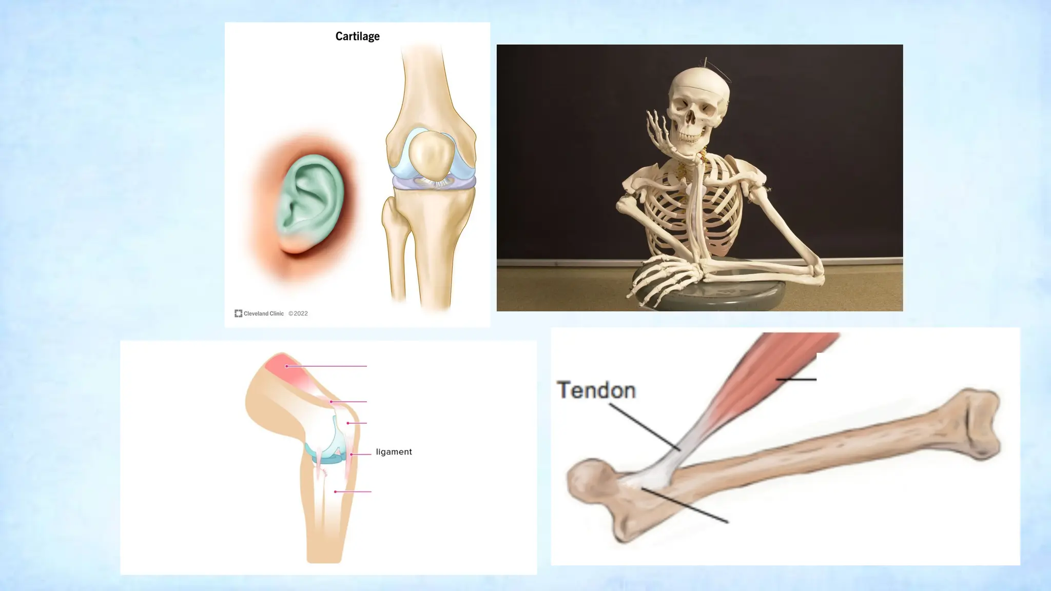
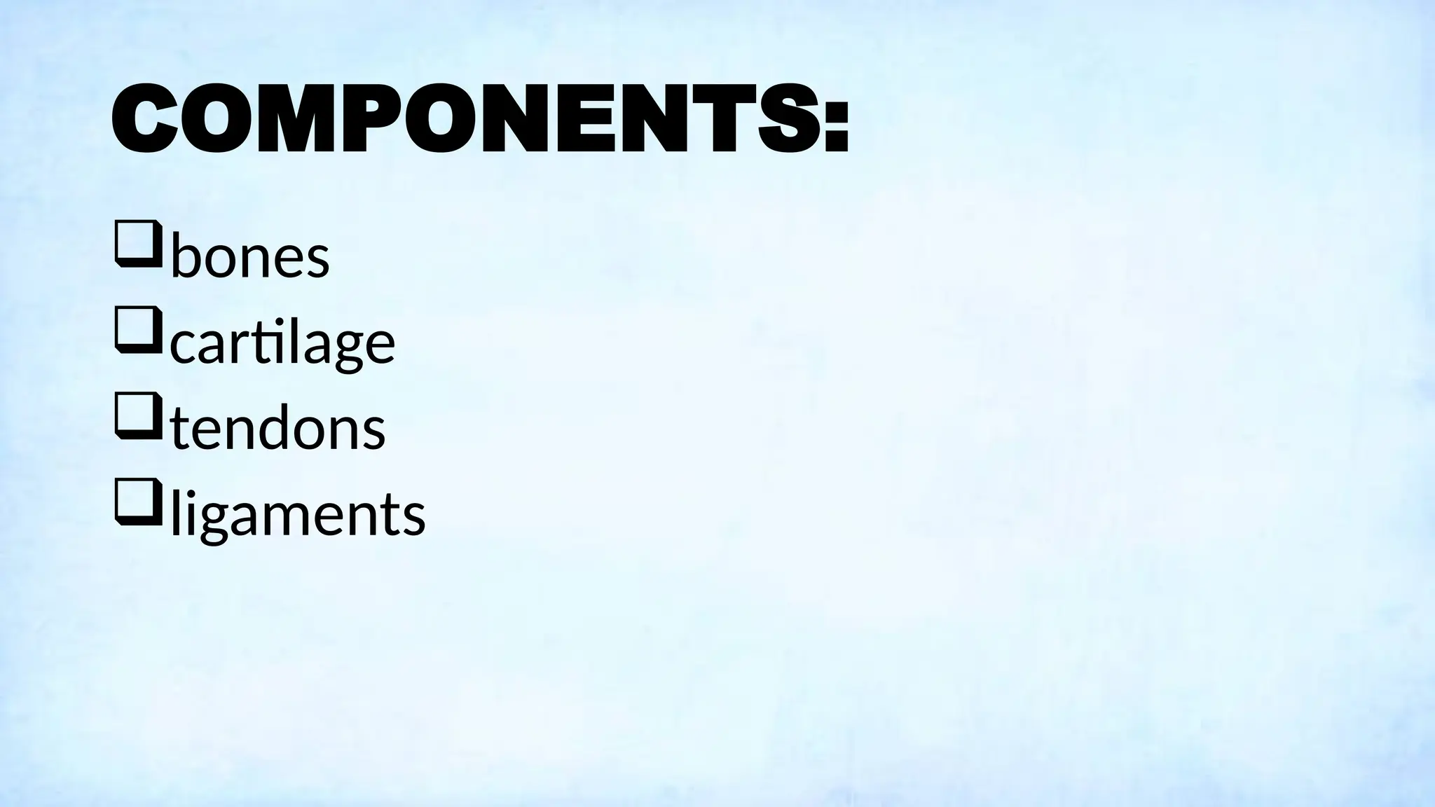
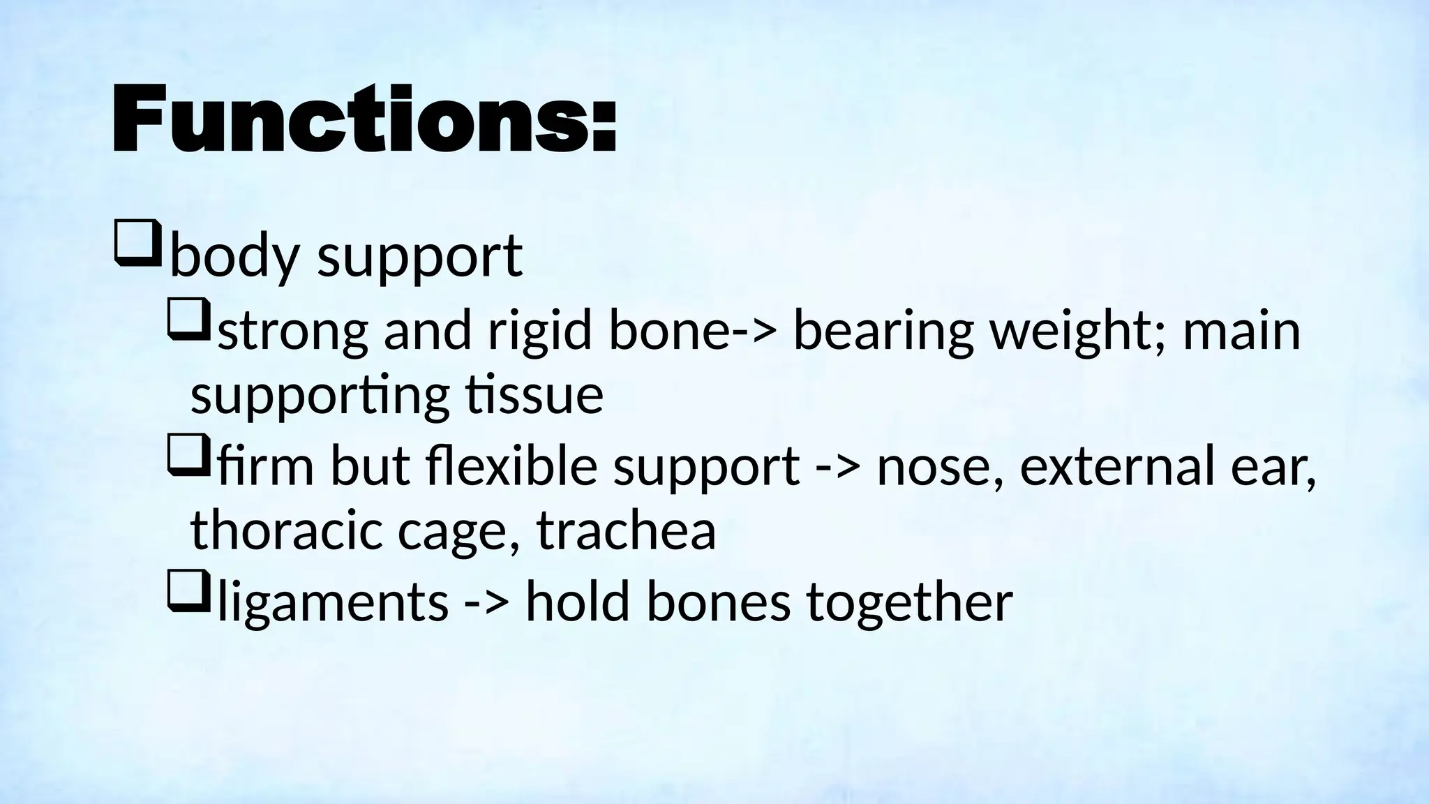
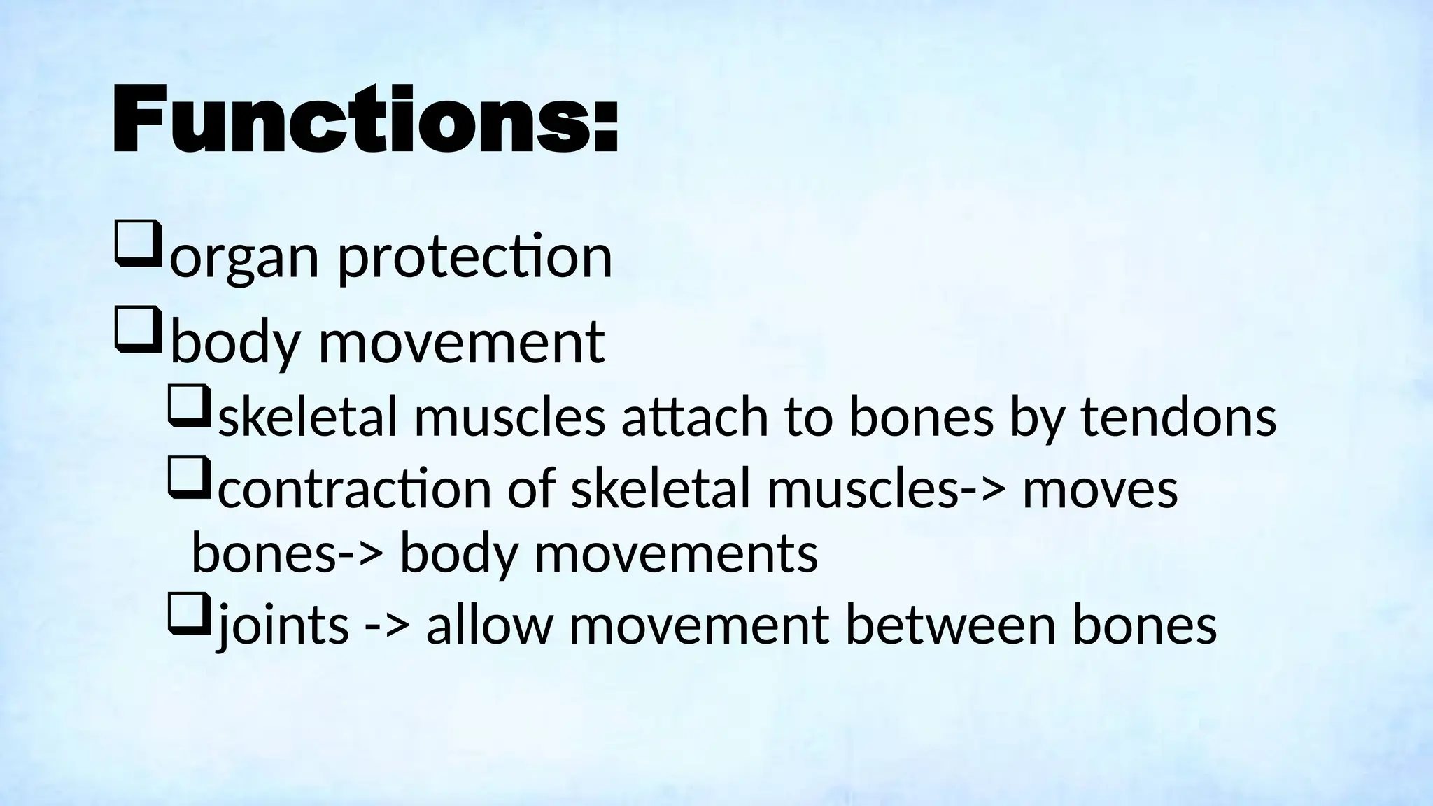
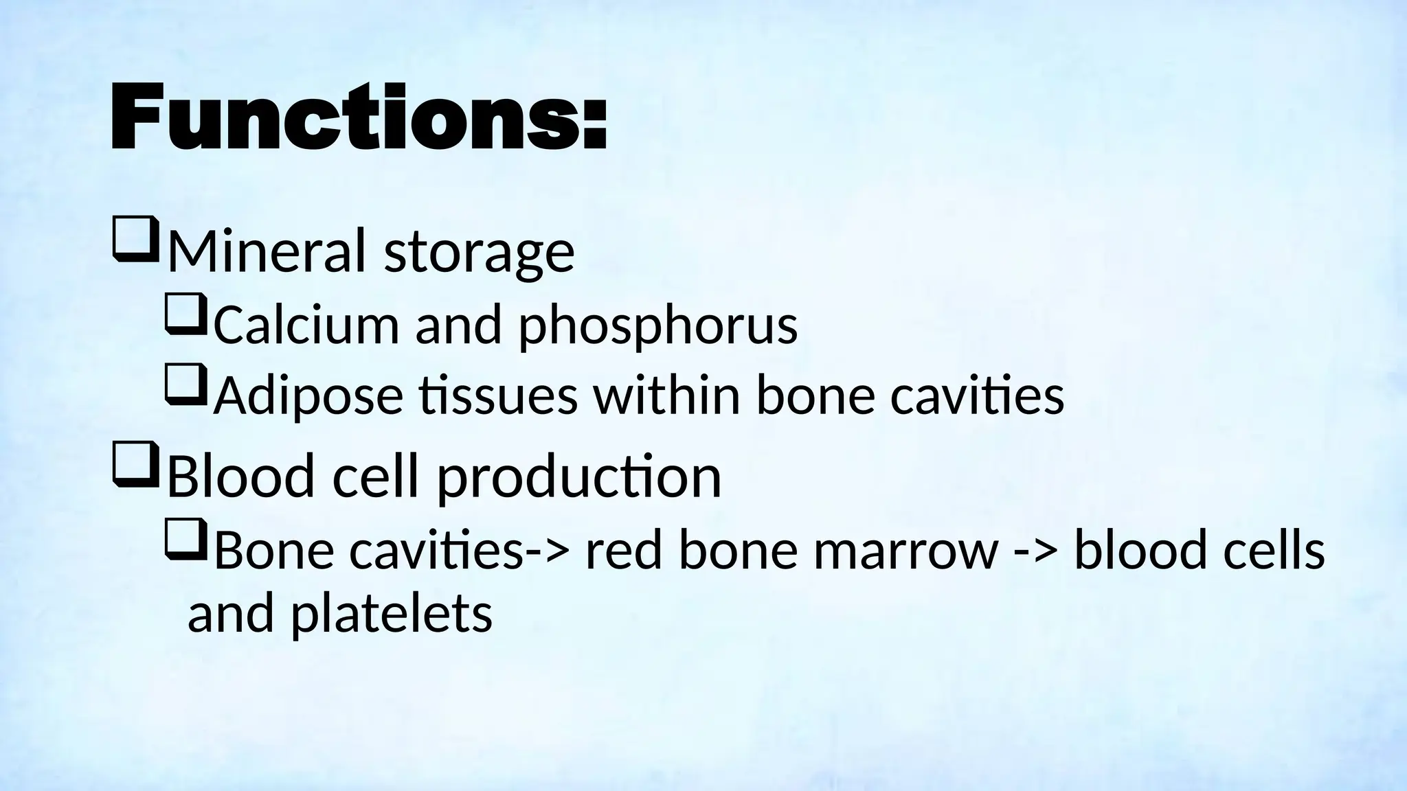
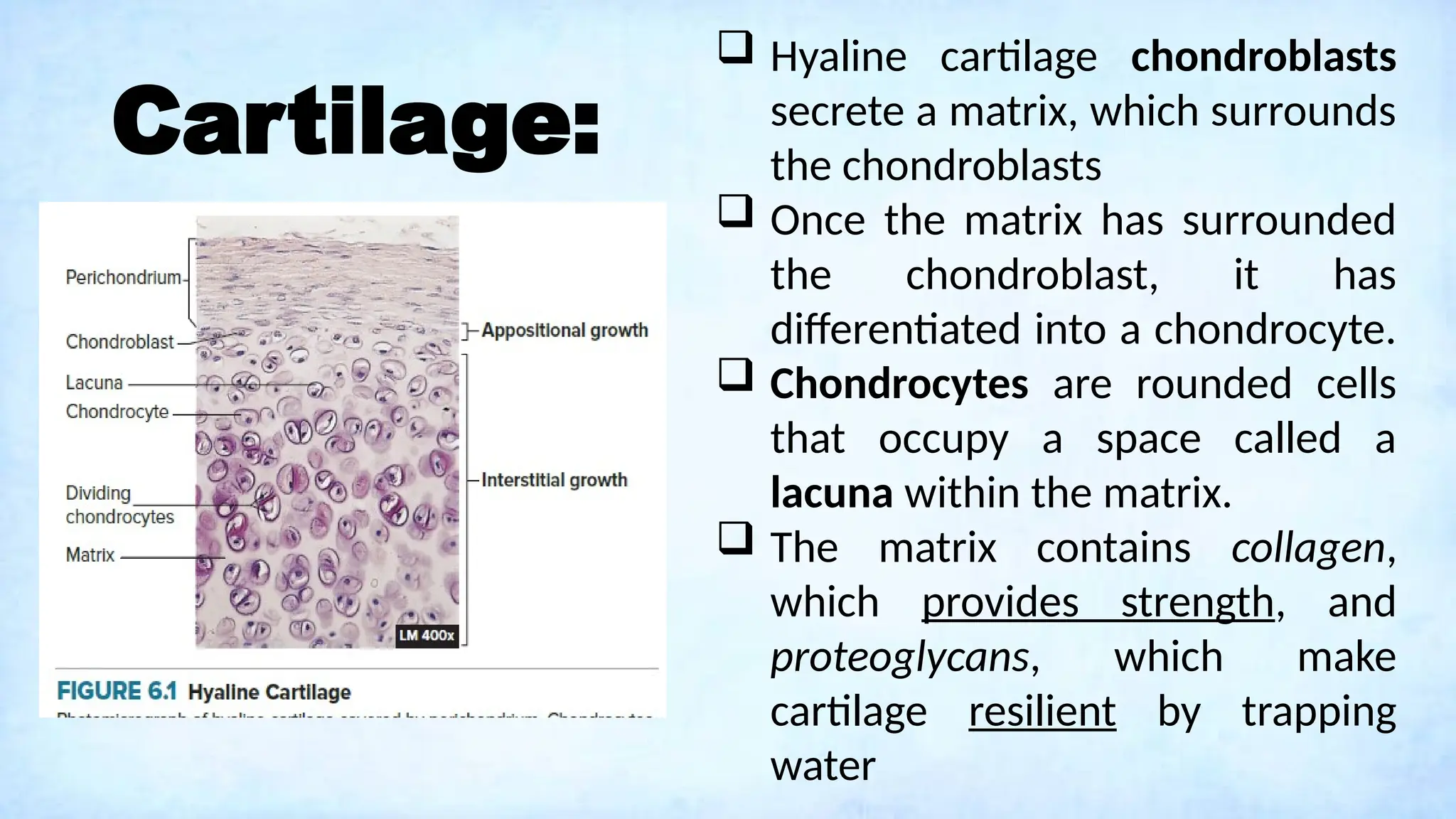
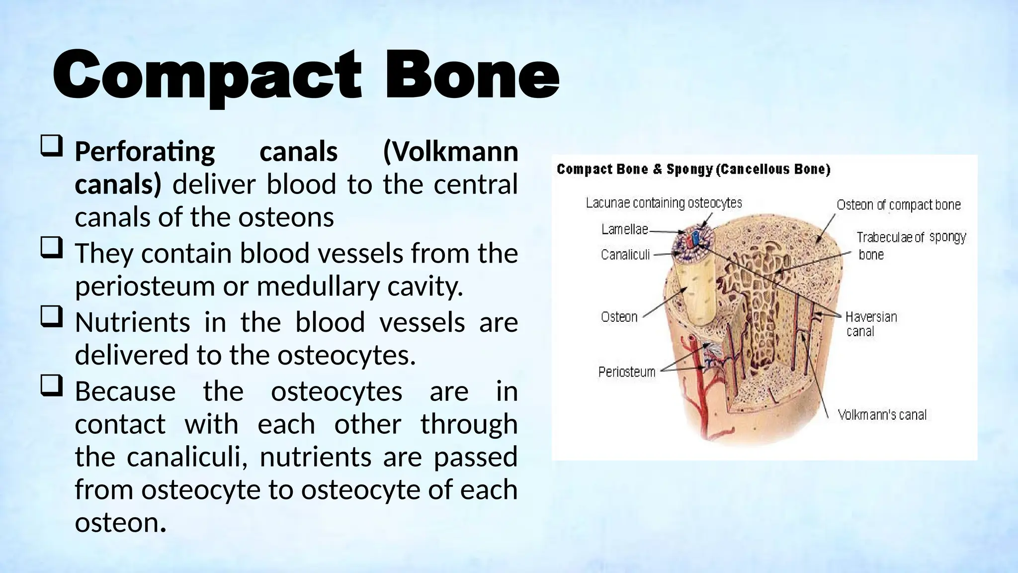
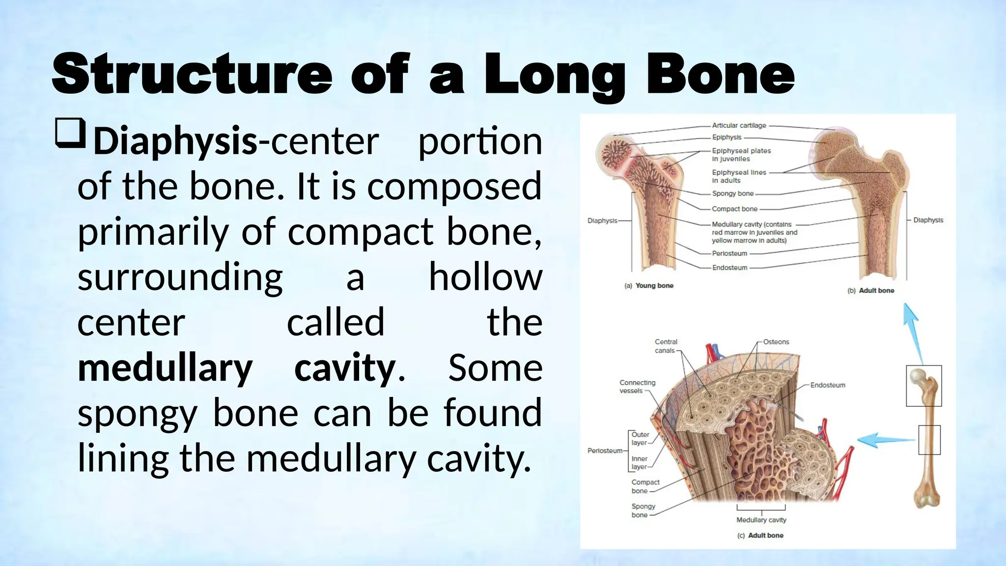
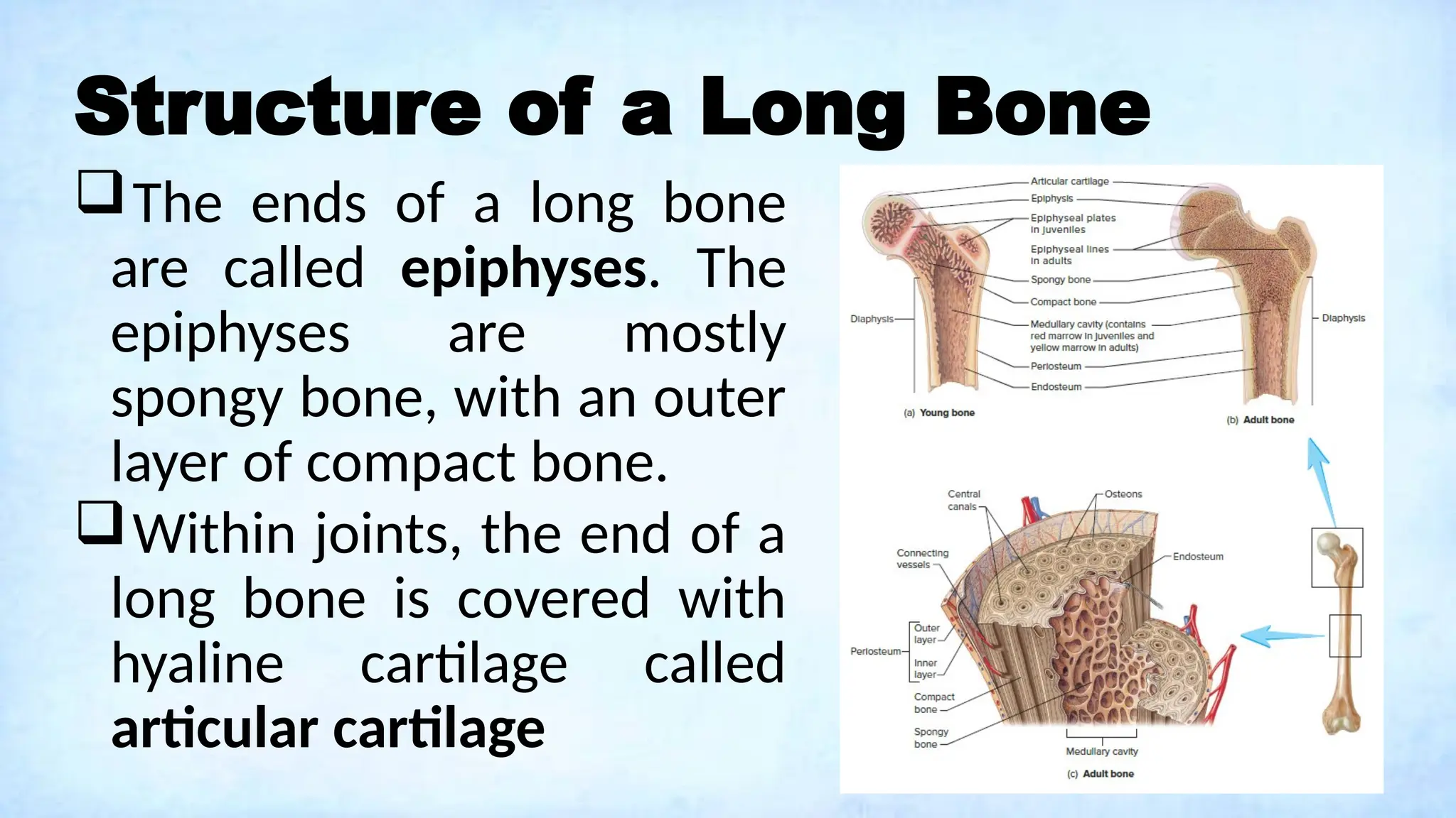
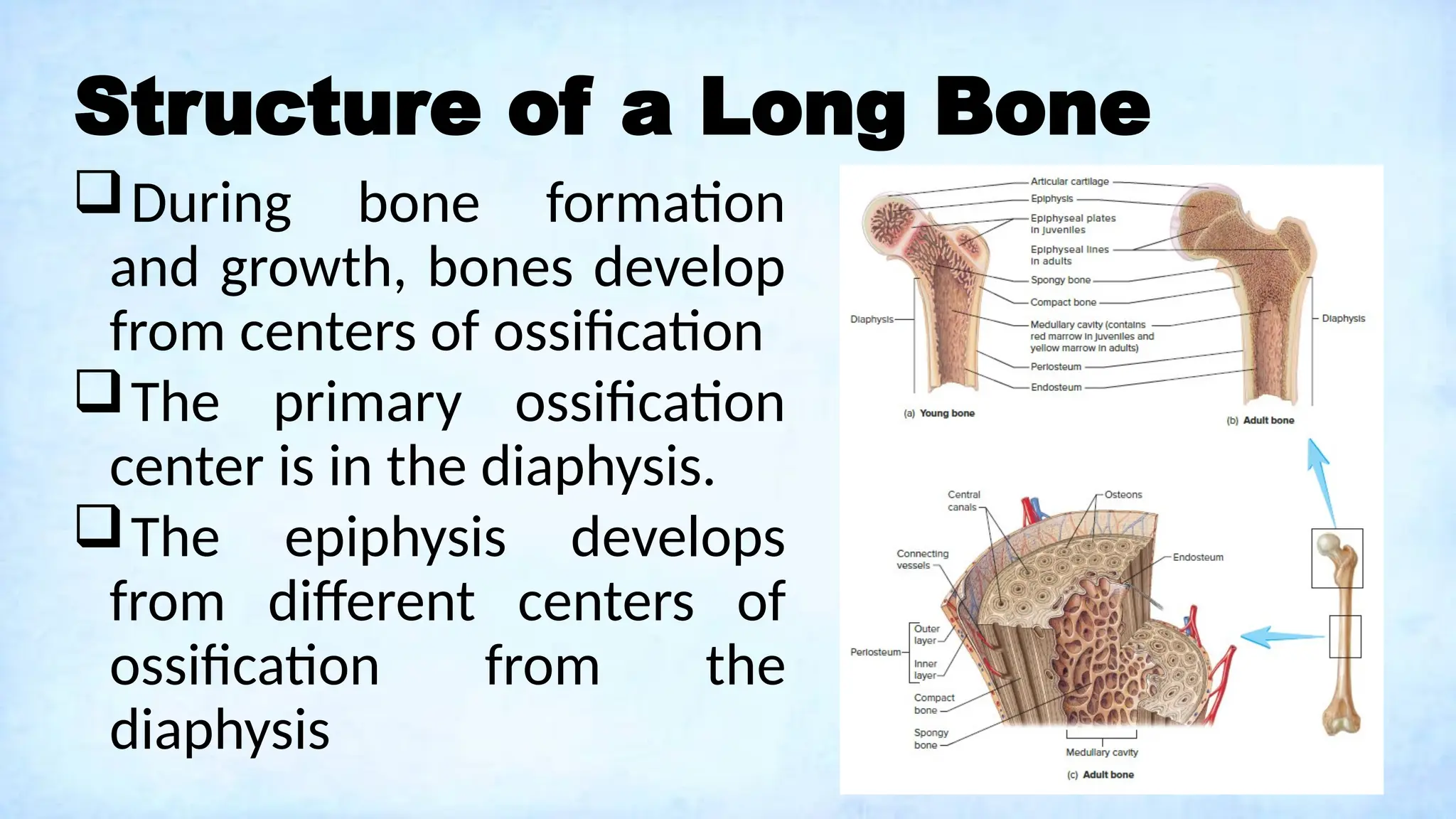
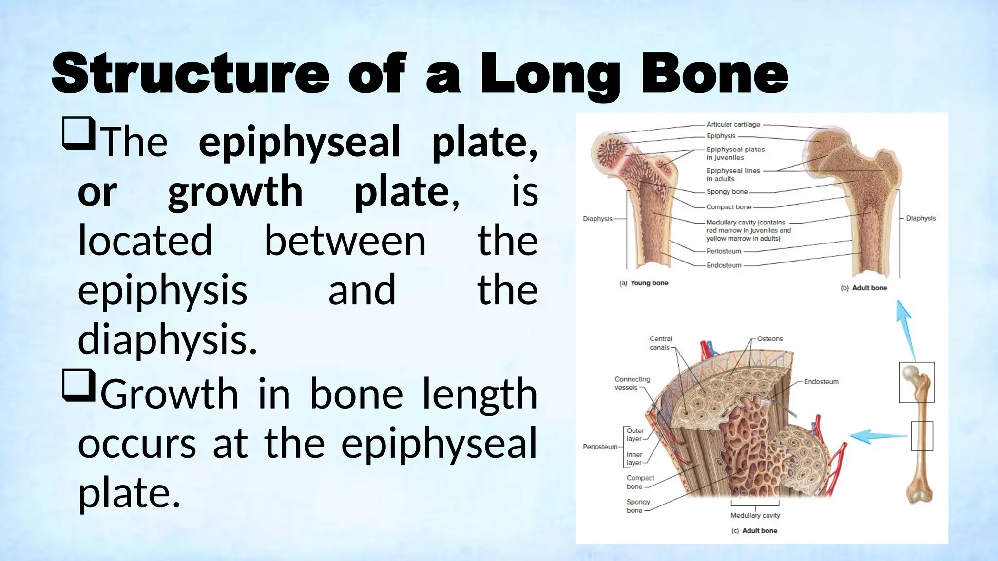
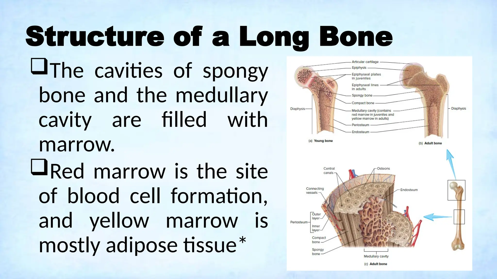
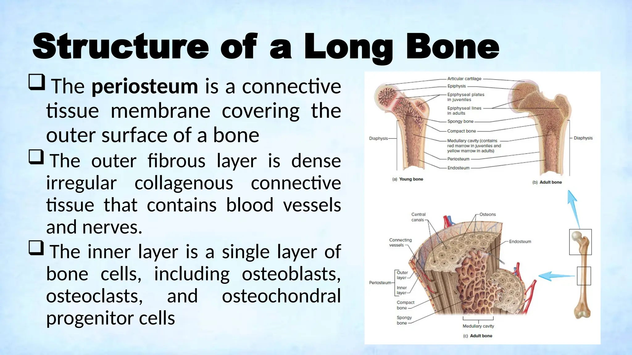
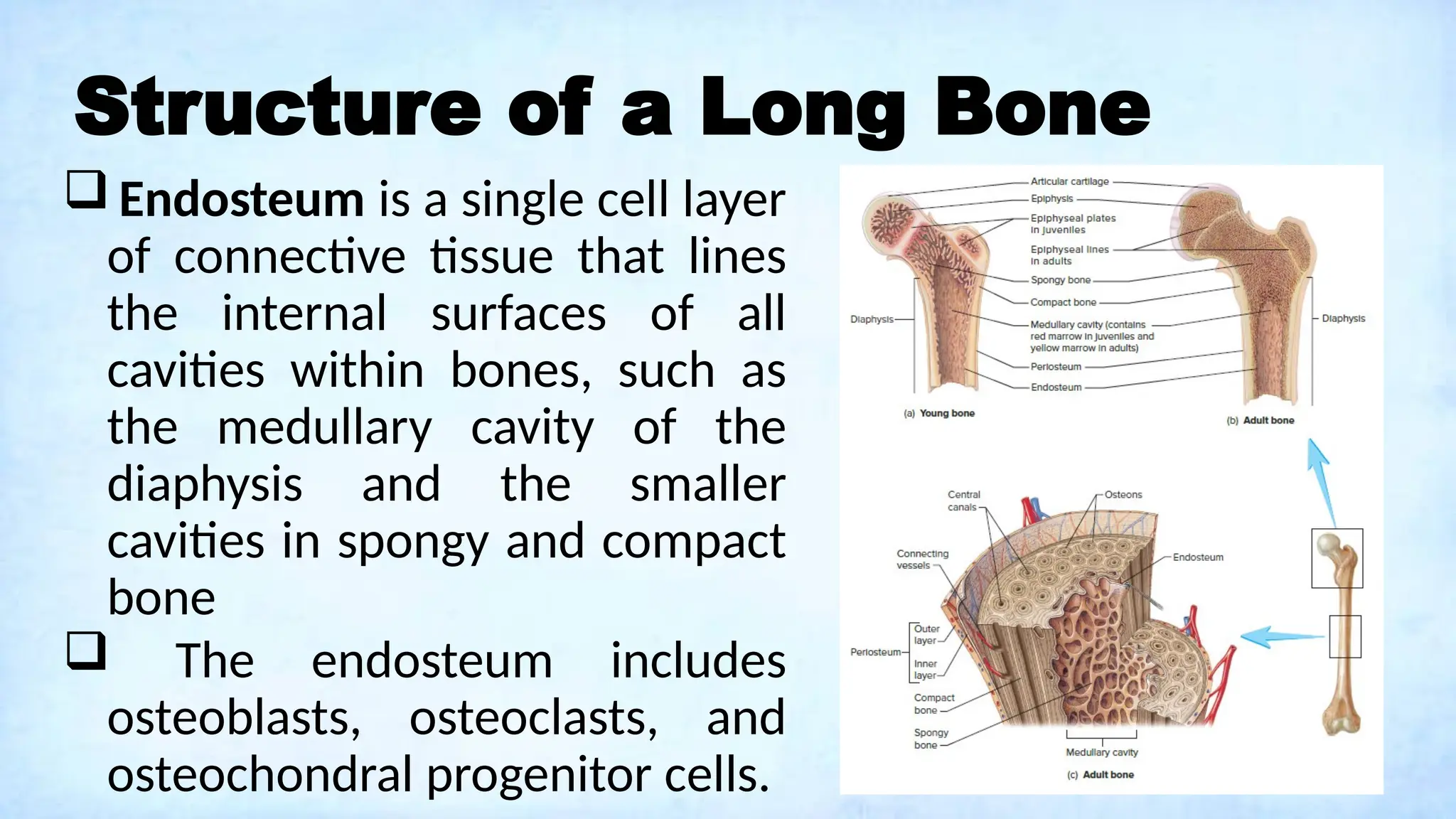
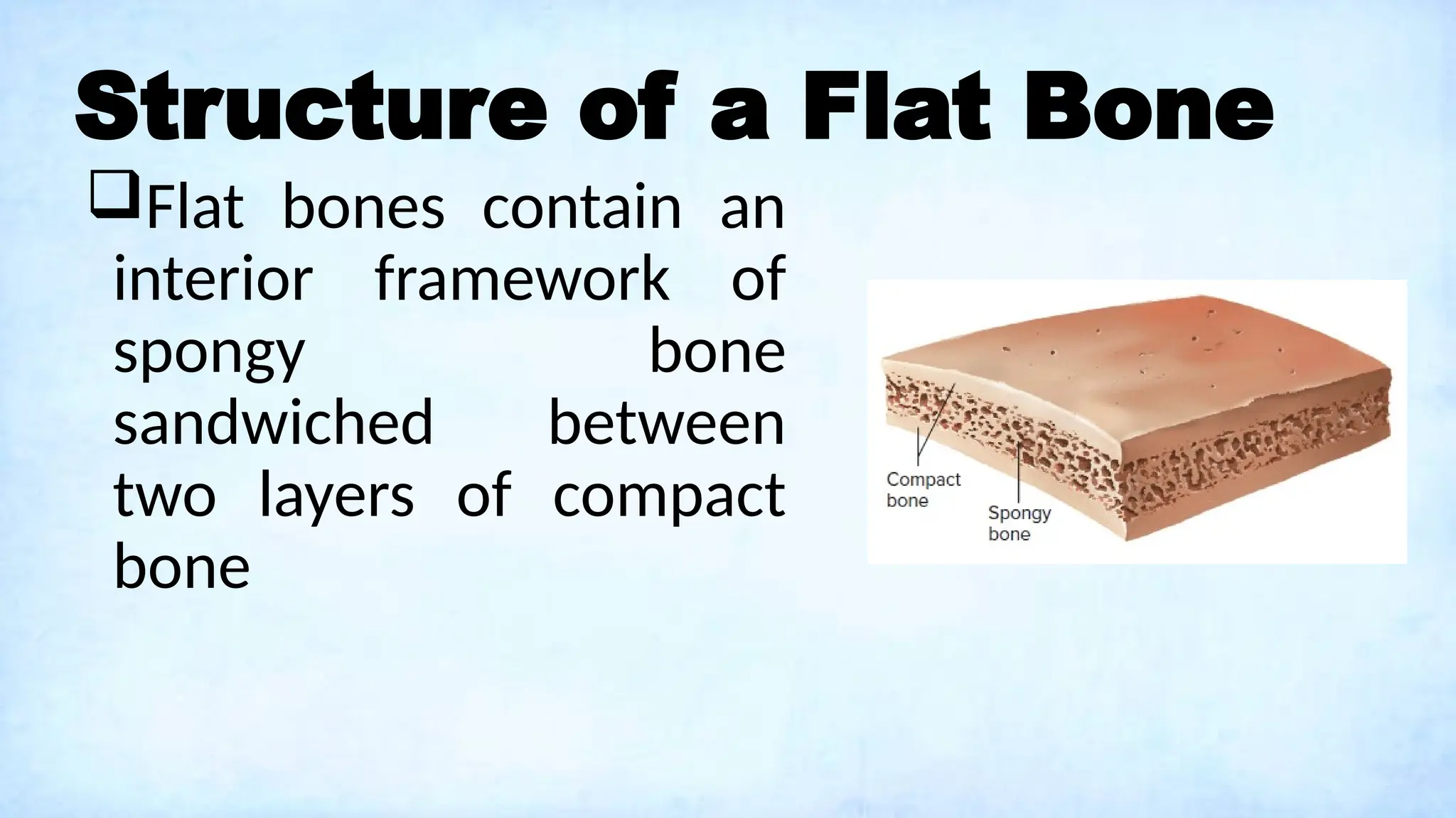
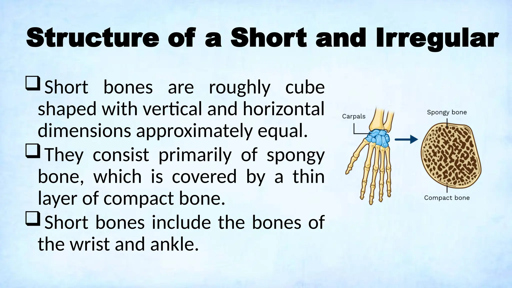
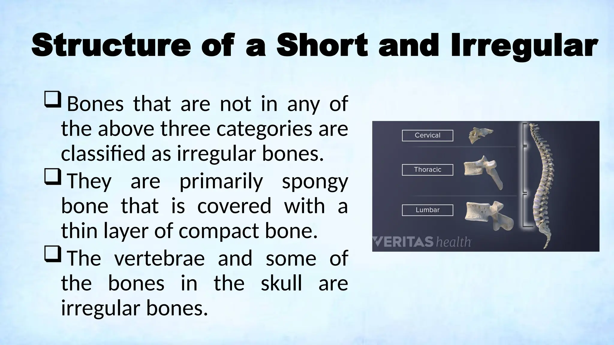
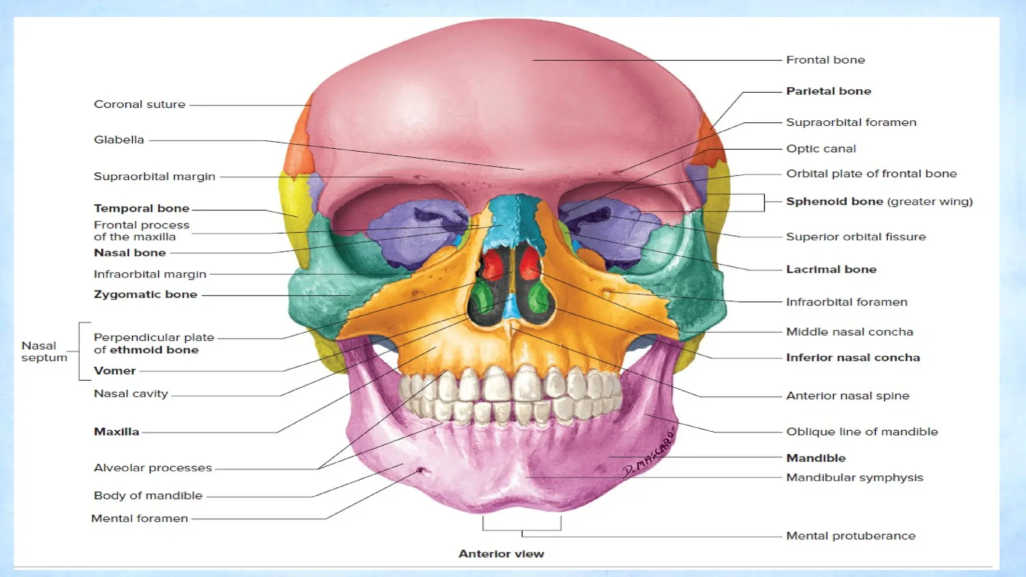
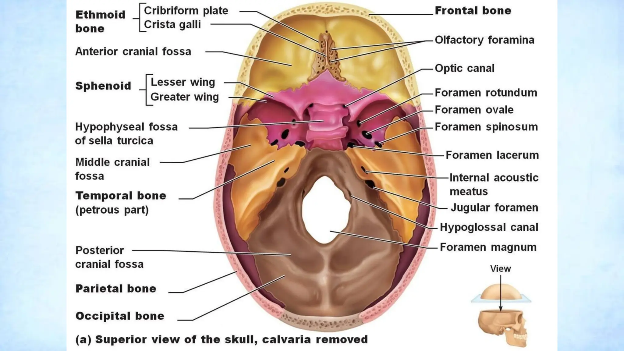
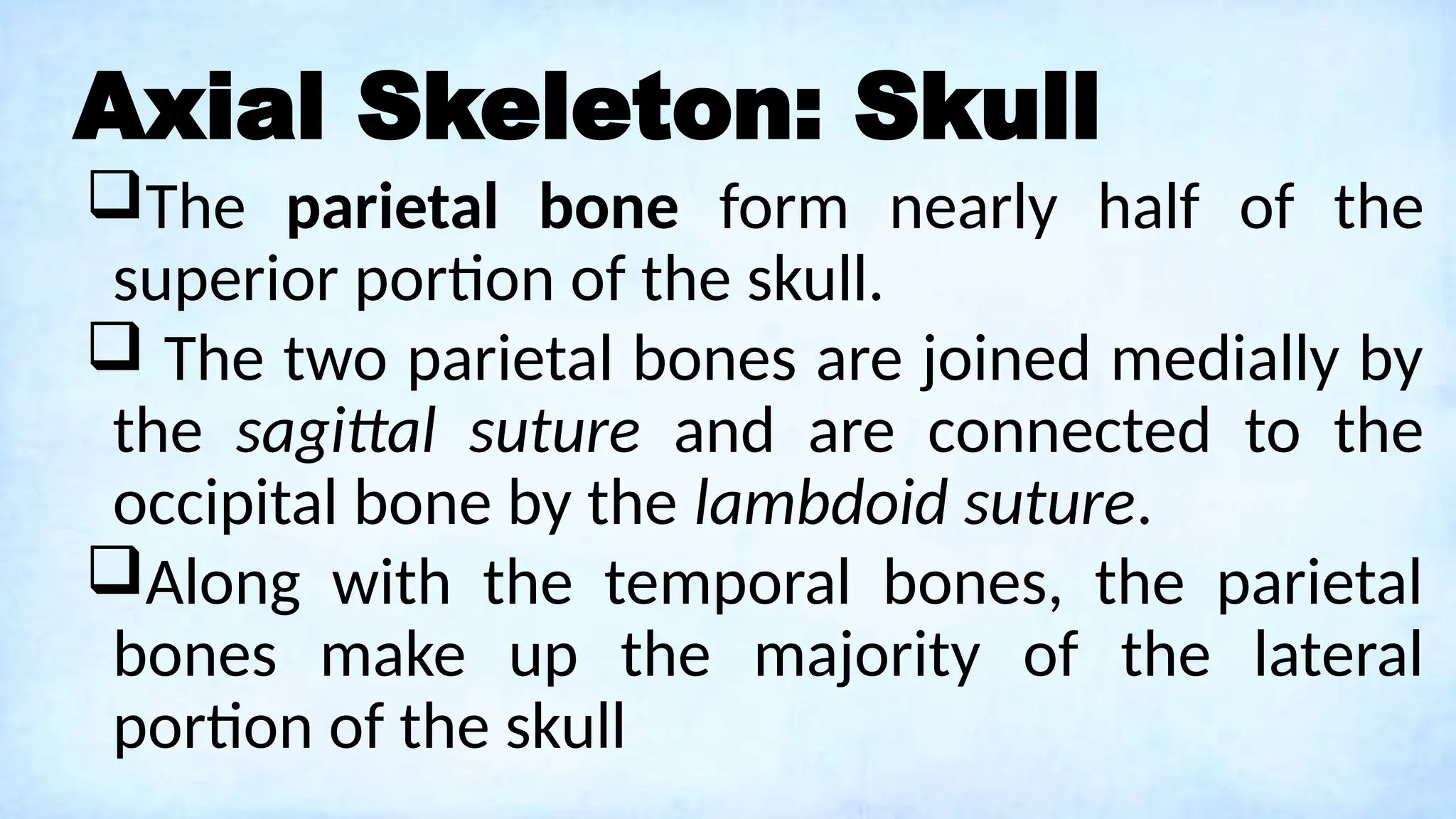
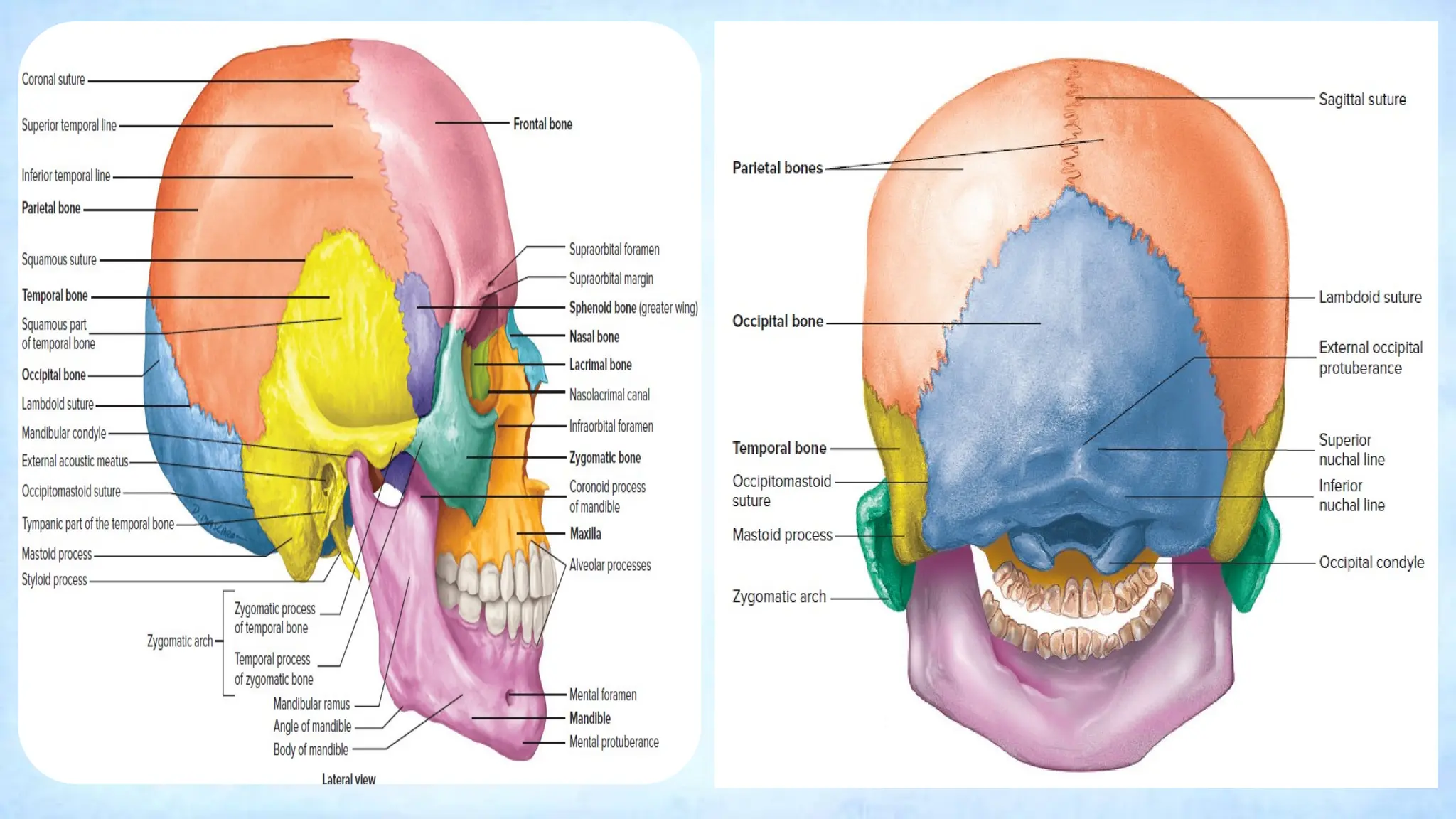
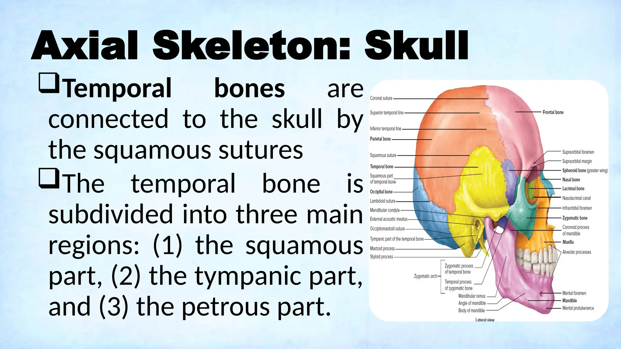
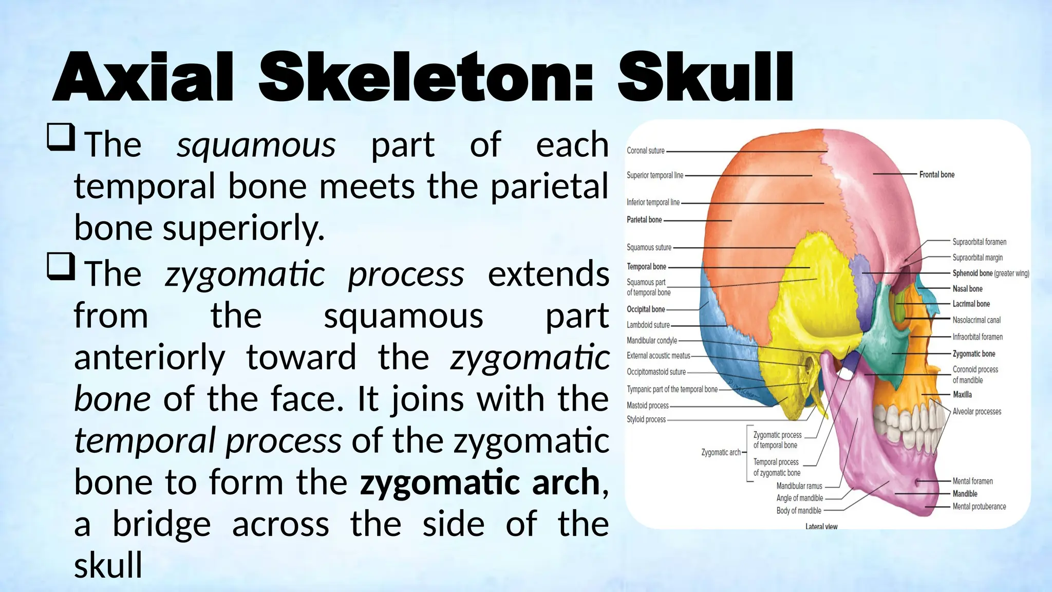
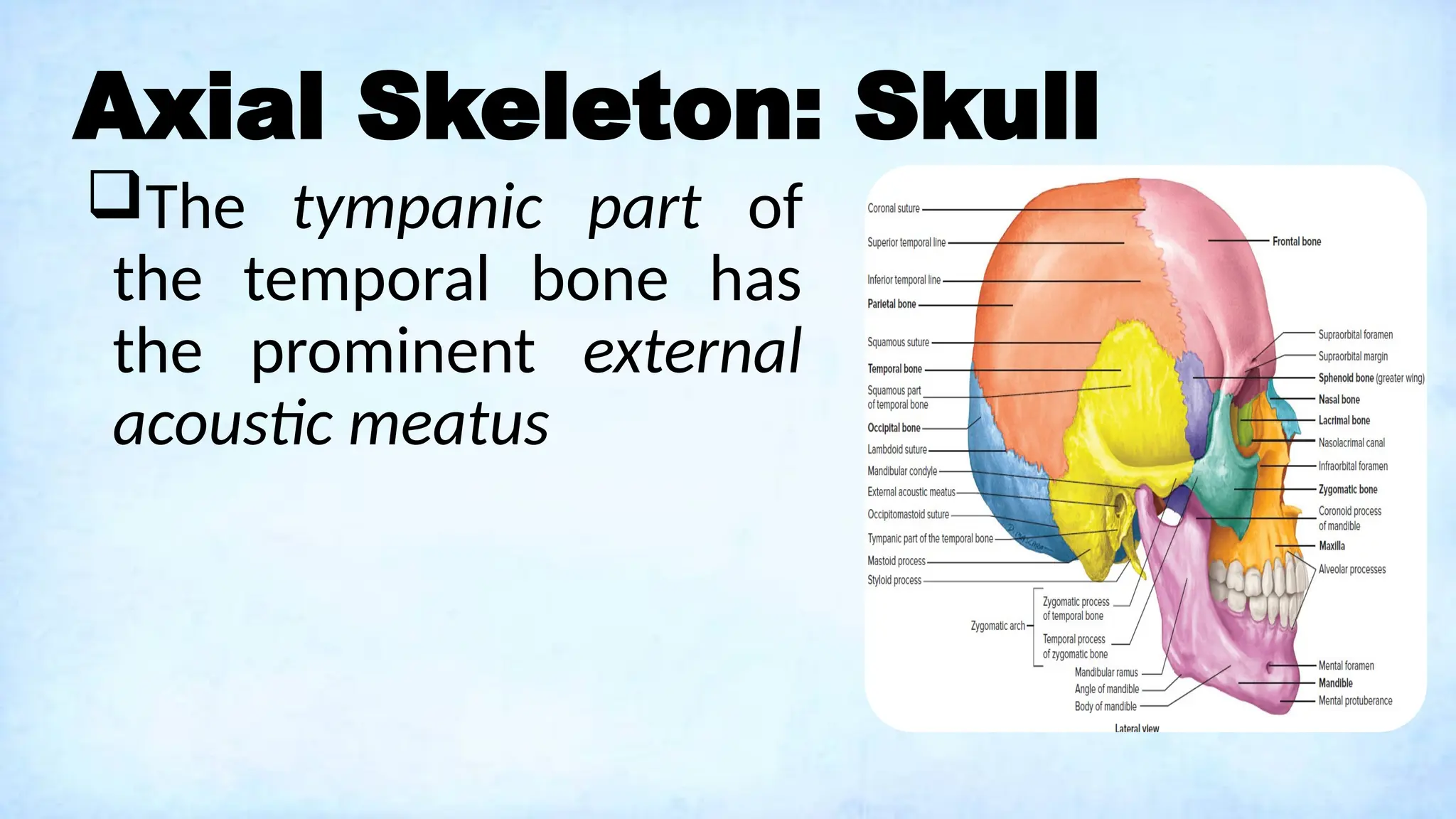
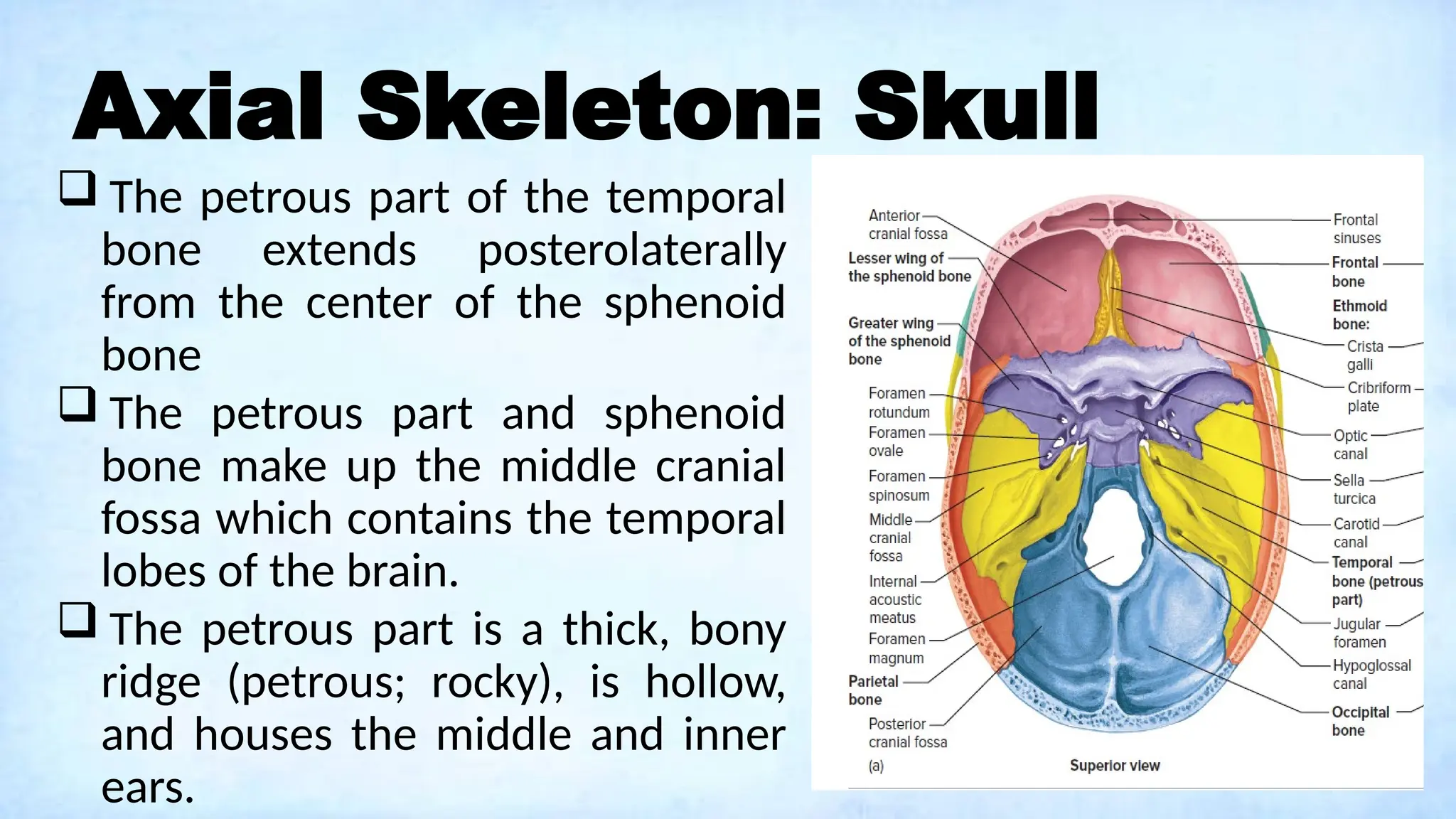
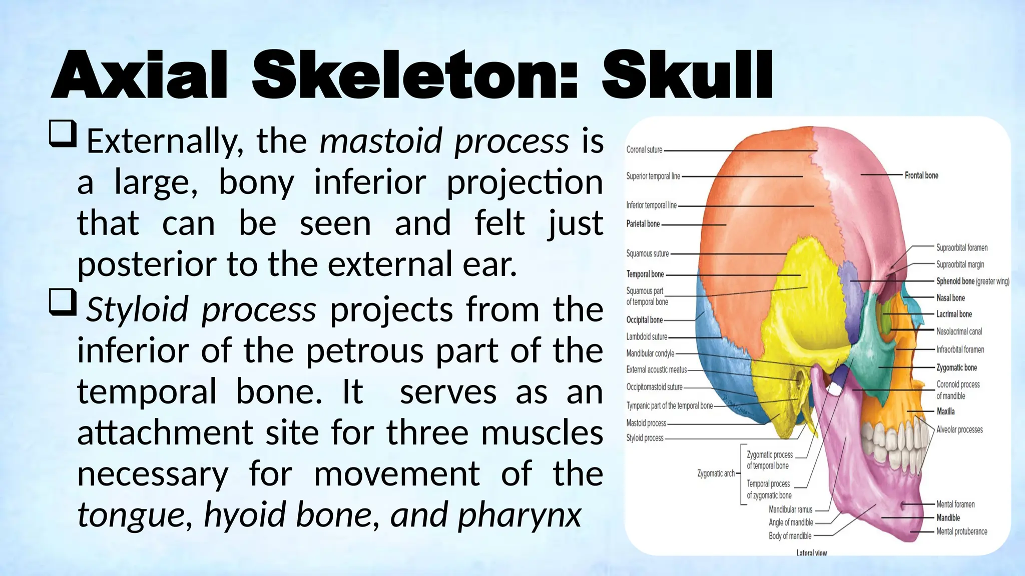
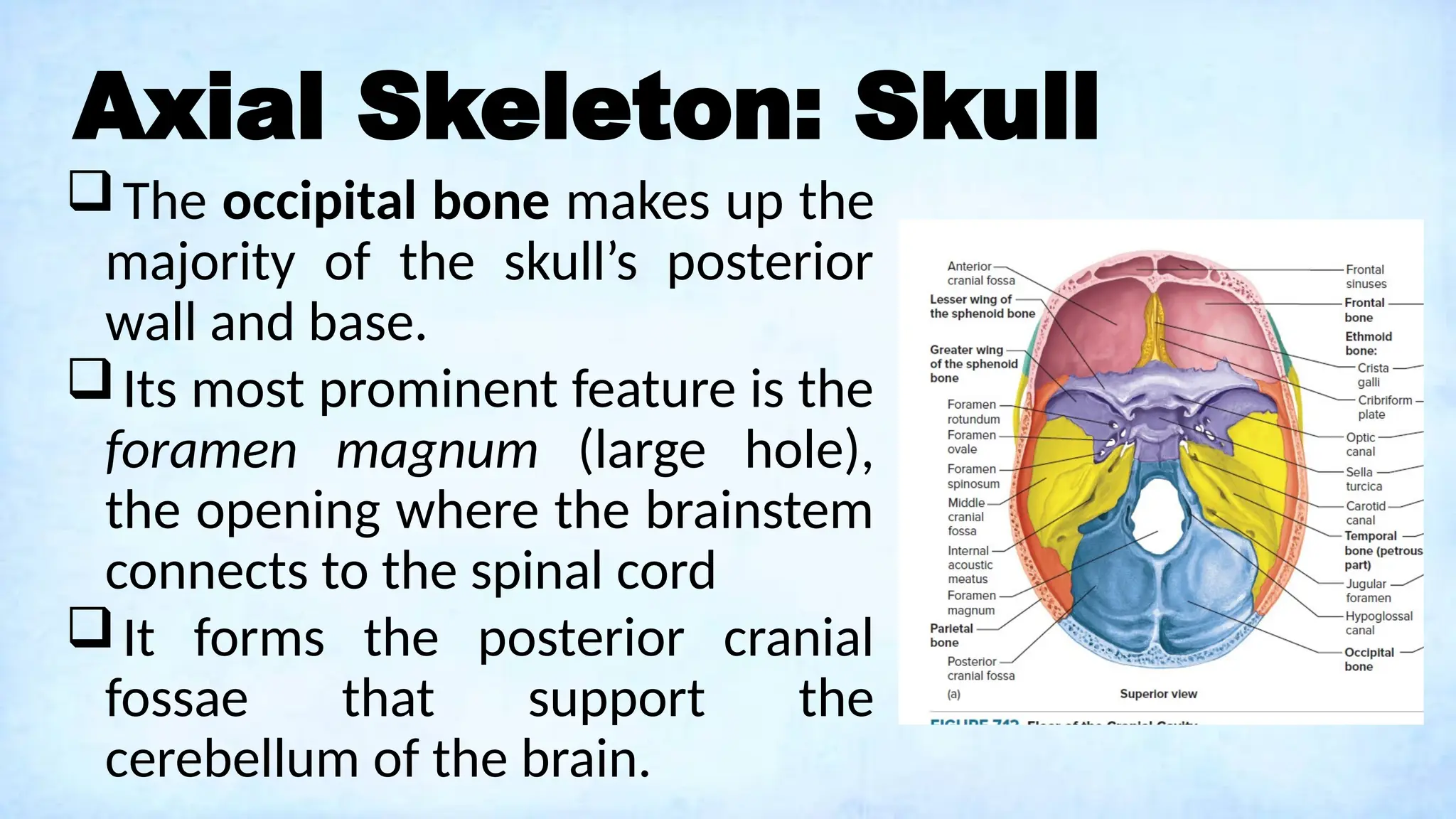
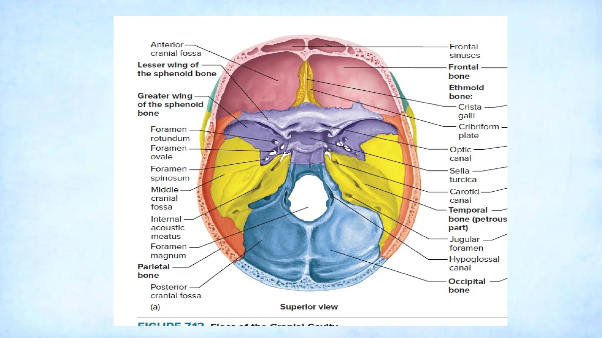
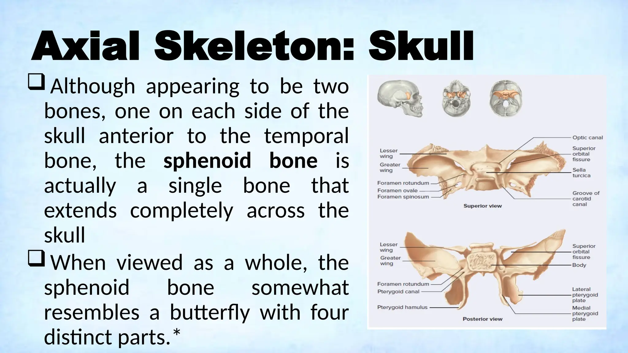
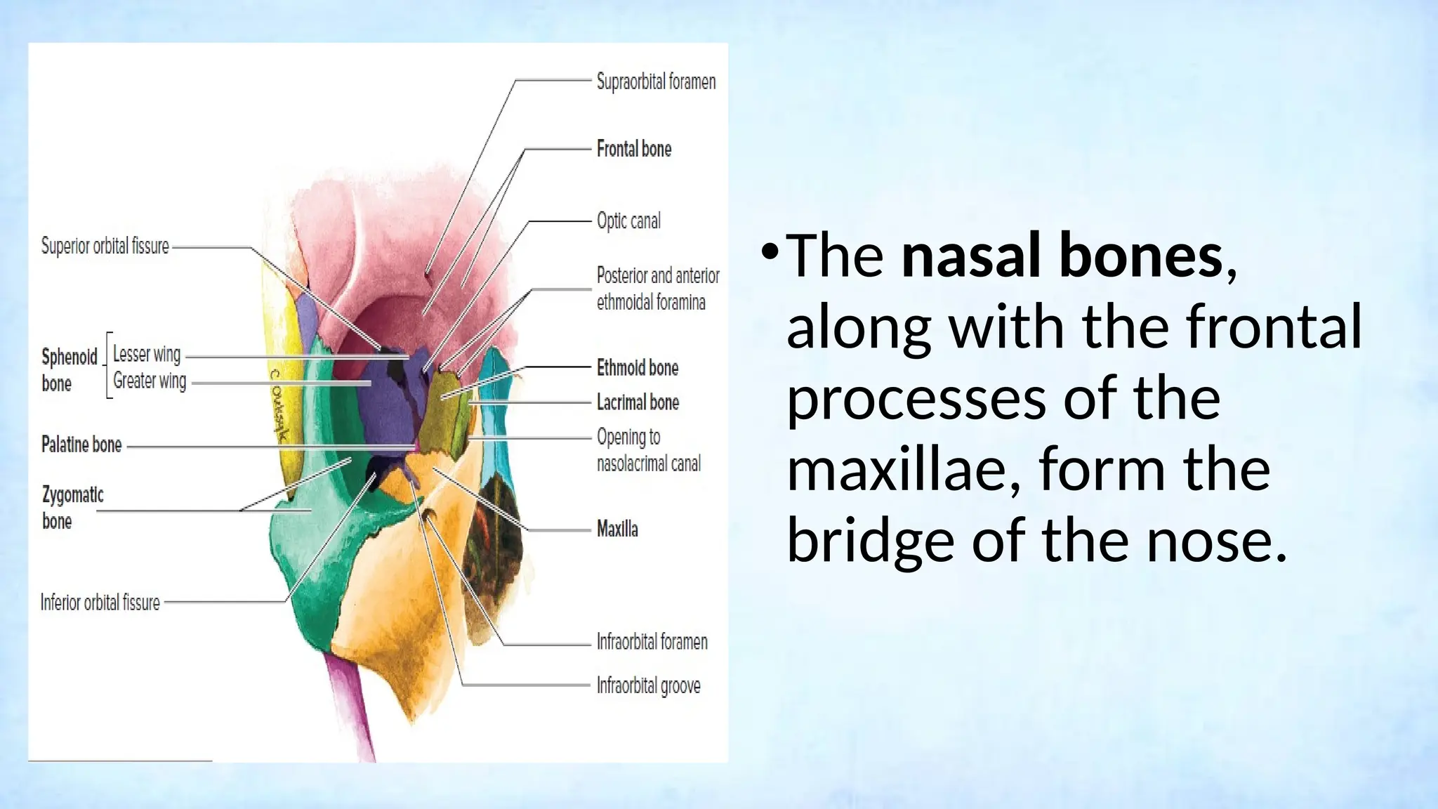
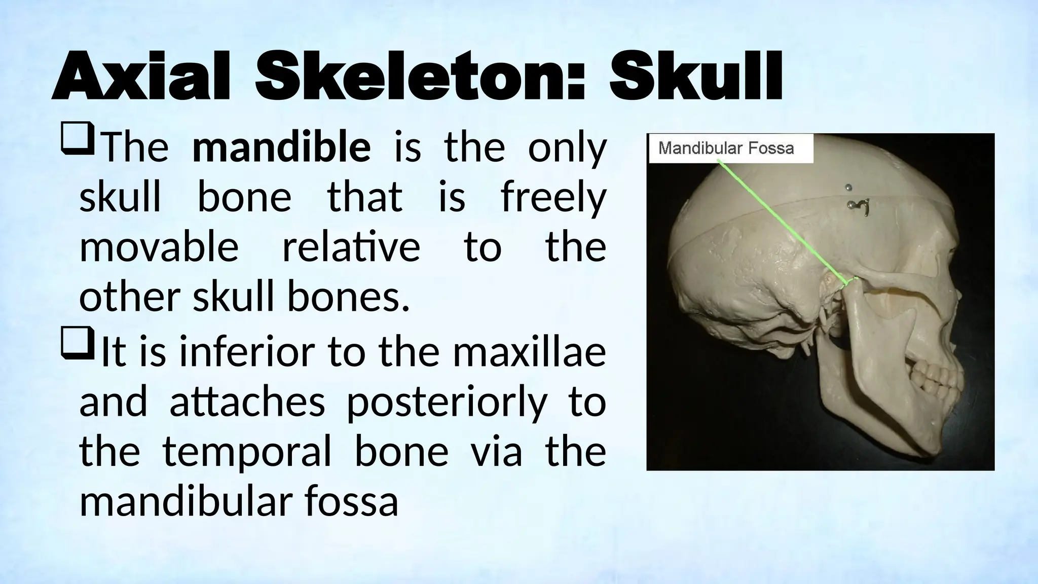
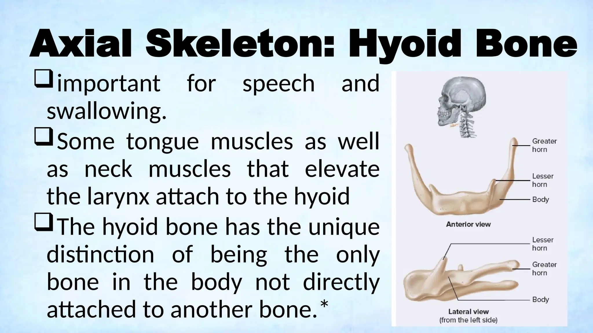
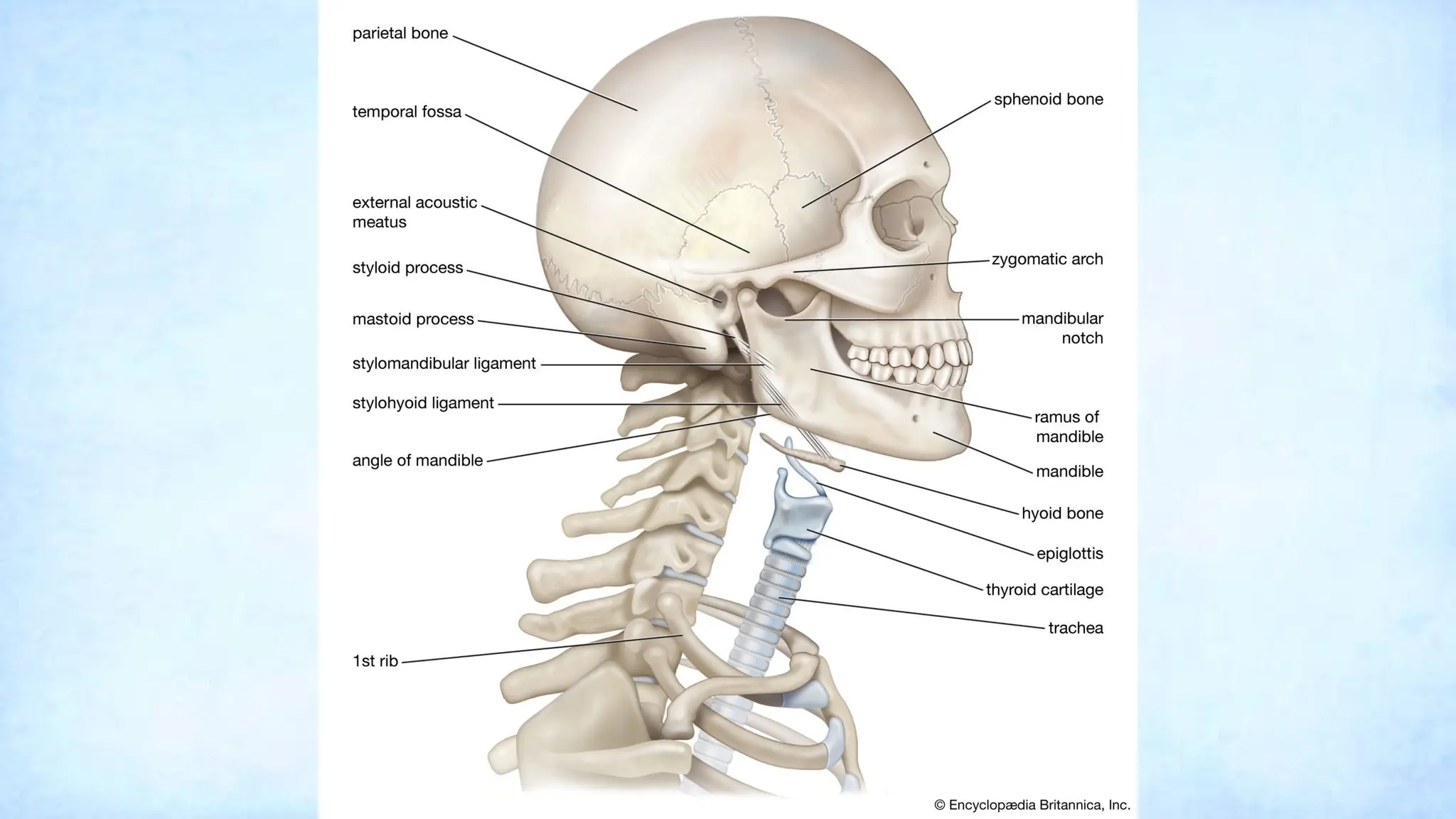
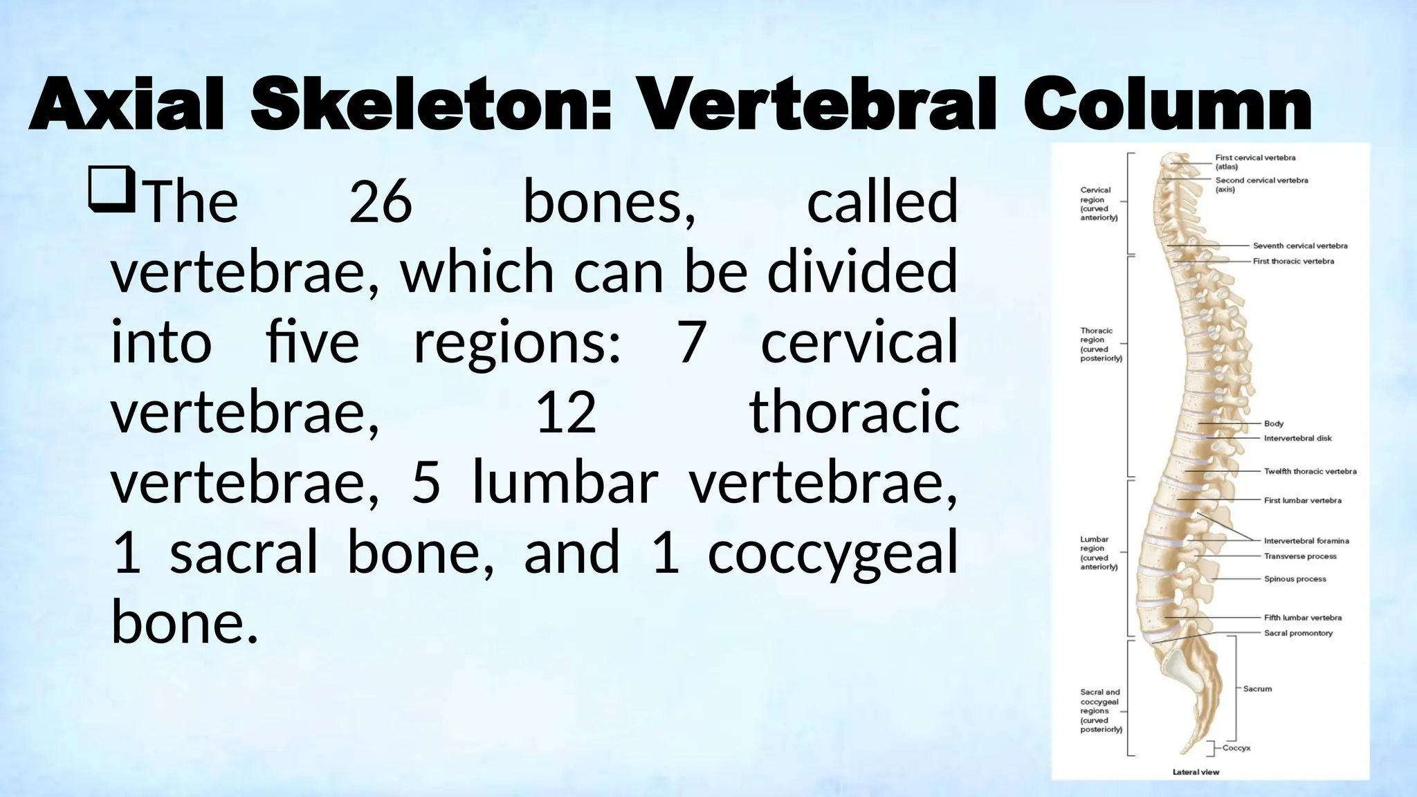
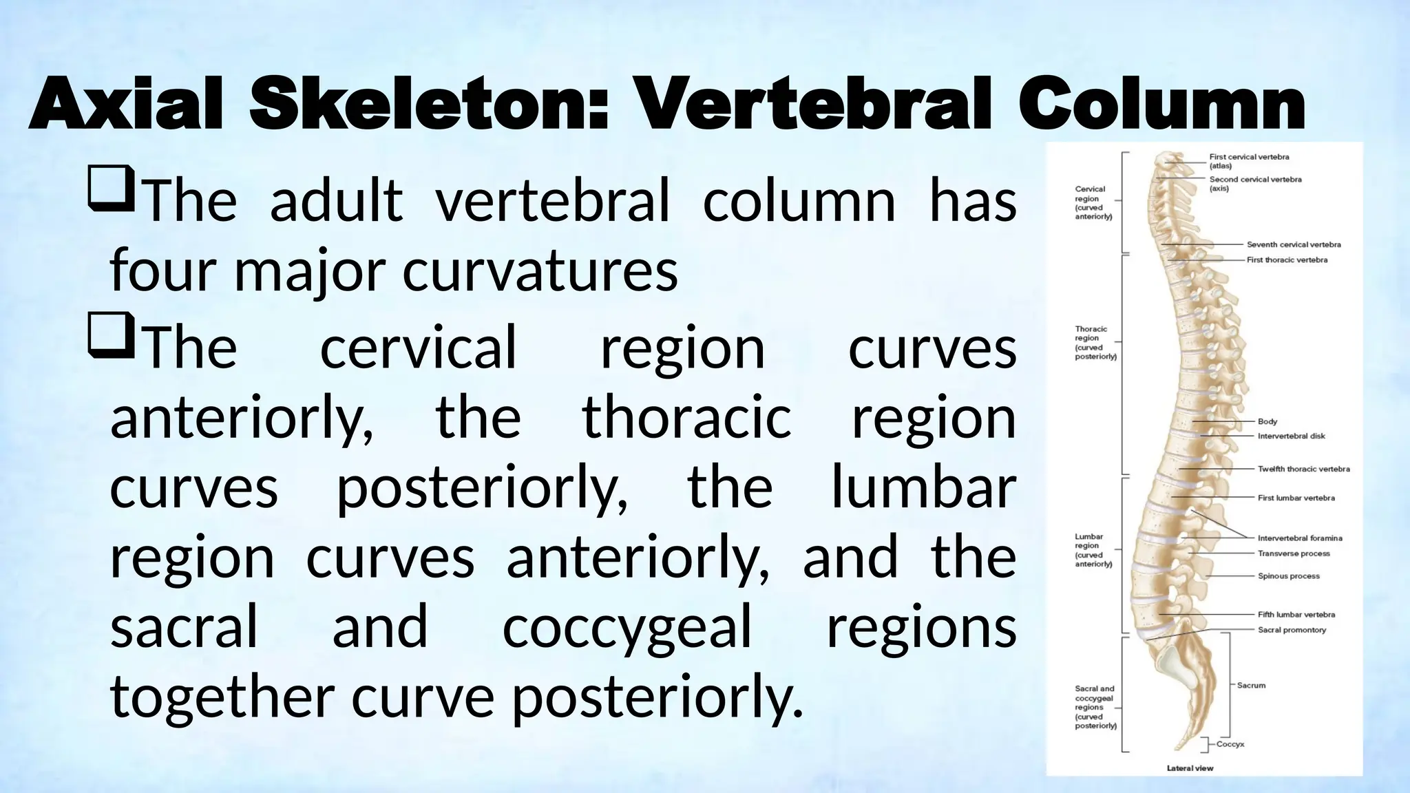
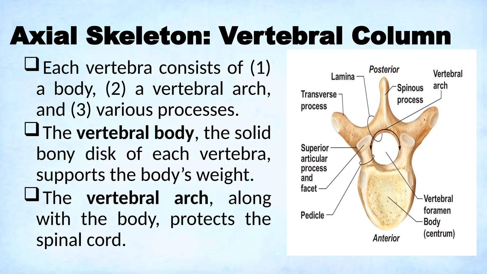
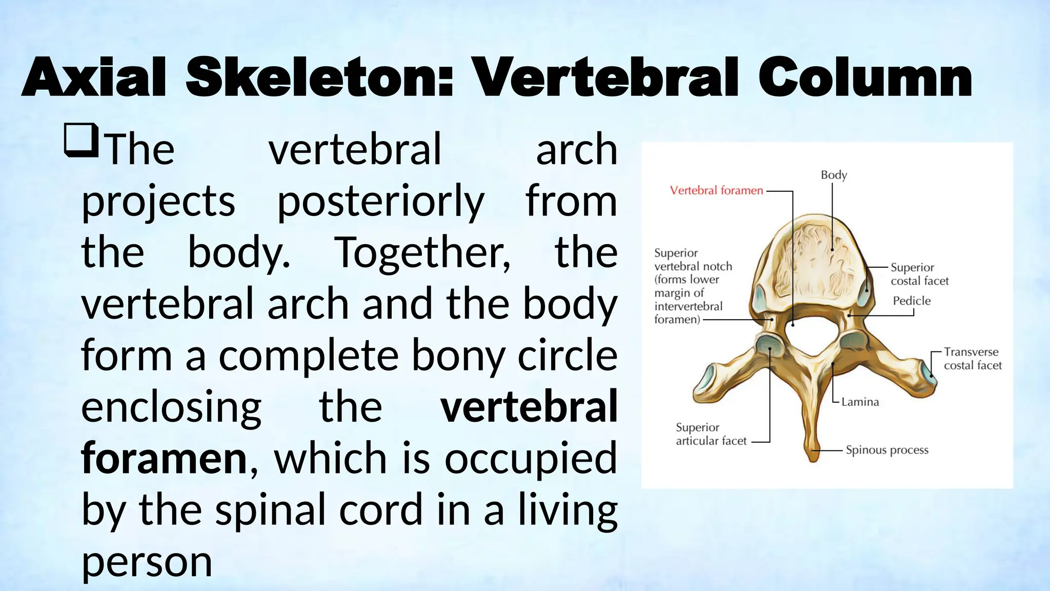
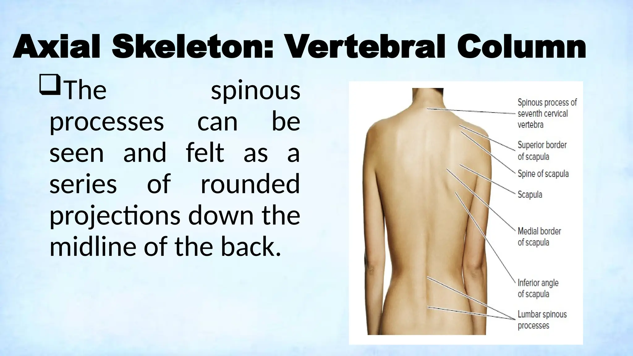
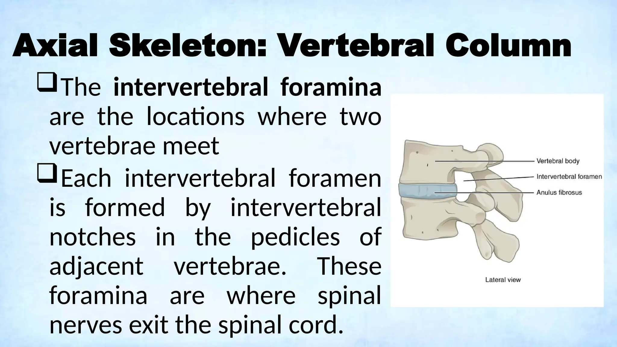
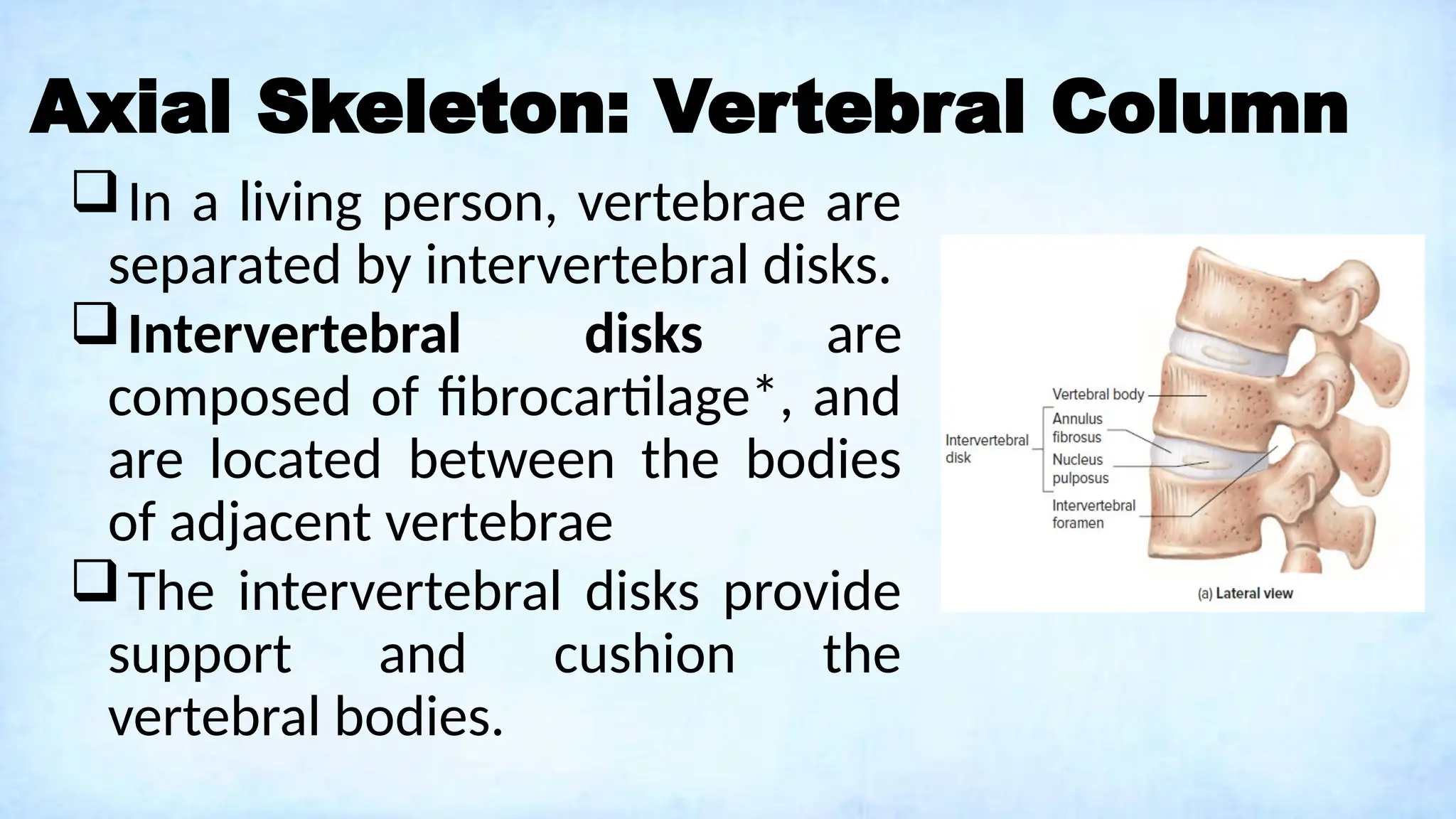
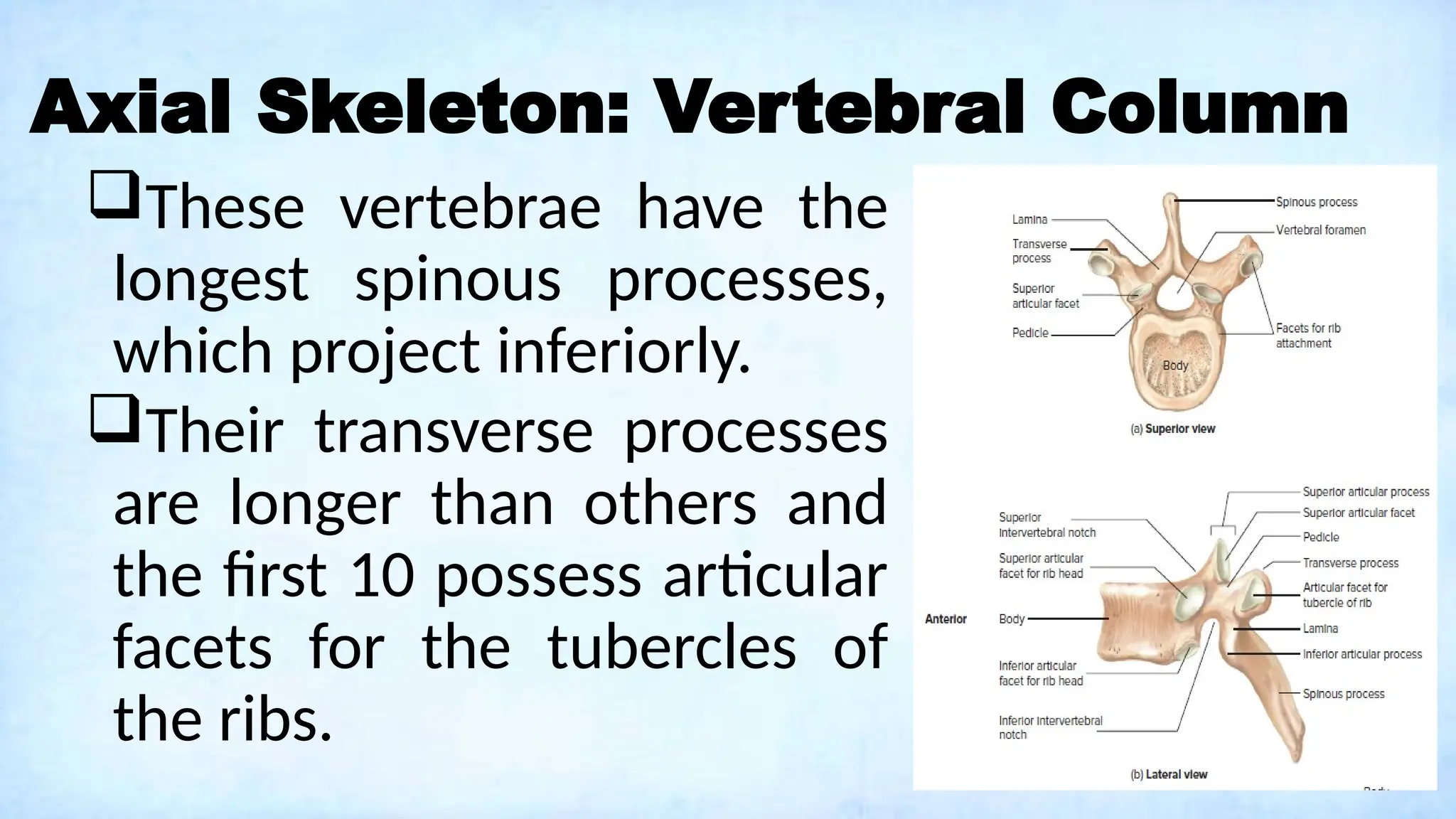
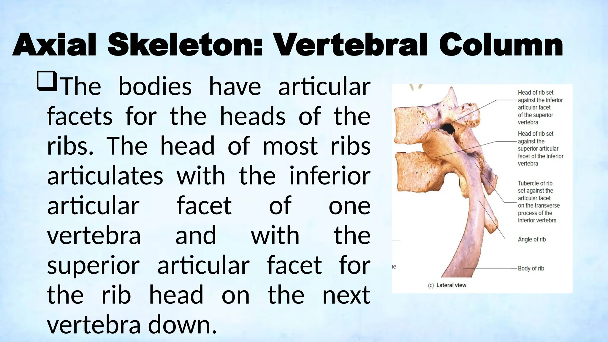
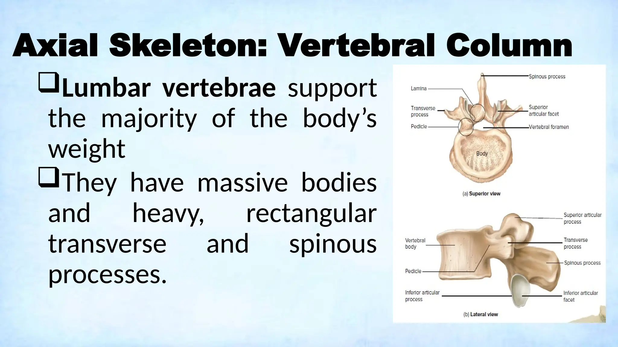
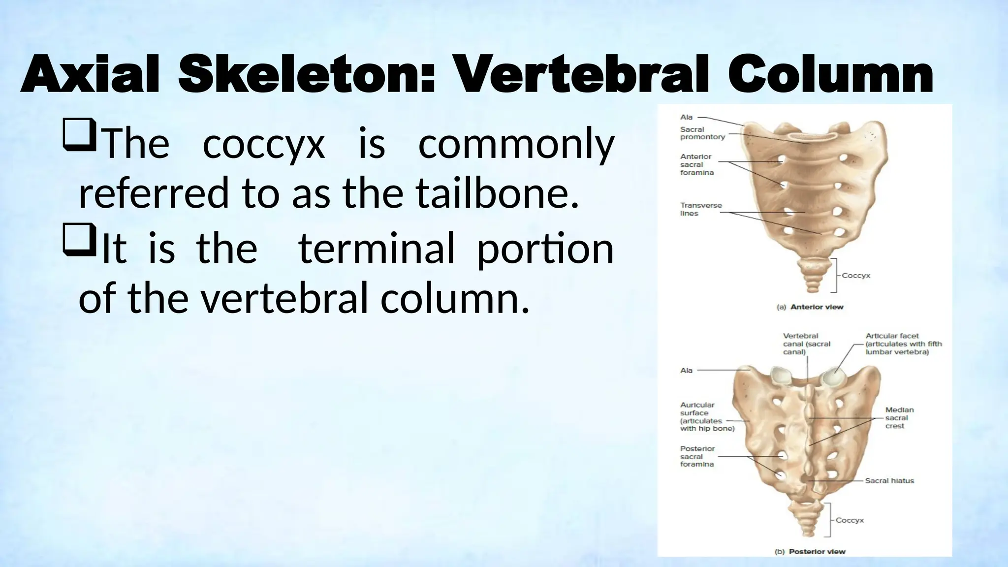
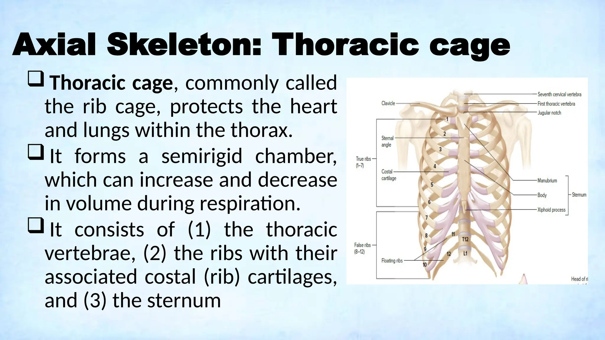
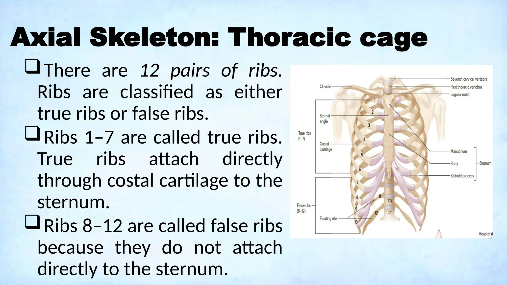
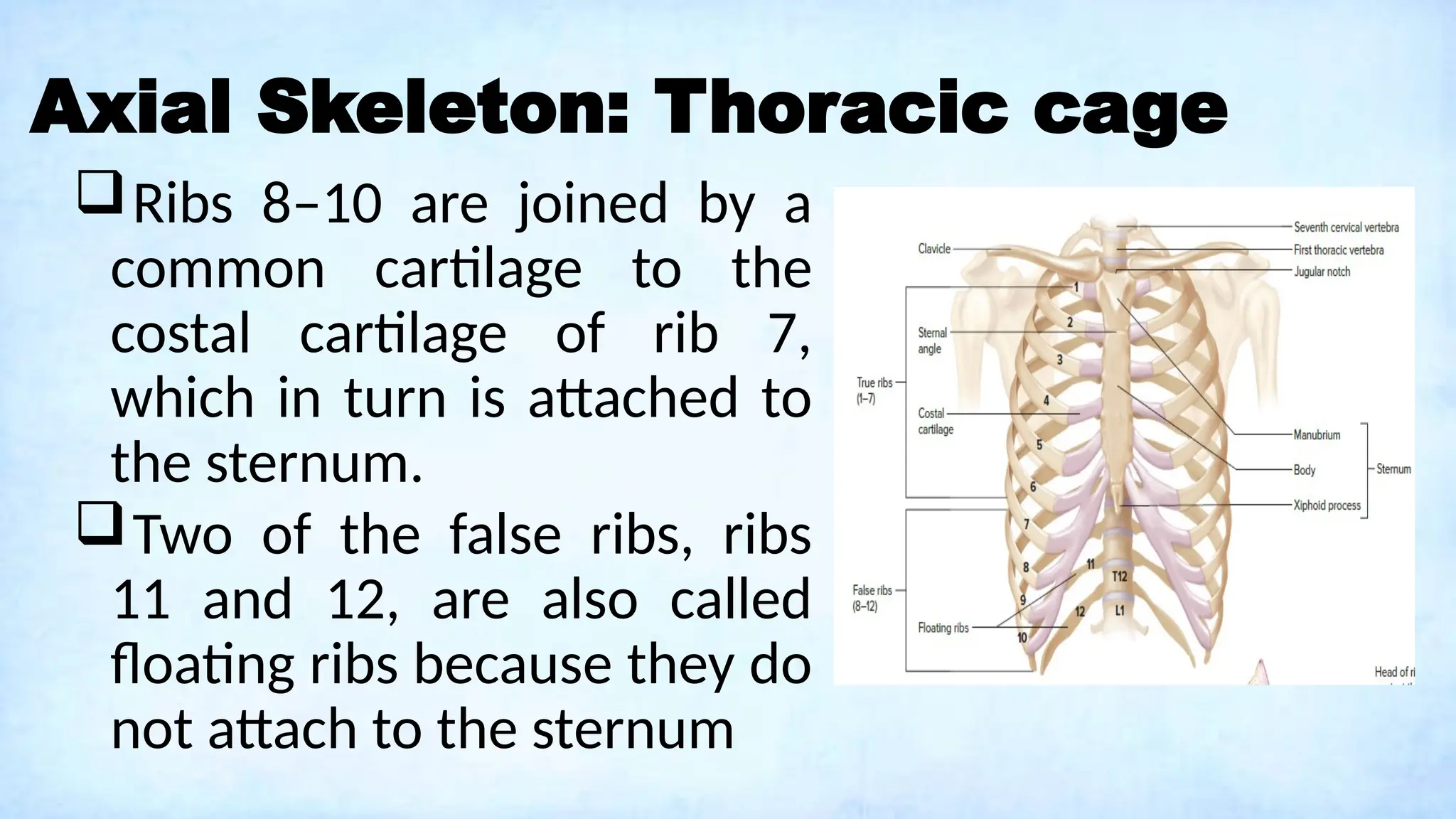
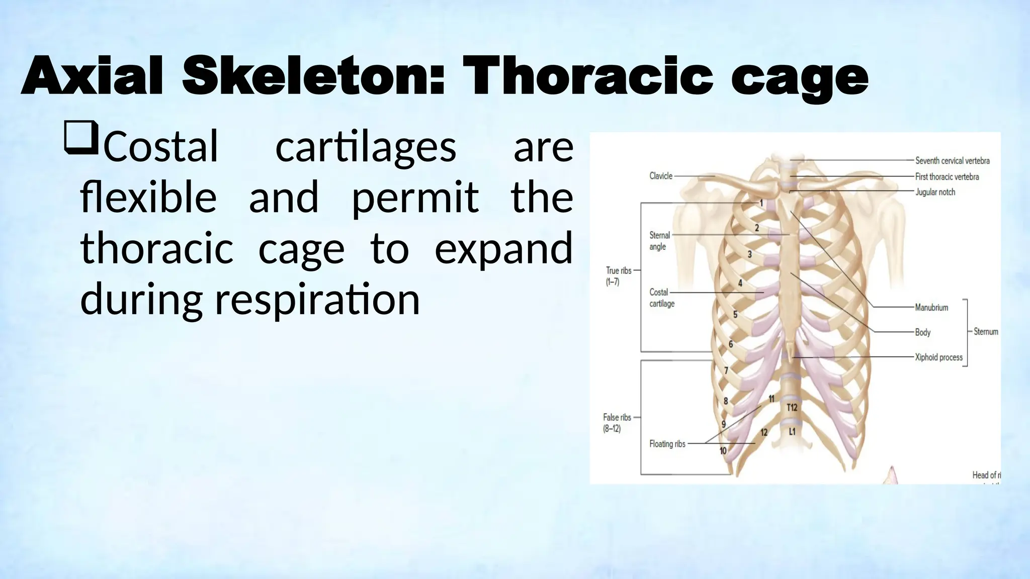
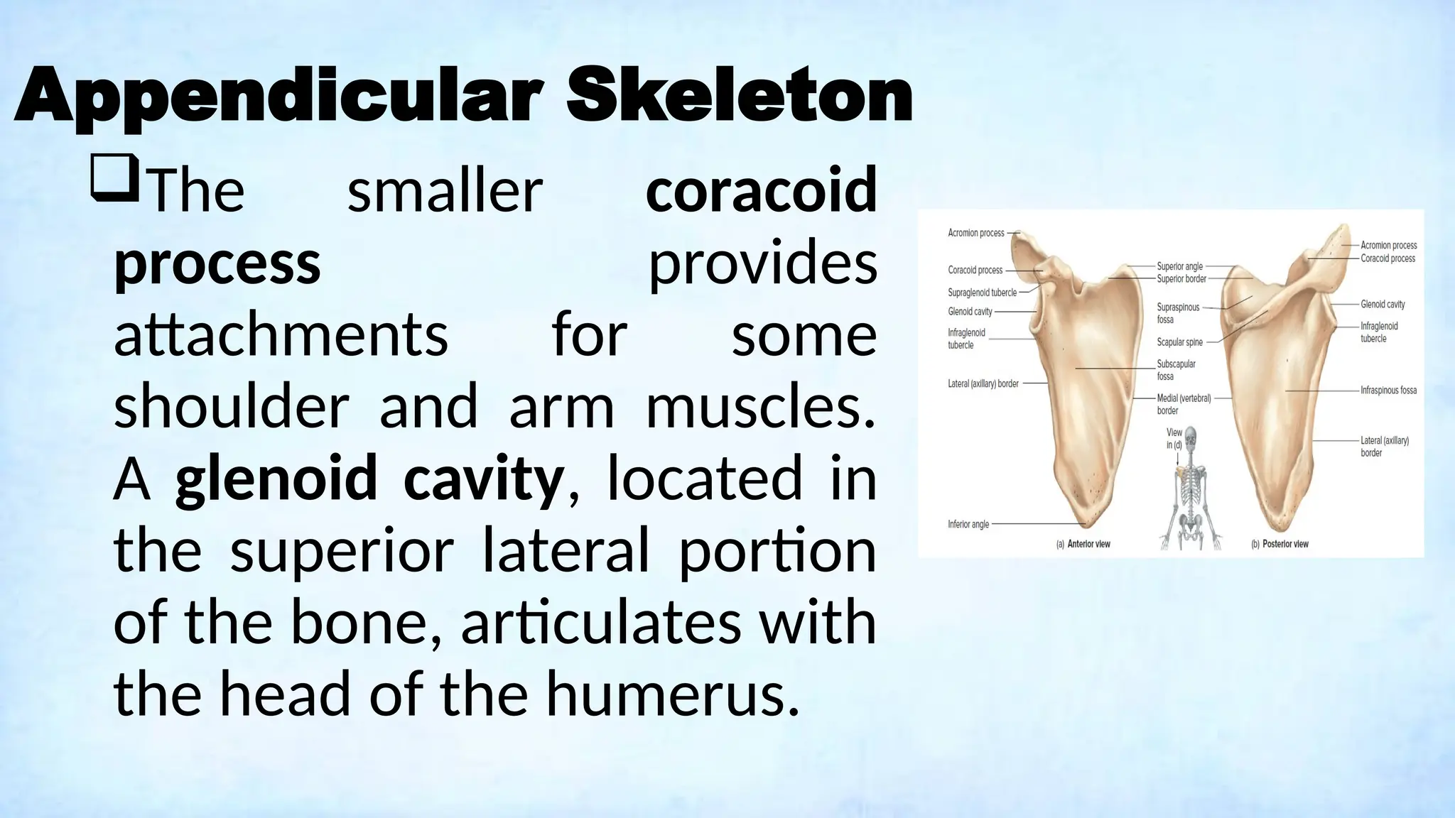
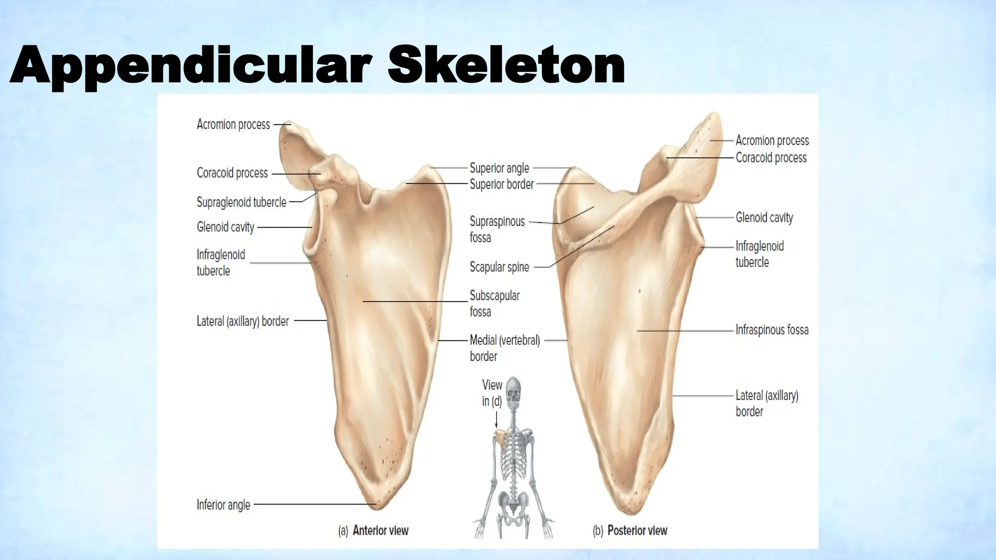
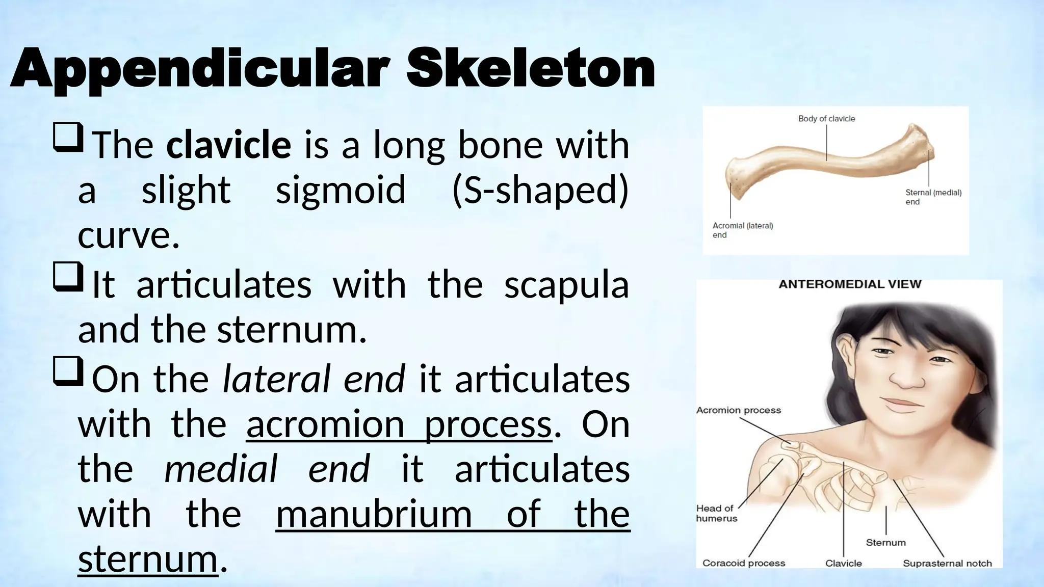
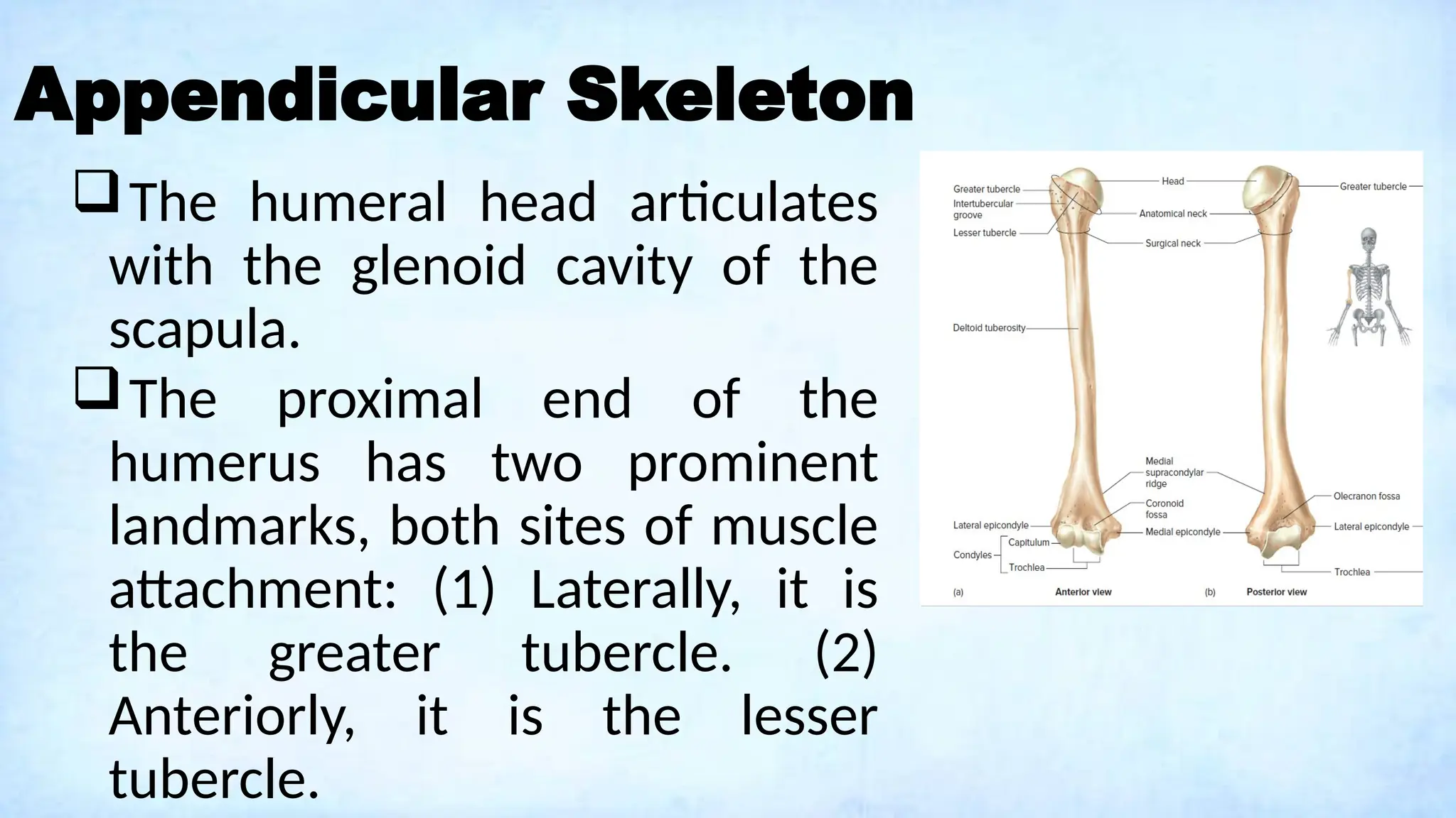
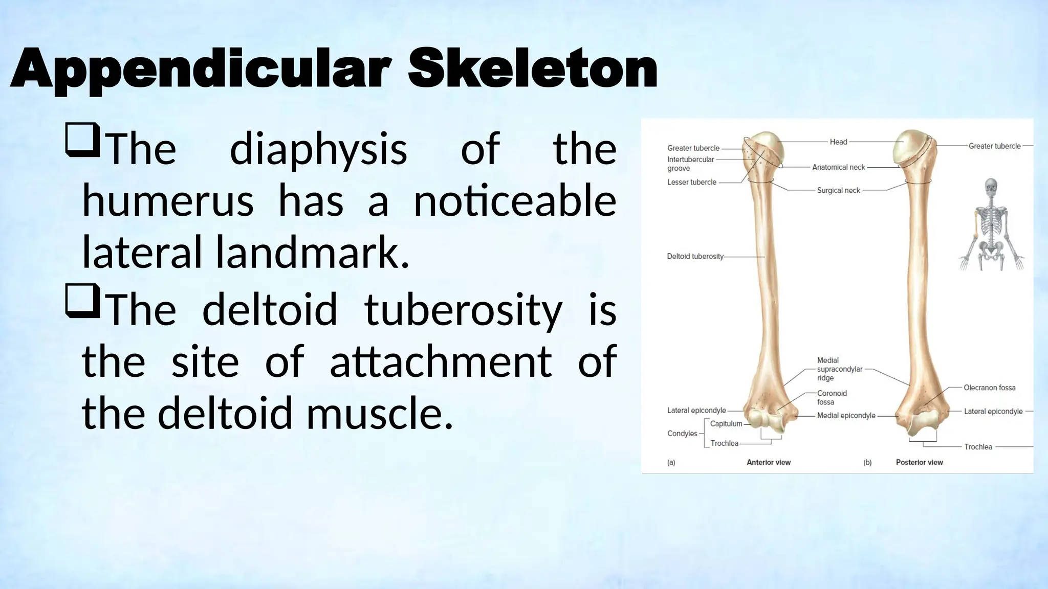
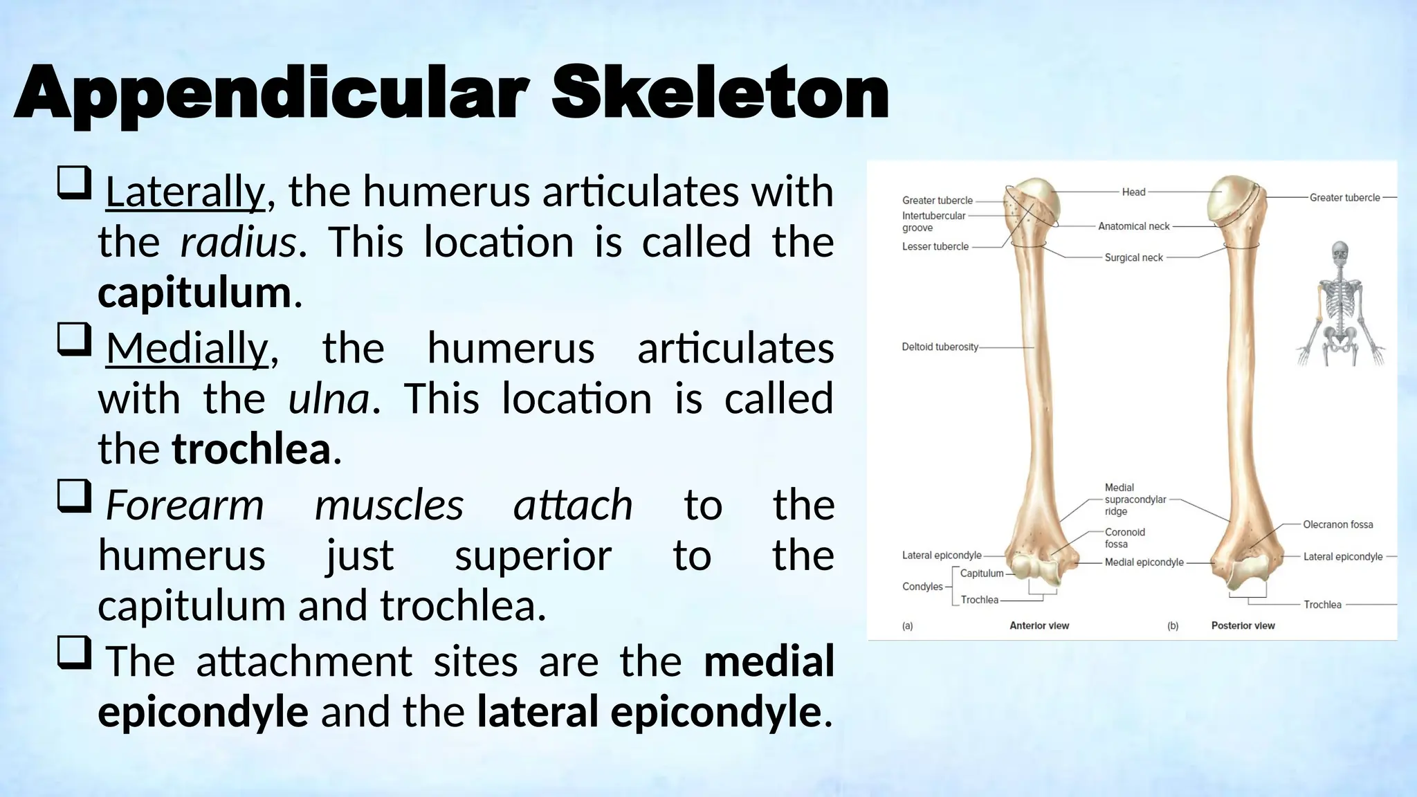
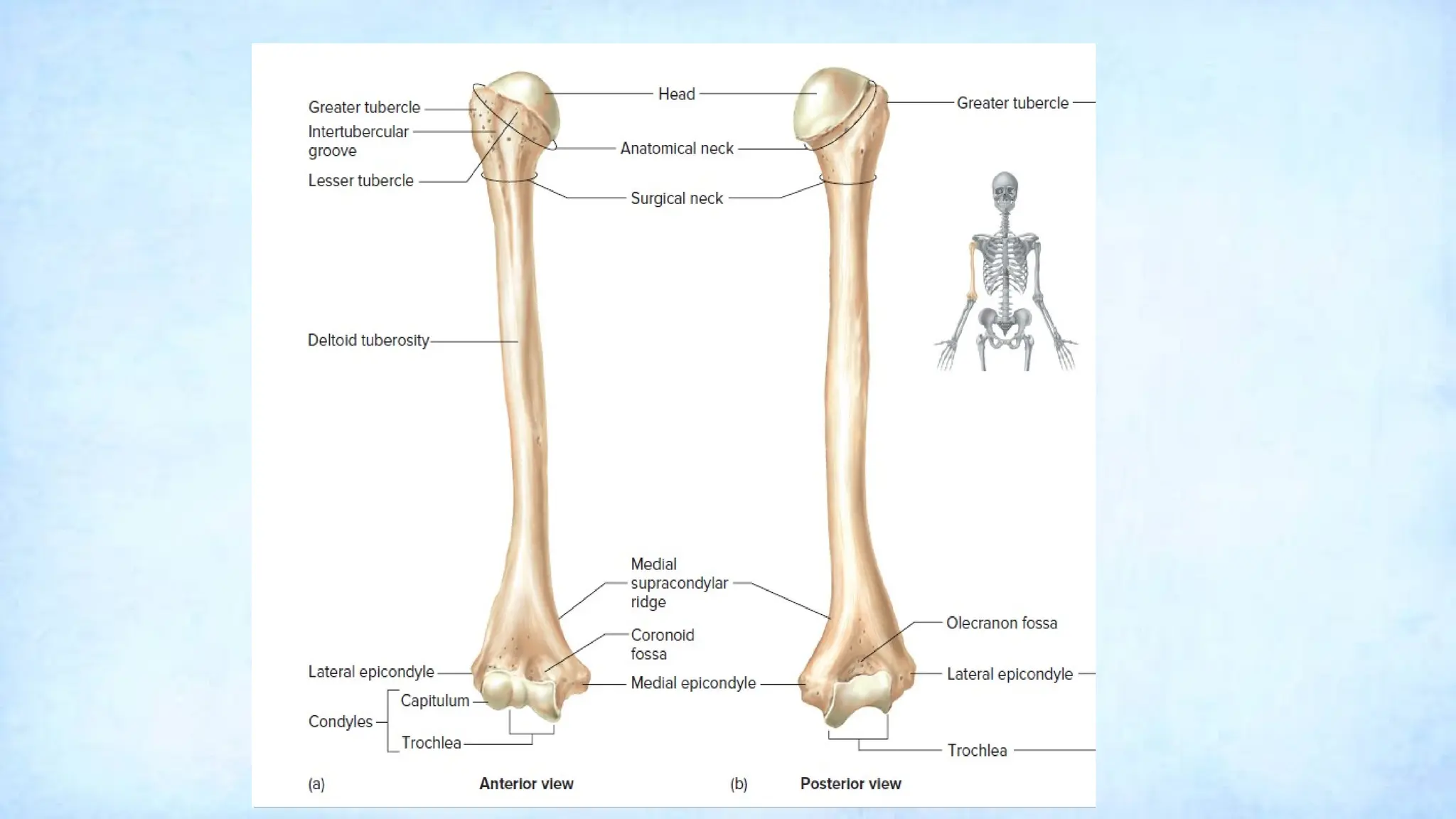
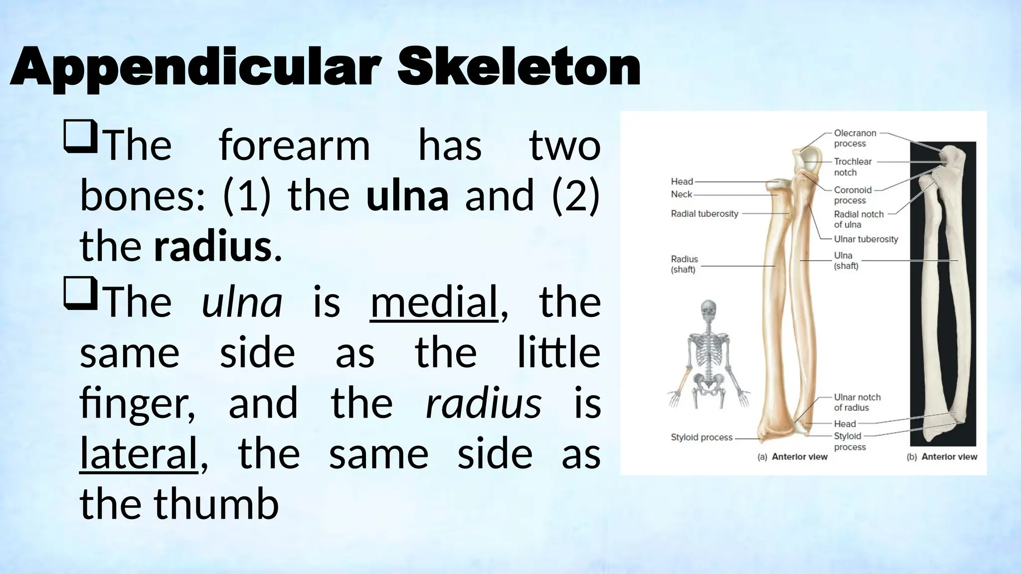
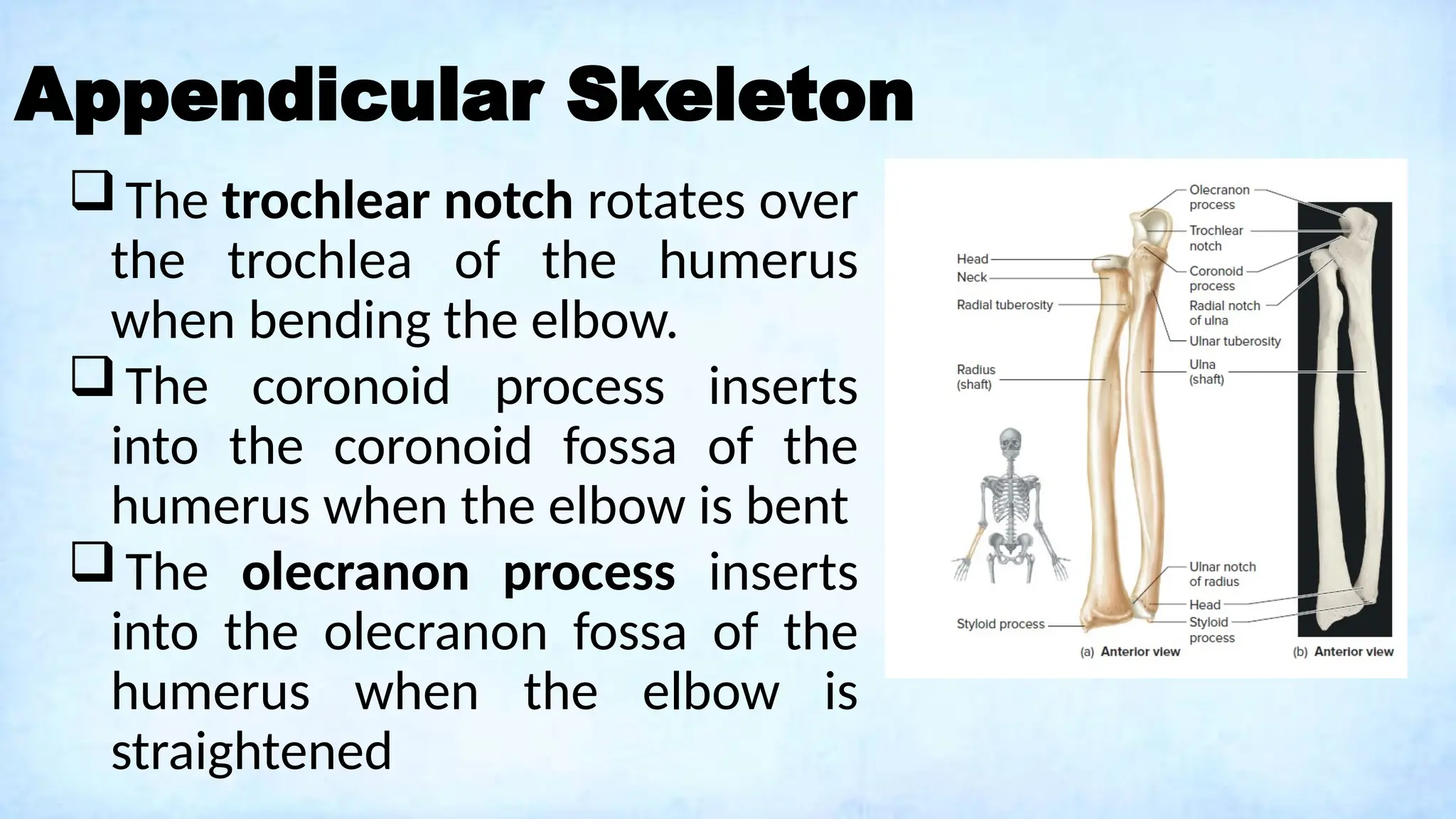
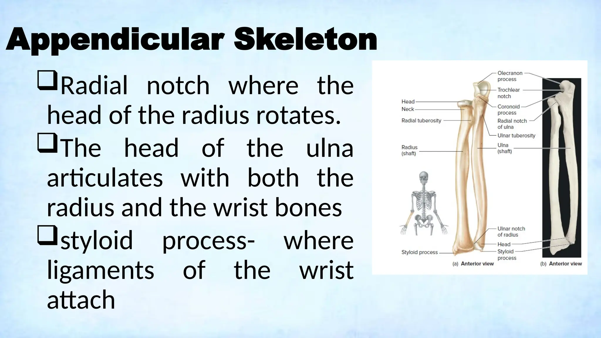
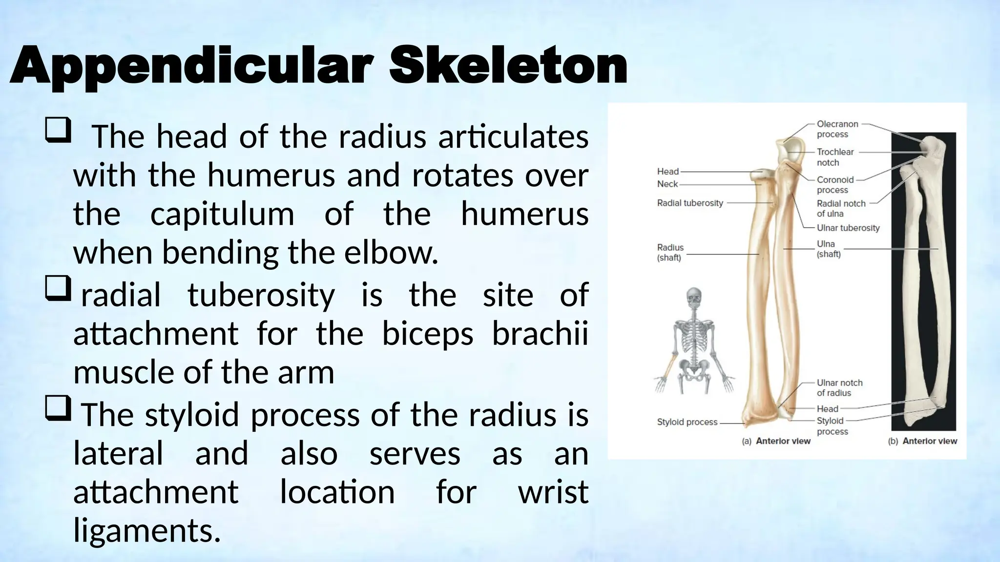
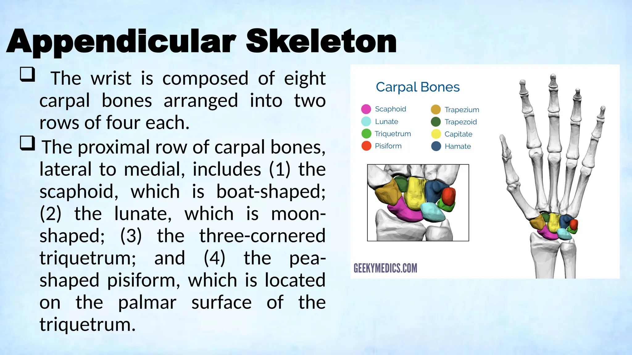
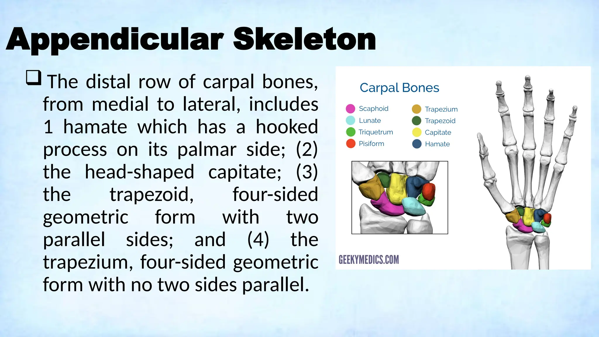
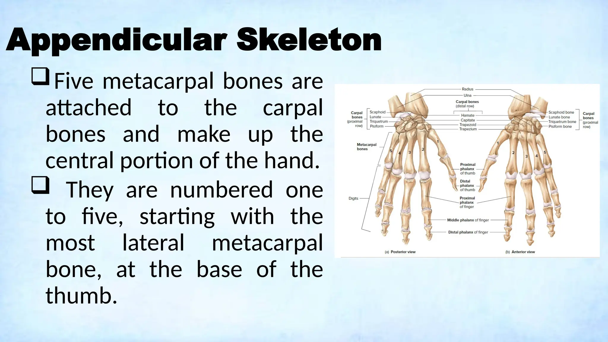
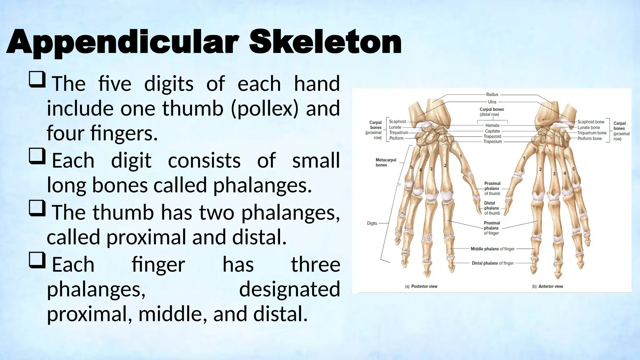
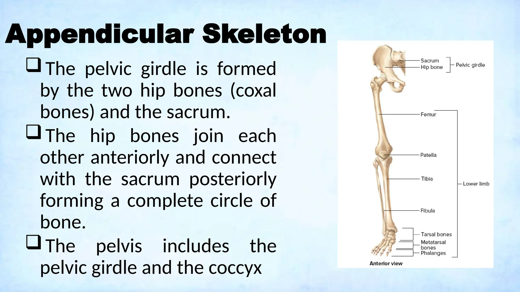
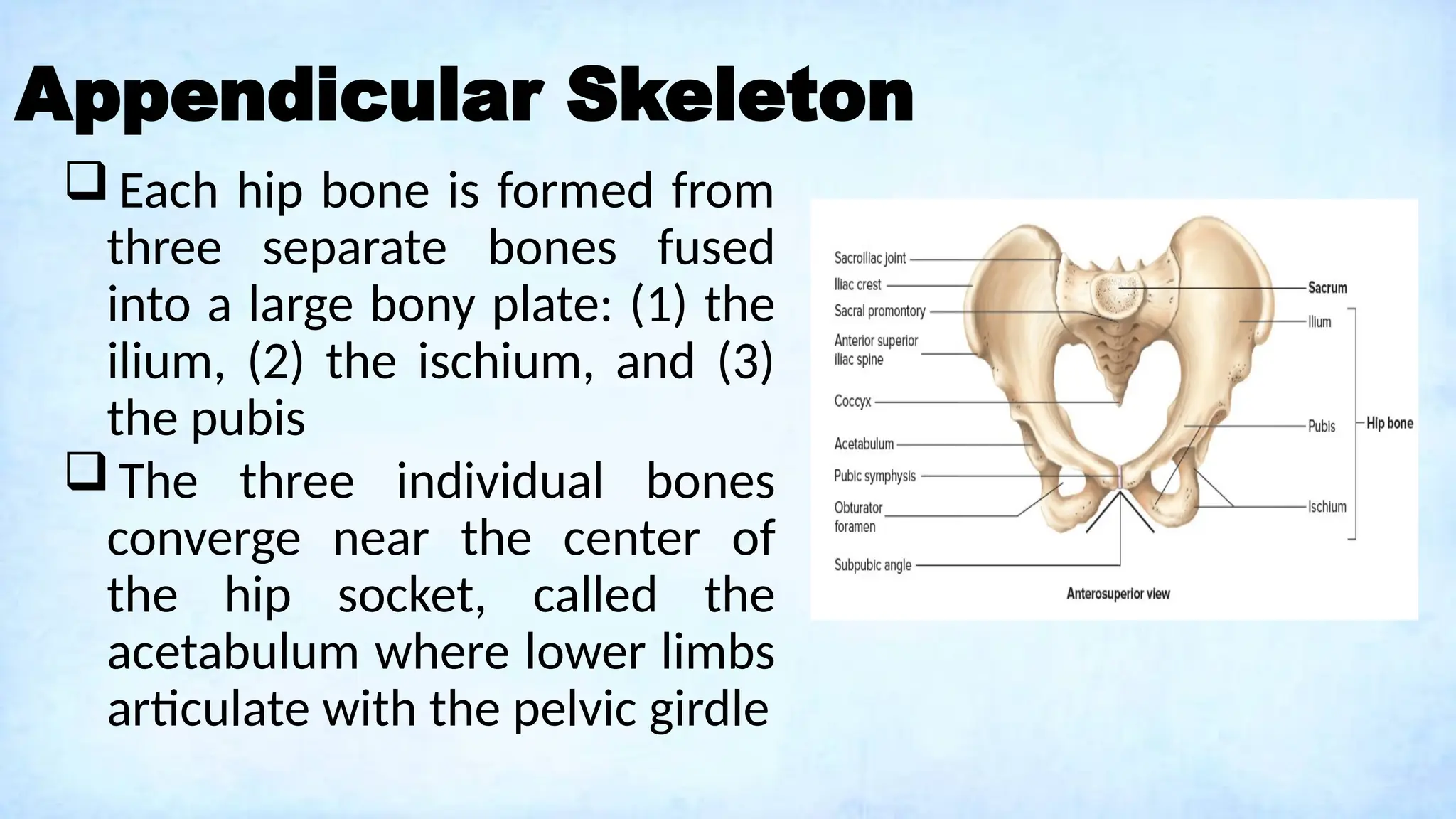
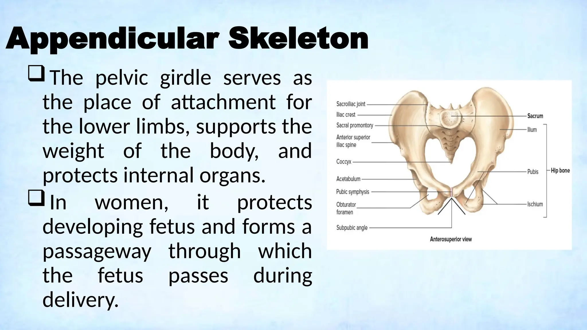
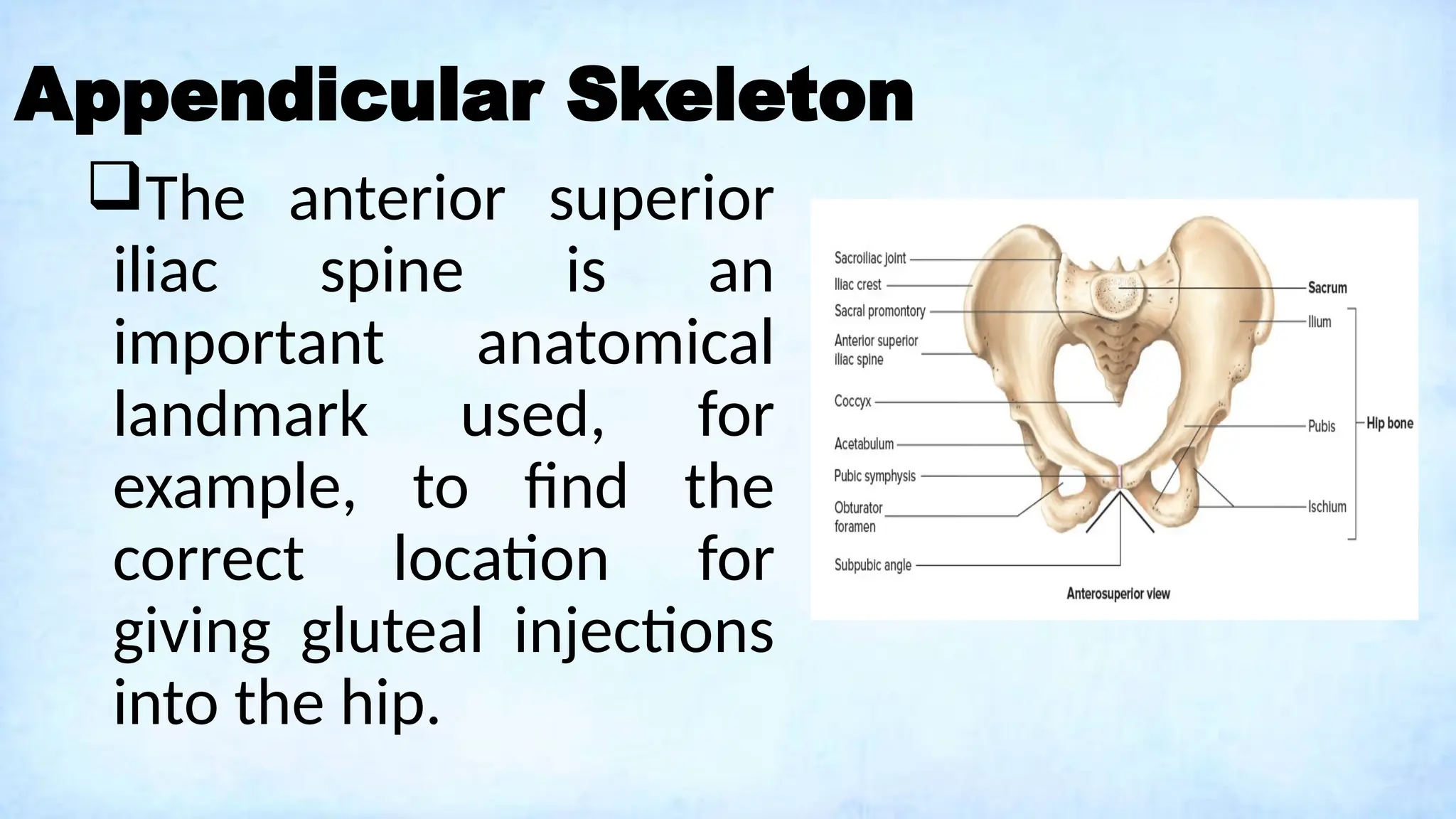
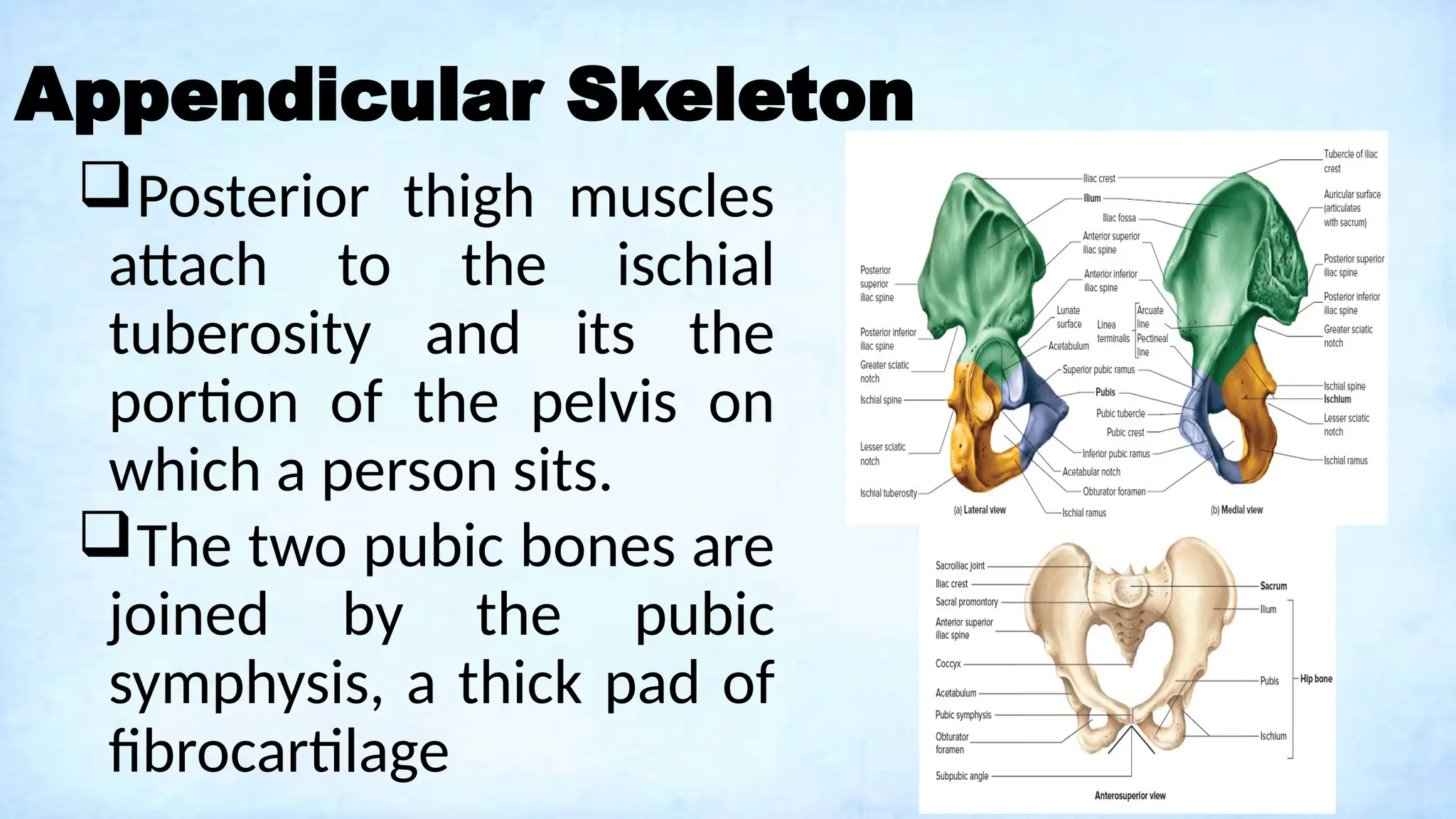
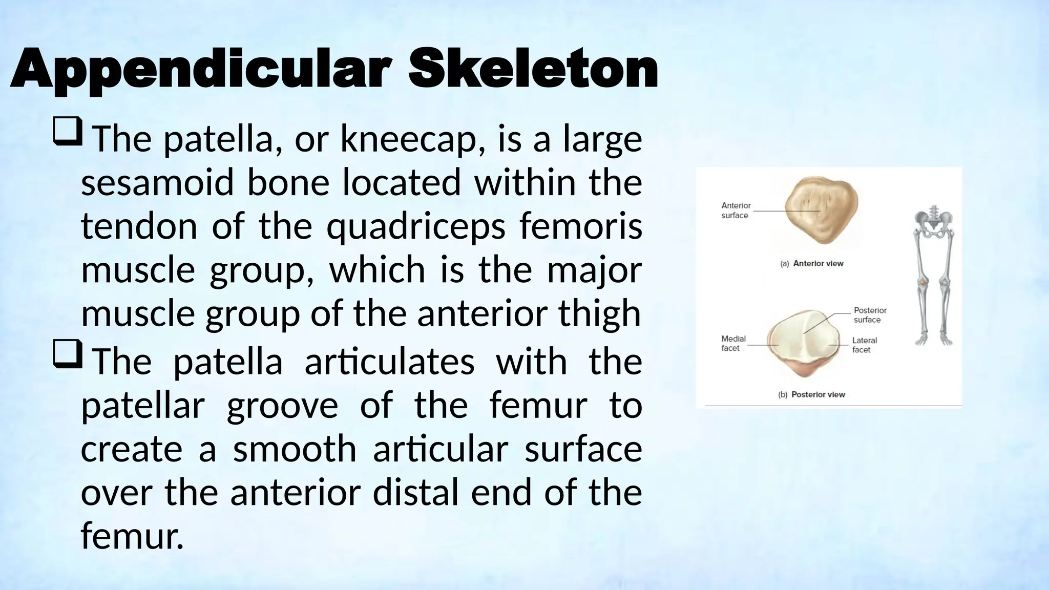
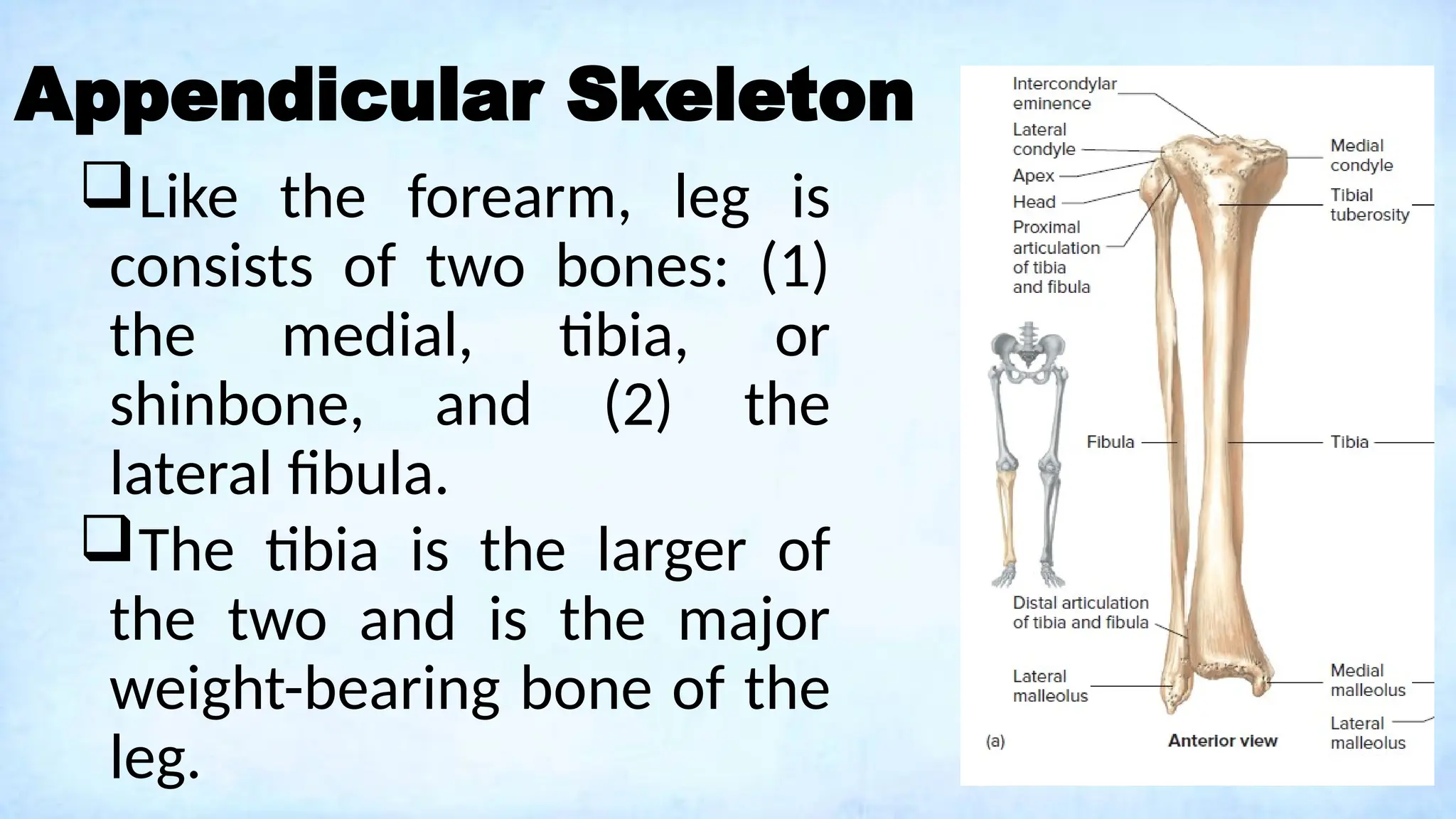
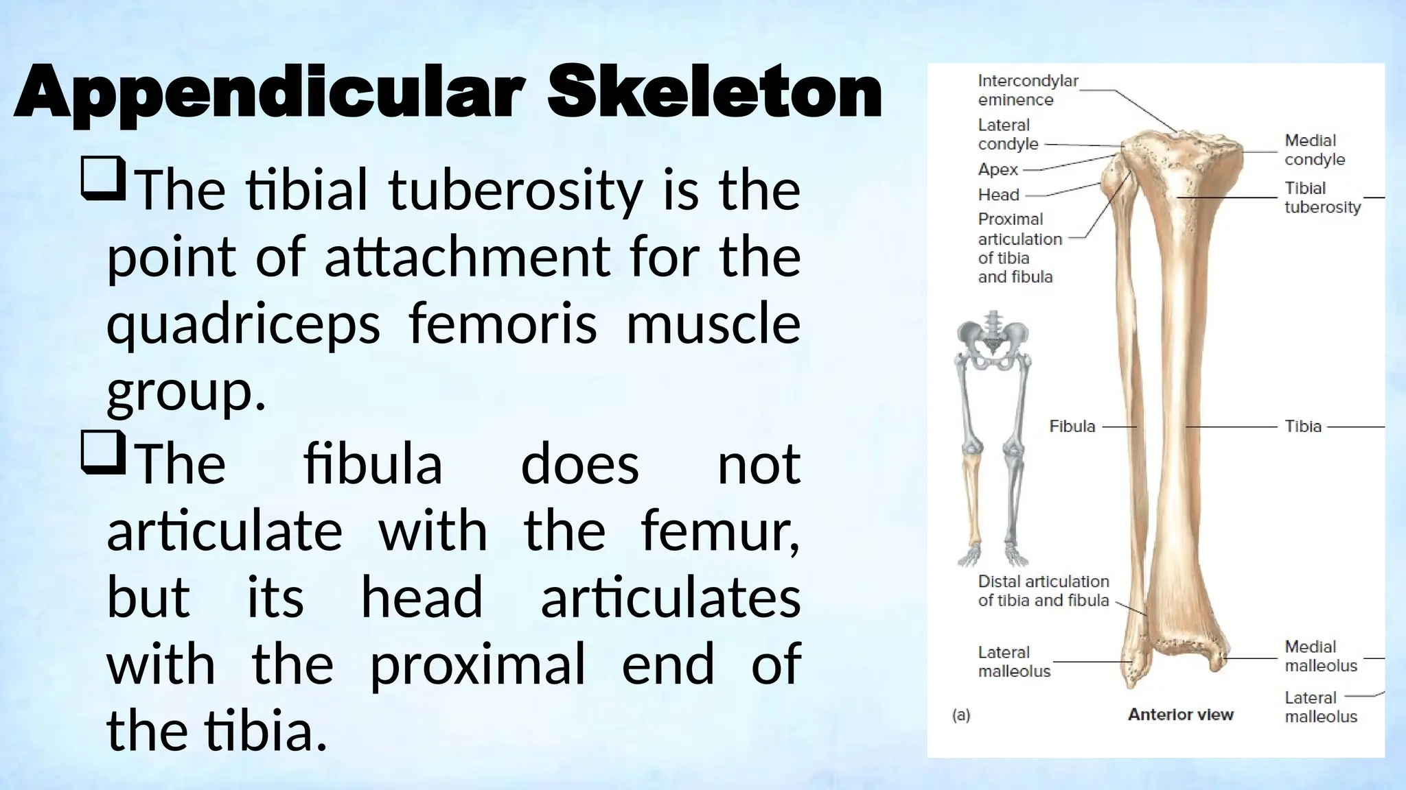
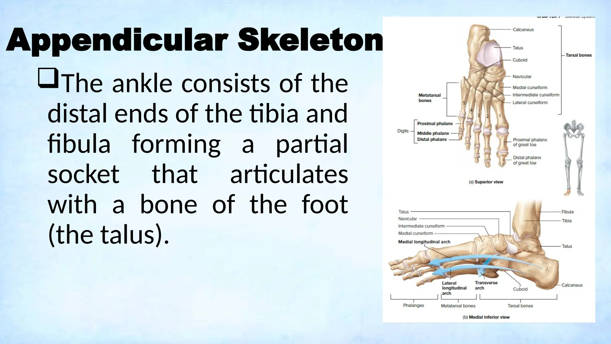
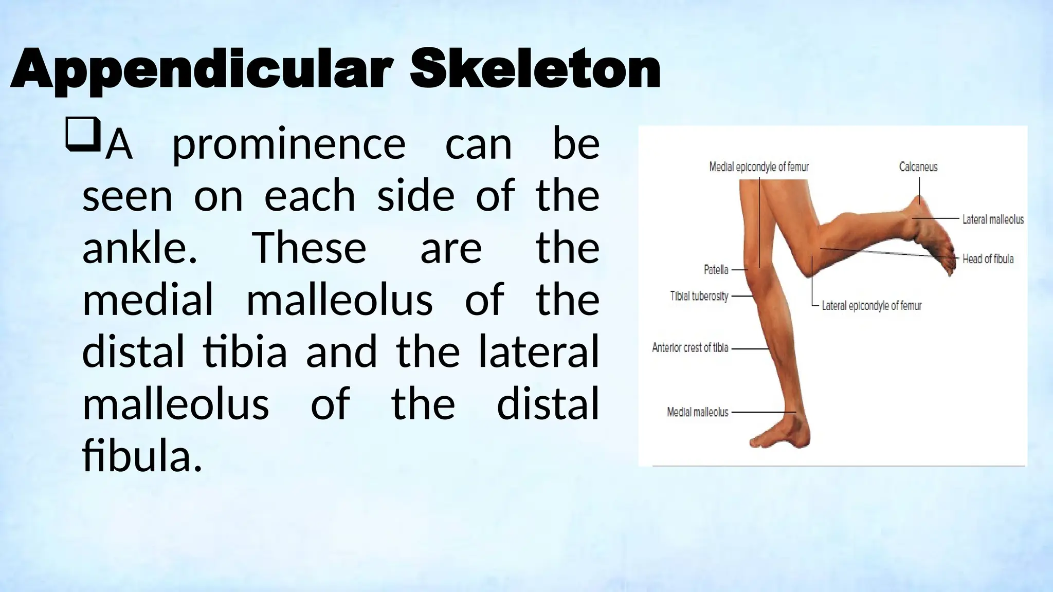
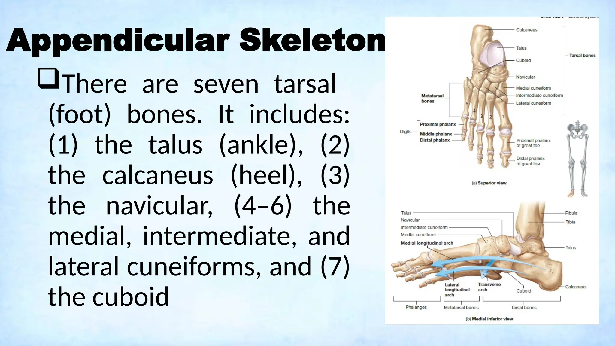
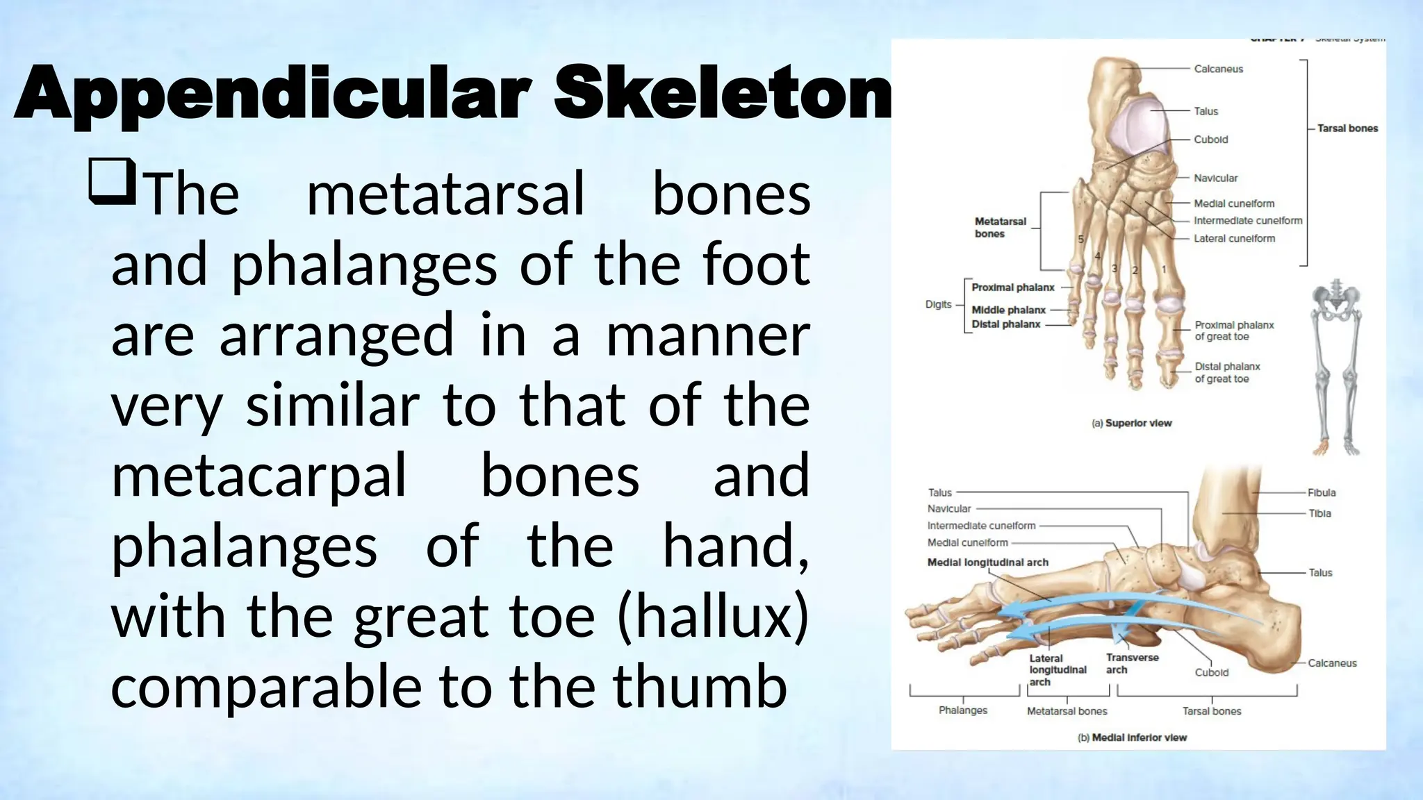
SKELETAL
SYSTEM
ERLISON LORENZ M. OGNILLA RN, LPT, MAT
INSTRUCTOR
COMPONENTS:
bones
cartilage
tendons
ligaments
Functions:
body support
strong and rigid bone-> bearing weight; main
supporting tissue
firm but flexible support -> nose, external ear,
thoracic cage, trachea
ligaments -> hold bones together
Functions:
organ protection
body movement
skeletal muscles attach to bones by tendons
contraction of skeletal muscles-> moves
bones-> body movements
joints -> allow movement between bones
Functions:
Mineral storage
Calcium and phosphorus
Adipose tissues within bone cavities
Blood cell production
Bone cavities-> red bone marrow -> blood cells
and platelets
Cartilage:
Three types:
 Hyaline -> most associated with bone*
 Fibrocartilage
Elastic
{
provide
support
Cartilage:
 Hyaline cartilage chondroblasts
secrete a matrix, which surrounds
the chondroblasts
 Once the matrix has surrounded
the chondroblast, it has
differentiated into a chondrocyte.
 Chondrocytes are rounded cells
that occupy a space called a
lacuna within the matrix.
 The matrix contains collagen,
which provides strength, and
proteoglycans, which make
cartilage resilient by trapping
water
Cartilage:
 Most cartilage is covered by a
protective connective tissue
sheath called the perichondrium
 The perichondrium is a double-
layered outer layer of dense
irregular connective tissue
containing fibroblasts. The inner,
more delicate layer has fewer
fibers and contains
chondroblasts.
 Blood vessels and nerves
penetrate the outer layer of the
perichondrium but do not enter
the cartilage matrix*
Cartilage:
 Articular cartilage, which is
hyaline cartilage that covers the
ends of bones where they come
together to form joints, has no
perichondrium, blood vessels, or
nerves.
There are two types of cartilage growth: (1)
appositional growth and (2) interstitial growth.
In appositional growth, chondroblasts
in the perichondrium add new cartilage to the
outside edge of the existing cartilage. In
interstitial growth, chondrocytes in the center of
the tissue divide and add more matrix in
between the existing cells
Bone Matrix:
Bone -> connective tissue = spread-out cells
separated by nonliving material (matrix)
Bone cells produce and get entrapped within
matrix
The cells also break down old matrix so that
new matrix can replace it.
Bone matrix composition is responsible for the
characteristics of bone.
Bone Matrix:
by weight, 35% organic (collagen and
proteoglycans) and 65% inorganic
(hydroxyapatite- calcium phosphate crystal)
collagen fibers = flexible strength*
 like concrete, the mineral components give
the matrix weight-bearing strength.
Bone Matrix:
(a) Normal bone. (b)
Demineralized bone, soaked in
acid, in which collagen is the
primary remaining component,
can be bent without breaking. (c)
When collagen is denatured,
mineral is the primary remaining
component, making the bone so
brittle that it is easilyshattered.
Bone Cells: Osteoblasts
Bone-building cells
 produce collagen and proteoglycans
secrete matrix vesicles -> high concentration
of calcium and phosphate ions -> formation of
hydroxyapatite crystal -> stimulate further
hydroxyapatite formation and mineralization of
the matrix
Bone Cells: Osteoblasts
The formation of new bone by osteoblasts is
called ossification or osteogenesis
a) On a preexisting surface, such as cartilage or bone, the cell extensions of different osteoblasts join
together. (b) Osteoblasts have produced bone matrix and are now osteocytes. (c) Photomicrograph of
an osteocyte in a lacuna
with cell extensions in the canaliculi. (c)
Bone Cells: Osteocytes
Osteoblasts become osteocytes
once the osteoblasts have
secreted sufficient bone matrix
They account for 90–95% of bone
cells and are very long-lived, with
a lifespan of up to 25 years
Osteocyte cell bodies are housed
within the bone matrix in spaces
called lacunae
Bone Cells: Osteoclasts
bone-destroying cells
break down old or damaged bone cells
Bone reabsorption*
This breakdown is important for mobilizing
crucial Ca2+ and phosphate ions for use in
many metabolic processes.
Spongy Bone
appears porous, has less bone
matrix and more space than
compact bone
consists of interconnecting rods
or plates of bone called
trabeculae
Between the trabeculae are
spaces, which in life are filled with
bone marrow and blood vessels.
Compact Bone
solid, outer layer
surrounding each
bone.
It has more matrix
and is denser with
fewer pores than
spongy bone.
Compact Bone
Osteon or haversian
system- functional unit
of long bone
composed of
concentric rings of
matrix which surround
a central tunnel and
contain osteocytes
Compact Bone
 Perforating canals (Volkmann
canals) deliver blood to the central
canals of the osteons
 They contain blood vessels from the
periosteum or medullary cavity.
 Nutrients in the blood vessels are
delivered to the osteocytes.
 Because the osteocytes are in
contact with each other through
the canaliculi, nutrients are passed
from osteocyte to osteocyte of each
osteon.
Structure of a Long Bone
Diaphysis-center portion
of the bone. It is composed
primarily of compact bone,
surrounding a hollow
center called the
medullary cavity. Some
spongy bone can be found
lining the medullary cavity.
Structure of a Long Bone
The ends of a long bone
are called epiphyses. The
epiphyses are mostly
spongy bone, with an outer
layer of compact bone.
Within joints, the end of a
long bone is covered with
hyaline cartilage called
articular cartilage
Structure of a Long Bone
During bone formation
and growth, bones develop
from centers of ossification
The primary ossification
center is in the diaphysis.
The epiphysis develops
from different centers of
ossification from the
diaphysis
Structure of a Long Bone
The epiphyseal plate,
or growth plate, is
located between the
epiphysis and the
diaphysis.
Growth in bone length
occurs at the epiphyseal
plate.
Structure of a Long Bone
The cavities of spongy
bone and the medullary
cavity are filled with
marrow.
Red marrow is the site
of blood cell formation,
and yellow marrow is
mostly adipose tissue*
Structure of a Long Bone
 The periosteum is a connective
tissue membrane covering the
outer surface of a bone
 The outer fibrous layer is dense
irregular collagenous connective
tissue that contains blood vessels
and nerves.
 The inner layer is a single layer of
bone cells, including osteoblasts,
osteoclasts, and osteochondral
progenitor cells
Structure of a Long Bone
 Endosteum is a single cell layer
of connective tissue that lines
the internal surfaces of all
cavities within bones, such as
the medullary cavity of the
diaphysis and the smaller
cavities in spongy and compact
bone
 The endosteum includes
osteoblasts, osteoclasts, and
osteochondral progenitor cells.
Structure of a Flat Bone
Flat bones contain an
interior framework of
spongy bone
sandwiched between
two layers of compact
bone
Structure of a Short and Irregular
Short and irregular bones have a composition
similar to the epiphyses of long bones—
compact bone surfaces surrounding a spongy
bone center with small spaces that are usually
filled with marrow.
Short and irregular bones are not elongated
and have no diaphyses.
Structure of a Short and Irregular
Short bones are roughly cube
shaped with vertical and horizontal
dimensions approximately equal.
They consist primarily of spongy
bone, which is covered by a thin
layer of compact bone.
Short bones include the bones of
the wrist and ankle.
Structure of a Short and Irregular
Bones that are not in any of
the above three categories are
classified as irregular bones.
They are primarily spongy
bone that is covered with a
thin layer of compact bone.
The vertebrae and some of
the bones in the skull are
irregular bones.
Axial Skeleton: Skull
The skull consists of 8 cranial bones and 14
facial bones, a total of 22 bones
The cranial bones, or cranium house and
protect the brain
The cranial bones are connected by
immovable joints called sutures. There are four
principal sutures: (1) coronal, (2) sagittal, (3)
lambdoid, and (4) squamous
Coronal
suture Sagittal
suture
Lambdoid
suture
Squamous
suture
Axial Skeleton: Skull
The 8 bones of the cranium include:
(1) the frontal bone,
(2) the 2 parietal bones,
(3) the 2 temporal bones,
(4) the occipital bone,
(5) the sphenoid bone, and
(6) the ethmoid bone.
Axial Skeleton: Skull
The frontal bone is connected to the two
parietal bones by the coronal suture
Forehead
 It forms the roof of both the orbit of the eye
and the nasal cavity
Internally, the frontal bone forms the anterior
cranial fossa, which supports the frontal lobes
of the brain
Axial Skeleton: Skull
The parietal bone form nearly half of the
superior portion of the skull.
 The two parietal bones are joined medially by
the sagittal suture and are connected to the
occipital bone by the lambdoid suture.
Along with the temporal bones, the parietal
bones make up the majority of the lateral
portion of the skull
Axial Skeleton: Skull
Temporal bones are
connected to the skull by
the squamous sutures
The temporal bone is
subdivided into three main
regions: (1) the squamous
part, (2) the tympanic part,
and (3) the petrous part.
Axial Skeleton: Skull
The squamous part of each
temporal bone meets the parietal
bone superiorly.
The zygomatic process extends
from the squamous part
anteriorly toward the zygomatic
bone of the face. It joins with the
temporal process of the zygomatic
bone to form the zygomatic arch,
a bridge across the side of the
skull
Axial Skeleton: Skull
The tympanic part of
the temporal bone has
the prominent external
acoustic meatus
Axial Skeleton: Skull
 The petrous part of the temporal
bone extends posterolaterally
from the center of the sphenoid
bone
 The petrous part and sphenoid
bone make up the middle cranial
fossa which contains the temporal
lobes of the brain.
 The petrous part is a thick, bony
ridge (petrous; rocky), is hollow,
and houses the middle and inner
ears.
Axial Skeleton: Skull
 Externally, the mastoid process is
a large, bony inferior projection
that can be seen and felt just
posterior to the external ear.
 Styloid process projects from the
inferior of the petrous part of the
temporal bone. It serves as an
attachment site for three muscles
necessary for movement of the
tongue, hyoid bone, and pharynx
Axial Skeleton: Skull
The occipital bone makes up the
majority of the skull’s posterior
wall and base.
Its most prominent feature is the
foramen magnum (large hole),
the opening where the brainstem
connects to the spinal cord
It forms the posterior cranial
fossae that support the
cerebellum of the brain.
Axial Skeleton: Skull
Although appearing to be two
bones, one on each side of the
skull anterior to the temporal
bone, the sphenoid bone is
actually a single bone that
extends completely across the
skull
When viewed as a whole, the
sphenoid bone somewhat
resembles a butterfly with four
distinct parts.*
Axial Skeleton: Skull
The ethmoid bone is a
porous, fragile bone.
It is centrally located
in the skull, forming the
nasal septum (wall), a
large portion of the
nasal cavity, as well as
the medial wall of the
orbits.
Axial Skeleton: Skull
The 14 facial bones of the skull include
(1) the 2 zygomatic bones,
(2) the 2 maxilla bones,
(3) the 2 palatine bones,
(4) the 2 lacrimal bones,
(5) the 2 nasal bones,
(6) the mandible,
(7) the vomer bone, and
(8) the 2 inferior nasal conchae
Axial Skeleton: Skull
The zygomatic bones, commonly known as the
cheekbones, are anterior to the sphenoid bone.
There they form part of the inferiolateral
border of the orbits
As previously discussed, the temporal process
of the zygomatic bone articulates with the
zygomatic process of the temporal bone to form
the zygomatic arch.
Axial Skeleton: Skull
Each maxilla is anterior and inferior to the
zygomatic bones and the two maxillae are
fused medially
The maxillae are two of the three jaw bones,
and form (1) the upper jaw, (2) the majority of
the roof of the mouth, and (3) the center
portion of the face
Axial Skeleton: Skull
The palatine bones
have horizontal plates
that fuse medially to
form the posterior
portion of the hard
palate as described
earlier with the
maxillae.
Axial Skeleton: Skull
The lacrimal bones are
the smallest of the skull
bones and house the
depression through which
the nasolacrimal duct
enters the nasolacrimal
canal, joining the orbits
and nasal cavity
•The nasal bones,
along with the frontal
processes of the
maxillae, form the
bridge of the nose.
Axial Skeleton: Skull
The mandible is the only
skull bone that is freely
movable relative to the
other skull bones.
It is inferior to the maxillae
and attaches posteriorly to
the temporal bone via the
mandibular fossa
Axial Skeleton: Hyoid Bone
important for speech and
swallowing.
Some tongue muscles as well
as neck muscles that elevate
the larynx attach to the hyoid
The hyoid bone has the unique
distinction of being the only
bone in the body not directly
attached to another bone.*
Axial Skeleton: Vertebral Column
performs five major functions:
Axial Skeleton: Vertebral Column
performs five major functions:
(1) It supports the weight of the head and
trunk, (2) it protects the spinal cord, (3) it
allows spinal nerves to exit the spinal cord, (4)
it provides a site for muscle attachment, and
(5) it permits movement of the head and
trunk.
Axial Skeleton: Vertebral Column
The 26 bones, called
vertebrae, which can be divided
into five regions: 7 cervical
vertebrae, 12 thoracic
vertebrae, 5 lumbar vertebrae,
1 sacral bone, and 1 coccygeal
bone.
Axial Skeleton: Vertebral Column
The adult vertebral column has
four major curvatures
The cervical region curves
anteriorly, the thoracic region
curves posteriorly, the lumbar
region curves anteriorly, and the
sacral and coccygeal regions
together curve posteriorly.
Axial Skeleton: Vertebral Column
Each vertebra consists of (1)
a body, (2) a vertebral arch,
and (3) various processes.
The vertebral body, the solid
bony disk of each vertebra,
supports the body’s weight.
The vertebral arch, along
with the body, protects the
spinal cord.
Axial Skeleton: Vertebral Column
The vertebral arch
projects posteriorly from
the body. Together, the
vertebral arch and the body
form a complete bony circle
enclosing the vertebral
foramen, which is occupied
by the spinal cord in a living
person
Axial Skeleton: Vertebral Column
The spinous
processes can be
seen and felt as a
series of rounded
projections down the
midline of the back.
Axial Skeleton: Vertebral Column
The intervertebral foramina
are the locations where two
vertebrae meet
Each intervertebral foramen
is formed by intervertebral
notches in the pedicles of
adjacent vertebrae. These
foramina are where spinal
nerves exit the spinal cord.
Axial Skeleton: Vertebral Column
In a living person, vertebrae are
separated by intervertebral disks.
Intervertebral disks are
composed of fibrocartilage*, and
are located between the bodies
of adjacent vertebrae
The intervertebral disks provide
support and cushion the
vertebral bodies.
Axial Skeleton: Vertebral Column
Cervical vertebrae are
located in the vertebral
column region with the
greatest range of motion.
These vertebrae support and
move the head.*
Most cervical vertebrae have
bifid (split) spinous processes.
Axial Skeleton: Vertebral Column
 The first two cervical vertebrae include
(1) the atlas (C1) and (2) the axis (C2).
 The atlas and axis are heftier than the
other five cervical vertebrae since they
directly support the weight of the head
and control its movements.
Axial Skeleton: Vertebral Column
The thoracic region is the
least moveable of the five
regions due to the
articulation of the thoracic
vertebrae with the ribs.
It supports the thoracic
cage, which houses and
protects the heart and lungs
Axial Skeleton: Vertebral Column
These vertebrae have the
longest spinous processes,
which project inferiorly.
Their transverse processes
are longer than others and
the first 10 possess articular
facets for the tubercles of
the ribs.
Axial Skeleton: Vertebral Column
The bodies have articular
facets for the heads of the
ribs. The head of most ribs
articulates with the inferior
articular facet of one
vertebra and with the
superior articular facet for
the rib head on the next
vertebra down.
Axial Skeleton: Vertebral Column
Lumbar vertebrae support
the majority of the body’s
weight
They have massive bodies
and heavy, rectangular
transverse and spinous
processes.
Axial Skeleton: Vertebral Column
The sacrum is located between
the two hip bones.
 It articulates with each hip
bone by way of the auricular
surfaces, forming the sacroiliac
joint.
The sacrum is part of the pelvic
girdle, which provides stable
support for our lower limbs.
Axial Skeleton: Vertebral Column
The coccyx is commonly
referred to as the tailbone.
It is the terminal portion
of the vertebral column.
Axial Skeleton: Thoracic cage
 Thoracic cage, commonly called
the rib cage, protects the heart
and lungs within the thorax.
 It forms a semirigid chamber,
which can increase and decrease
in volume during respiration.
 It consists of (1) the thoracic
vertebrae, (2) the ribs with their
associated costal (rib) cartilages,
and (3) the sternum
Axial Skeleton: Thoracic cage
There are 12 pairs of ribs.
Ribs are classified as either
true ribs or false ribs.
Ribs 1–7 are called true ribs.
True ribs attach directly
through costal cartilage to the
sternum.
Ribs 8–12 are called false ribs
because they do not attach
directly to the sternum.
Axial Skeleton: Thoracic cage
Ribs 8–10 are joined by a
common cartilage to the
costal cartilage of rib 7,
which in turn is attached to
the sternum.
Two of the false ribs, ribs
11 and 12, are also called
floating ribs because they do
not attach to the sternum
Axial Skeleton: Thoracic cage
Costal cartilages are
flexible and permit the
thoracic cage to expand
during respiration
Axial Skeleton: Thoracic cage
 Most ribs attach to the thoracic
vertebrae in two locations.
 First, the head of the rib articulates
with the bodies and intervertebral
disks of two adjacent vertebrae.
 These sites of articulation are called
the inferior articular facet on the
superior vertebra and the superior
articular facet on the inferior vertebra.
 Second, the tubercle of the rib
articulates with the transverse process
of the inferior vertebra
Axial Skeleton: Thoracic cage
The sternum, or
breastbone, has been
described as sword
shaped and has three
parts: (1) The
manubrium; (2) the body,
and (3) the xiphoid
process
Appendicular Skeleton
The pectoral girdle consists
of two pairs of bones that
attach each of the upper
limbs to the body:
Each pair is composed of a
scapula, commonly known
as the shoulder blade and a
clavicle, commonly known as
the collarbone
Appendicular Skeleton
The acromion process has
three functions: (1) to form
a protective cover for the
shoulder joint, (2) to form
the attachment site for the
clavicle, and (3) to provide
attachment points for some
of the shoulder muscles.
Appendicular Skeleton
The smaller coracoid
process provides
attachments for some
shoulder and arm muscles.
A glenoid cavity, located in
the superior lateral portion
of the bone, articulates with
the head of the humerus.
Appendicular Skeleton
The clavicle is a long bone with
a slight sigmoid (S-shaped)
curve.
It articulates with the scapula
and the sternum.
On the lateral end it articulates
with the acromion process. On
the medial end it articulates
with the manubrium of the
sternum.
Appendicular Skeleton
The humeral head articulates
with the glenoid cavity of the
scapula.
The proximal end of the
humerus has two prominent
landmarks, both sites of muscle
attachment: (1) Laterally, it is
the greater tubercle. (2)
Anteriorly, it is the lesser
tubercle.
Appendicular Skeleton
The diaphysis of the
humerus has a noticeable
lateral landmark.
The deltoid tuberosity is
the site of attachment of
the deltoid muscle.
Appendicular Skeleton
 Laterally, the humerus articulates with
the radius. This location is called the
capitulum.
 Medially, the humerus articulates
with the ulna. This location is called
the trochlea.
 Forearm muscles attach to the
humerus just superior to the
capitulum and trochlea.
 The attachment sites are the medial
epicondyle and the lateral epicondyle.
Appendicular Skeleton
The forearm has two
bones: (1) the ulna and (2)
the radius.
The ulna is medial, the
same side as the little
finger, and the radius is
lateral, the same side as
the thumb
Appendicular Skeleton
The trochlear notch rotates over
the trochlea of the humerus
when bending the elbow.
The coronoid process inserts
into the coronoid fossa of the
humerus when the elbow is bent
The olecranon process inserts
into the olecranon fossa of the
humerus when the elbow is
straightened
Appendicular Skeleton
Radial notch where the
head of the radius rotates.
The head of the ulna
articulates with both the
radius and the wrist bones
styloid process- where
ligaments of the wrist
attach
Appendicular Skeleton
 The head of the radius articulates
with the humerus and rotates over
the capitulum of the humerus
when bending the elbow.
 radial tuberosity is the site of
attachment for the biceps brachii
muscle of the arm
 The styloid process of the radius is
lateral and also serves as an
attachment location for wrist
ligaments.
Appendicular Skeleton
 The wrist is composed of eight
carpal bones arranged into two
rows of four each.
 The proximal row of carpal bones,
lateral to medial, includes (1) the
scaphoid, which is boat-shaped;
(2) the lunate, which is moon-
shaped; (3) the three-cornered
triquetrum; and (4) the pea-
shaped pisiform, which is located
on the palmar surface of the
triquetrum.
Appendicular Skeleton
 The distal row of carpal bones,
from medial to lateral, includes
1 hamate which has a hooked
process on its palmar side; (2)
the head-shaped capitate; (3)
the trapezoid, four-sided
geometric form with two
parallel sides; and (4) the
trapezium, four-sided geometric
form with no two sides parallel.
Appendicular Skeleton
Five metacarpal bones are
attached to the carpal
bones and make up the
central portion of the hand.
 They are numbered one
to five, starting with the
most lateral metacarpal
bone, at the base of the
thumb.
Appendicular Skeleton
 The five digits of each hand
include one thumb (pollex) and
four fingers.
 Each digit consists of small
long bones called phalanges.
 The thumb has two phalanges,
called proximal and distal.
 Each finger has three
phalanges, designated
proximal, middle, and distal.
Appendicular Skeleton
The pelvic girdle is formed
by the two hip bones (coxal
bones) and the sacrum.
The hip bones join each
other anteriorly and connect
with the sacrum posteriorly
forming a complete circle of
bone.
The pelvis includes the
pelvic girdle and the coccyx
Appendicular Skeleton
The pelvic girdle is formed
by the two hip bones (coxal
bones) and the sacrum.
The hip bones join each
other anteriorly and connect
with the sacrum posteriorly
forming a complete circle of
bone.
The pelvis includes the
pelvic girdle and the coccyx
Appendicular Skeleton
 Each hip bone is formed from
three separate bones fused
into a large bony plate: (1) the
ilium, (2) the ischium, and (3)
the pubis
 The three individual bones
converge near the center of
the hip socket, called the
acetabulum where lower limbs
articulate with the pelvic girdle
Appendicular Skeleton
The pelvic girdle serves as
the place of attachment for
the lower limbs, supports the
weight of the body, and
protects internal organs.
In women, it protects
developing fetus and forms a
passageway through which
the fetus passes during
delivery.
Appendicular Skeleton
The anterior superior
iliac spine is an
important anatomical
landmark used, for
example, to find the
correct location for
giving gluteal injections
into the hip.
Appendicular Skeleton
Posterior thigh muscles
attach to the ischial
tuberosity and its the
portion of the pelvis on
which a person sits.
The two pubic bones are
joined by the pubic
symphysis, a thick pad of
fibrocartilage
Appendicular Skeleton
The thigh, like the arm,
contains a single bone,
the femur.
The femur has a
prominent, rounded
head, where it
articulates with the
acetabulum
Appendicular Skeleton
The proximal shaft
exhibits two projections:
a greater trochanter and
lesser trochanter.
Both trochanters are
attachment sites for
muscles that fasten the
hip to the thigh.
Appendicular Skeleton
 The femur articulates with the tibia to
form the knee.
 There are two smooth rounded
projections that rotate on the superior
surface of the tibia when we bend our
knee. These two rounded projections
are (1) the medial condyle and (2) the
lateral condyle.
 Just superior to the medial and lateral
condyles are two large ligament
attachment sites. These are (1) the
medial epicondyle and (2) the lateral
epicondyle
Appendicular Skeleton
The patella, or kneecap, is a large
sesamoid bone located within the
tendon of the quadriceps femoris
muscle group, which is the major
muscle group of the anterior thigh
The patella articulates with the
patellar groove of the femur to
create a smooth articular surface
over the anterior distal end of the
femur.
Appendicular Skeleton
Like the forearm, leg is
consists of two bones: (1)
the medial, tibia, or
shinbone, and (2) the
lateral fibula.
The tibia is the larger of
the two and is the major
weight-bearing bone of the
leg.
Appendicular Skeleton
The tibial tuberosity is the
point of attachment for the
quadriceps femoris muscle
group.
The fibula does not
articulate with the femur,
but its head articulates
with the proximal end of
the tibia.
Appendicular Skeleton
The ankle consists of the
distal ends of the tibia and
fibula forming a partial
socket that articulates
with a bone of the foot
(the talus).
Appendicular Skeleton
A prominence can be
seen on each side of the
ankle. These are the
medial malleolus of the
distal tibia and the lateral
malleolus of the distal
fibula.
Appendicular Skeleton
There are seven tarsal
(foot) bones. It includes:
(1) the talus (ankle), (2)
the calcaneus (heel), (3)
the navicular, (4–6) the
medial, intermediate, and
lateral cuneiforms, and (7)
the cuboid
Appendicular Skeleton
The metatarsal bones
and phalanges of the foot
are arranged in a manner
very similar to that of the
metacarpal bones and
phalanges of the hand,
with the great toe (hallux)
comparable to the thumb

A Lecture on Skeletal System: Components, Structures, and Functions.pptx

  • 1.
    SKELETAL SYSTEM ERLISON LORENZ M.OGNILLA RN, LPT, MAT INSTRUCTOR
  • 4.
  • 6.
    Functions: body support strong andrigid bone-> bearing weight; main supporting tissue firm but flexible support -> nose, external ear, thoracic cage, trachea ligaments -> hold bones together
  • 7.
    Functions: organ protection body movement skeletalmuscles attach to bones by tendons contraction of skeletal muscles-> moves bones-> body movements joints -> allow movement between bones
  • 8.
    Functions: Mineral storage Calcium andphosphorus Adipose tissues within bone cavities Blood cell production Bone cavities-> red bone marrow -> blood cells and platelets
  • 9.
    Cartilage: Three types:  Hyaline-> most associated with bone*  Fibrocartilage Elastic { provide support
  • 10.
    Cartilage:  Hyaline cartilagechondroblasts secrete a matrix, which surrounds the chondroblasts  Once the matrix has surrounded the chondroblast, it has differentiated into a chondrocyte.  Chondrocytes are rounded cells that occupy a space called a lacuna within the matrix.  The matrix contains collagen, which provides strength, and proteoglycans, which make cartilage resilient by trapping water
  • 11.
    Cartilage:  Most cartilageis covered by a protective connective tissue sheath called the perichondrium  The perichondrium is a double- layered outer layer of dense irregular connective tissue containing fibroblasts. The inner, more delicate layer has fewer fibers and contains chondroblasts.  Blood vessels and nerves penetrate the outer layer of the perichondrium but do not enter the cartilage matrix*
  • 12.
    Cartilage:  Articular cartilage,which is hyaline cartilage that covers the ends of bones where they come together to form joints, has no perichondrium, blood vessels, or nerves. There are two types of cartilage growth: (1) appositional growth and (2) interstitial growth. In appositional growth, chondroblasts in the perichondrium add new cartilage to the outside edge of the existing cartilage. In interstitial growth, chondrocytes in the center of the tissue divide and add more matrix in between the existing cells
  • 13.
    Bone Matrix: Bone ->connective tissue = spread-out cells separated by nonliving material (matrix) Bone cells produce and get entrapped within matrix The cells also break down old matrix so that new matrix can replace it. Bone matrix composition is responsible for the characteristics of bone.
  • 14.
    Bone Matrix: by weight,35% organic (collagen and proteoglycans) and 65% inorganic (hydroxyapatite- calcium phosphate crystal) collagen fibers = flexible strength*  like concrete, the mineral components give the matrix weight-bearing strength.
  • 15.
    Bone Matrix: (a) Normalbone. (b) Demineralized bone, soaked in acid, in which collagen is the primary remaining component, can be bent without breaking. (c) When collagen is denatured, mineral is the primary remaining component, making the bone so brittle that it is easilyshattered.
  • 16.
    Bone Cells: Osteoblasts Bone-buildingcells  produce collagen and proteoglycans secrete matrix vesicles -> high concentration of calcium and phosphate ions -> formation of hydroxyapatite crystal -> stimulate further hydroxyapatite formation and mineralization of the matrix
  • 17.
    Bone Cells: Osteoblasts Theformation of new bone by osteoblasts is called ossification or osteogenesis a) On a preexisting surface, such as cartilage or bone, the cell extensions of different osteoblasts join together. (b) Osteoblasts have produced bone matrix and are now osteocytes. (c) Photomicrograph of an osteocyte in a lacuna with cell extensions in the canaliculi. (c)
  • 18.
    Bone Cells: Osteocytes Osteoblastsbecome osteocytes once the osteoblasts have secreted sufficient bone matrix They account for 90–95% of bone cells and are very long-lived, with a lifespan of up to 25 years Osteocyte cell bodies are housed within the bone matrix in spaces called lacunae
  • 19.
    Bone Cells: Osteoclasts bone-destroyingcells break down old or damaged bone cells Bone reabsorption* This breakdown is important for mobilizing crucial Ca2+ and phosphate ions for use in many metabolic processes.
  • 20.
    Spongy Bone appears porous,has less bone matrix and more space than compact bone consists of interconnecting rods or plates of bone called trabeculae Between the trabeculae are spaces, which in life are filled with bone marrow and blood vessels.
  • 21.
    Compact Bone solid, outerlayer surrounding each bone. It has more matrix and is denser with fewer pores than spongy bone.
  • 22.
    Compact Bone Osteon orhaversian system- functional unit of long bone composed of concentric rings of matrix which surround a central tunnel and contain osteocytes
  • 23.
    Compact Bone  Perforatingcanals (Volkmann canals) deliver blood to the central canals of the osteons  They contain blood vessels from the periosteum or medullary cavity.  Nutrients in the blood vessels are delivered to the osteocytes.  Because the osteocytes are in contact with each other through the canaliculi, nutrients are passed from osteocyte to osteocyte of each osteon.
  • 24.
    Structure of aLong Bone Diaphysis-center portion of the bone. It is composed primarily of compact bone, surrounding a hollow center called the medullary cavity. Some spongy bone can be found lining the medullary cavity.
  • 25.
    Structure of aLong Bone The ends of a long bone are called epiphyses. The epiphyses are mostly spongy bone, with an outer layer of compact bone. Within joints, the end of a long bone is covered with hyaline cartilage called articular cartilage
  • 26.
    Structure of aLong Bone During bone formation and growth, bones develop from centers of ossification The primary ossification center is in the diaphysis. The epiphysis develops from different centers of ossification from the diaphysis
  • 27.
    Structure of aLong Bone The epiphyseal plate, or growth plate, is located between the epiphysis and the diaphysis. Growth in bone length occurs at the epiphyseal plate.
  • 28.
    Structure of aLong Bone The cavities of spongy bone and the medullary cavity are filled with marrow. Red marrow is the site of blood cell formation, and yellow marrow is mostly adipose tissue*
  • 29.
    Structure of aLong Bone  The periosteum is a connective tissue membrane covering the outer surface of a bone  The outer fibrous layer is dense irregular collagenous connective tissue that contains blood vessels and nerves.  The inner layer is a single layer of bone cells, including osteoblasts, osteoclasts, and osteochondral progenitor cells
  • 30.
    Structure of aLong Bone  Endosteum is a single cell layer of connective tissue that lines the internal surfaces of all cavities within bones, such as the medullary cavity of the diaphysis and the smaller cavities in spongy and compact bone  The endosteum includes osteoblasts, osteoclasts, and osteochondral progenitor cells.
  • 31.
    Structure of aFlat Bone Flat bones contain an interior framework of spongy bone sandwiched between two layers of compact bone
  • 32.
    Structure of aShort and Irregular Short and irregular bones have a composition similar to the epiphyses of long bones— compact bone surfaces surrounding a spongy bone center with small spaces that are usually filled with marrow. Short and irregular bones are not elongated and have no diaphyses.
  • 33.
    Structure of aShort and Irregular Short bones are roughly cube shaped with vertical and horizontal dimensions approximately equal. They consist primarily of spongy bone, which is covered by a thin layer of compact bone. Short bones include the bones of the wrist and ankle.
  • 34.
    Structure of aShort and Irregular Bones that are not in any of the above three categories are classified as irregular bones. They are primarily spongy bone that is covered with a thin layer of compact bone. The vertebrae and some of the bones in the skull are irregular bones.
  • 35.
    Axial Skeleton: Skull Theskull consists of 8 cranial bones and 14 facial bones, a total of 22 bones The cranial bones, or cranium house and protect the brain The cranial bones are connected by immovable joints called sutures. There are four principal sutures: (1) coronal, (2) sagittal, (3) lambdoid, and (4) squamous
  • 36.
  • 37.
    Axial Skeleton: Skull The8 bones of the cranium include: (1) the frontal bone, (2) the 2 parietal bones, (3) the 2 temporal bones, (4) the occipital bone, (5) the sphenoid bone, and (6) the ethmoid bone.
  • 38.
    Axial Skeleton: Skull Thefrontal bone is connected to the two parietal bones by the coronal suture Forehead  It forms the roof of both the orbit of the eye and the nasal cavity Internally, the frontal bone forms the anterior cranial fossa, which supports the frontal lobes of the brain
  • 41.
    Axial Skeleton: Skull Theparietal bone form nearly half of the superior portion of the skull.  The two parietal bones are joined medially by the sagittal suture and are connected to the occipital bone by the lambdoid suture. Along with the temporal bones, the parietal bones make up the majority of the lateral portion of the skull
  • 43.
    Axial Skeleton: Skull Temporalbones are connected to the skull by the squamous sutures The temporal bone is subdivided into three main regions: (1) the squamous part, (2) the tympanic part, and (3) the petrous part.
  • 44.
    Axial Skeleton: Skull Thesquamous part of each temporal bone meets the parietal bone superiorly. The zygomatic process extends from the squamous part anteriorly toward the zygomatic bone of the face. It joins with the temporal process of the zygomatic bone to form the zygomatic arch, a bridge across the side of the skull
  • 45.
    Axial Skeleton: Skull Thetympanic part of the temporal bone has the prominent external acoustic meatus
  • 46.
    Axial Skeleton: Skull The petrous part of the temporal bone extends posterolaterally from the center of the sphenoid bone  The petrous part and sphenoid bone make up the middle cranial fossa which contains the temporal lobes of the brain.  The petrous part is a thick, bony ridge (petrous; rocky), is hollow, and houses the middle and inner ears.
  • 47.
    Axial Skeleton: Skull Externally, the mastoid process is a large, bony inferior projection that can be seen and felt just posterior to the external ear.  Styloid process projects from the inferior of the petrous part of the temporal bone. It serves as an attachment site for three muscles necessary for movement of the tongue, hyoid bone, and pharynx
  • 48.
    Axial Skeleton: Skull Theoccipital bone makes up the majority of the skull’s posterior wall and base. Its most prominent feature is the foramen magnum (large hole), the opening where the brainstem connects to the spinal cord It forms the posterior cranial fossae that support the cerebellum of the brain.
  • 50.
    Axial Skeleton: Skull Althoughappearing to be two bones, one on each side of the skull anterior to the temporal bone, the sphenoid bone is actually a single bone that extends completely across the skull When viewed as a whole, the sphenoid bone somewhat resembles a butterfly with four distinct parts.*
  • 51.
    Axial Skeleton: Skull Theethmoid bone is a porous, fragile bone. It is centrally located in the skull, forming the nasal septum (wall), a large portion of the nasal cavity, as well as the medial wall of the orbits.
  • 52.
    Axial Skeleton: Skull The14 facial bones of the skull include (1) the 2 zygomatic bones, (2) the 2 maxilla bones, (3) the 2 palatine bones, (4) the 2 lacrimal bones, (5) the 2 nasal bones, (6) the mandible, (7) the vomer bone, and (8) the 2 inferior nasal conchae
  • 54.
    Axial Skeleton: Skull Thezygomatic bones, commonly known as the cheekbones, are anterior to the sphenoid bone. There they form part of the inferiolateral border of the orbits As previously discussed, the temporal process of the zygomatic bone articulates with the zygomatic process of the temporal bone to form the zygomatic arch.
  • 55.
    Axial Skeleton: Skull Eachmaxilla is anterior and inferior to the zygomatic bones and the two maxillae are fused medially The maxillae are two of the three jaw bones, and form (1) the upper jaw, (2) the majority of the roof of the mouth, and (3) the center portion of the face
  • 57.
    Axial Skeleton: Skull Thepalatine bones have horizontal plates that fuse medially to form the posterior portion of the hard palate as described earlier with the maxillae.
  • 58.
    Axial Skeleton: Skull Thelacrimal bones are the smallest of the skull bones and house the depression through which the nasolacrimal duct enters the nasolacrimal canal, joining the orbits and nasal cavity
  • 59.
    •The nasal bones, alongwith the frontal processes of the maxillae, form the bridge of the nose.
  • 60.
    Axial Skeleton: Skull Themandible is the only skull bone that is freely movable relative to the other skull bones. It is inferior to the maxillae and attaches posteriorly to the temporal bone via the mandibular fossa
  • 61.
    Axial Skeleton: HyoidBone important for speech and swallowing. Some tongue muscles as well as neck muscles that elevate the larynx attach to the hyoid The hyoid bone has the unique distinction of being the only bone in the body not directly attached to another bone.*
  • 63.
    Axial Skeleton: VertebralColumn performs five major functions:
  • 64.
    Axial Skeleton: VertebralColumn performs five major functions: (1) It supports the weight of the head and trunk, (2) it protects the spinal cord, (3) it allows spinal nerves to exit the spinal cord, (4) it provides a site for muscle attachment, and (5) it permits movement of the head and trunk.
  • 65.
    Axial Skeleton: VertebralColumn The 26 bones, called vertebrae, which can be divided into five regions: 7 cervical vertebrae, 12 thoracic vertebrae, 5 lumbar vertebrae, 1 sacral bone, and 1 coccygeal bone.
  • 66.
    Axial Skeleton: VertebralColumn The adult vertebral column has four major curvatures The cervical region curves anteriorly, the thoracic region curves posteriorly, the lumbar region curves anteriorly, and the sacral and coccygeal regions together curve posteriorly.
  • 67.
    Axial Skeleton: VertebralColumn Each vertebra consists of (1) a body, (2) a vertebral arch, and (3) various processes. The vertebral body, the solid bony disk of each vertebra, supports the body’s weight. The vertebral arch, along with the body, protects the spinal cord.
  • 68.
    Axial Skeleton: VertebralColumn The vertebral arch projects posteriorly from the body. Together, the vertebral arch and the body form a complete bony circle enclosing the vertebral foramen, which is occupied by the spinal cord in a living person
  • 69.
    Axial Skeleton: VertebralColumn The spinous processes can be seen and felt as a series of rounded projections down the midline of the back.
  • 70.
    Axial Skeleton: VertebralColumn The intervertebral foramina are the locations where two vertebrae meet Each intervertebral foramen is formed by intervertebral notches in the pedicles of adjacent vertebrae. These foramina are where spinal nerves exit the spinal cord.
  • 71.
    Axial Skeleton: VertebralColumn In a living person, vertebrae are separated by intervertebral disks. Intervertebral disks are composed of fibrocartilage*, and are located between the bodies of adjacent vertebrae The intervertebral disks provide support and cushion the vertebral bodies.
  • 73.
    Axial Skeleton: VertebralColumn Cervical vertebrae are located in the vertebral column region with the greatest range of motion. These vertebrae support and move the head.* Most cervical vertebrae have bifid (split) spinous processes.
  • 74.
    Axial Skeleton: VertebralColumn  The first two cervical vertebrae include (1) the atlas (C1) and (2) the axis (C2).  The atlas and axis are heftier than the other five cervical vertebrae since they directly support the weight of the head and control its movements.
  • 75.
    Axial Skeleton: VertebralColumn The thoracic region is the least moveable of the five regions due to the articulation of the thoracic vertebrae with the ribs. It supports the thoracic cage, which houses and protects the heart and lungs
  • 76.
    Axial Skeleton: VertebralColumn These vertebrae have the longest spinous processes, which project inferiorly. Their transverse processes are longer than others and the first 10 possess articular facets for the tubercles of the ribs.
  • 77.
    Axial Skeleton: VertebralColumn The bodies have articular facets for the heads of the ribs. The head of most ribs articulates with the inferior articular facet of one vertebra and with the superior articular facet for the rib head on the next vertebra down.
  • 78.
    Axial Skeleton: VertebralColumn Lumbar vertebrae support the majority of the body’s weight They have massive bodies and heavy, rectangular transverse and spinous processes.
  • 79.
    Axial Skeleton: VertebralColumn The sacrum is located between the two hip bones.  It articulates with each hip bone by way of the auricular surfaces, forming the sacroiliac joint. The sacrum is part of the pelvic girdle, which provides stable support for our lower limbs.
  • 80.
    Axial Skeleton: VertebralColumn The coccyx is commonly referred to as the tailbone. It is the terminal portion of the vertebral column.
  • 81.
    Axial Skeleton: Thoraciccage  Thoracic cage, commonly called the rib cage, protects the heart and lungs within the thorax.  It forms a semirigid chamber, which can increase and decrease in volume during respiration.  It consists of (1) the thoracic vertebrae, (2) the ribs with their associated costal (rib) cartilages, and (3) the sternum
  • 82.
    Axial Skeleton: Thoraciccage There are 12 pairs of ribs. Ribs are classified as either true ribs or false ribs. Ribs 1–7 are called true ribs. True ribs attach directly through costal cartilage to the sternum. Ribs 8–12 are called false ribs because they do not attach directly to the sternum.
  • 83.
    Axial Skeleton: Thoraciccage Ribs 8–10 are joined by a common cartilage to the costal cartilage of rib 7, which in turn is attached to the sternum. Two of the false ribs, ribs 11 and 12, are also called floating ribs because they do not attach to the sternum
  • 84.
    Axial Skeleton: Thoraciccage Costal cartilages are flexible and permit the thoracic cage to expand during respiration
  • 85.
    Axial Skeleton: Thoraciccage  Most ribs attach to the thoracic vertebrae in two locations.  First, the head of the rib articulates with the bodies and intervertebral disks of two adjacent vertebrae.  These sites of articulation are called the inferior articular facet on the superior vertebra and the superior articular facet on the inferior vertebra.  Second, the tubercle of the rib articulates with the transverse process of the inferior vertebra
  • 86.
    Axial Skeleton: Thoraciccage The sternum, or breastbone, has been described as sword shaped and has three parts: (1) The manubrium; (2) the body, and (3) the xiphoid process
  • 87.
    Appendicular Skeleton The pectoralgirdle consists of two pairs of bones that attach each of the upper limbs to the body: Each pair is composed of a scapula, commonly known as the shoulder blade and a clavicle, commonly known as the collarbone
  • 88.
    Appendicular Skeleton The acromionprocess has three functions: (1) to form a protective cover for the shoulder joint, (2) to form the attachment site for the clavicle, and (3) to provide attachment points for some of the shoulder muscles.
  • 89.
    Appendicular Skeleton The smallercoracoid process provides attachments for some shoulder and arm muscles. A glenoid cavity, located in the superior lateral portion of the bone, articulates with the head of the humerus.
  • 90.
  • 91.
    Appendicular Skeleton The clavicleis a long bone with a slight sigmoid (S-shaped) curve. It articulates with the scapula and the sternum. On the lateral end it articulates with the acromion process. On the medial end it articulates with the manubrium of the sternum.
  • 92.
    Appendicular Skeleton The humeralhead articulates with the glenoid cavity of the scapula. The proximal end of the humerus has two prominent landmarks, both sites of muscle attachment: (1) Laterally, it is the greater tubercle. (2) Anteriorly, it is the lesser tubercle.
  • 93.
    Appendicular Skeleton The diaphysisof the humerus has a noticeable lateral landmark. The deltoid tuberosity is the site of attachment of the deltoid muscle.
  • 94.
    Appendicular Skeleton  Laterally,the humerus articulates with the radius. This location is called the capitulum.  Medially, the humerus articulates with the ulna. This location is called the trochlea.  Forearm muscles attach to the humerus just superior to the capitulum and trochlea.  The attachment sites are the medial epicondyle and the lateral epicondyle.
  • 96.
    Appendicular Skeleton The forearmhas two bones: (1) the ulna and (2) the radius. The ulna is medial, the same side as the little finger, and the radius is lateral, the same side as the thumb
  • 97.
    Appendicular Skeleton The trochlearnotch rotates over the trochlea of the humerus when bending the elbow. The coronoid process inserts into the coronoid fossa of the humerus when the elbow is bent The olecranon process inserts into the olecranon fossa of the humerus when the elbow is straightened
  • 98.
    Appendicular Skeleton Radial notchwhere the head of the radius rotates. The head of the ulna articulates with both the radius and the wrist bones styloid process- where ligaments of the wrist attach
  • 99.
    Appendicular Skeleton  Thehead of the radius articulates with the humerus and rotates over the capitulum of the humerus when bending the elbow.  radial tuberosity is the site of attachment for the biceps brachii muscle of the arm  The styloid process of the radius is lateral and also serves as an attachment location for wrist ligaments.
  • 100.
    Appendicular Skeleton  Thewrist is composed of eight carpal bones arranged into two rows of four each.  The proximal row of carpal bones, lateral to medial, includes (1) the scaphoid, which is boat-shaped; (2) the lunate, which is moon- shaped; (3) the three-cornered triquetrum; and (4) the pea- shaped pisiform, which is located on the palmar surface of the triquetrum.
  • 101.
    Appendicular Skeleton  Thedistal row of carpal bones, from medial to lateral, includes 1 hamate which has a hooked process on its palmar side; (2) the head-shaped capitate; (3) the trapezoid, four-sided geometric form with two parallel sides; and (4) the trapezium, four-sided geometric form with no two sides parallel.
  • 102.
    Appendicular Skeleton Five metacarpalbones are attached to the carpal bones and make up the central portion of the hand.  They are numbered one to five, starting with the most lateral metacarpal bone, at the base of the thumb.
  • 103.
    Appendicular Skeleton  Thefive digits of each hand include one thumb (pollex) and four fingers.  Each digit consists of small long bones called phalanges.  The thumb has two phalanges, called proximal and distal.  Each finger has three phalanges, designated proximal, middle, and distal.
  • 104.
    Appendicular Skeleton The pelvicgirdle is formed by the two hip bones (coxal bones) and the sacrum. The hip bones join each other anteriorly and connect with the sacrum posteriorly forming a complete circle of bone. The pelvis includes the pelvic girdle and the coccyx
  • 105.
    Appendicular Skeleton The pelvicgirdle is formed by the two hip bones (coxal bones) and the sacrum. The hip bones join each other anteriorly and connect with the sacrum posteriorly forming a complete circle of bone. The pelvis includes the pelvic girdle and the coccyx
  • 106.
    Appendicular Skeleton  Eachhip bone is formed from three separate bones fused into a large bony plate: (1) the ilium, (2) the ischium, and (3) the pubis  The three individual bones converge near the center of the hip socket, called the acetabulum where lower limbs articulate with the pelvic girdle
  • 107.
    Appendicular Skeleton The pelvicgirdle serves as the place of attachment for the lower limbs, supports the weight of the body, and protects internal organs. In women, it protects developing fetus and forms a passageway through which the fetus passes during delivery.
  • 108.
    Appendicular Skeleton The anteriorsuperior iliac spine is an important anatomical landmark used, for example, to find the correct location for giving gluteal injections into the hip.
  • 109.
    Appendicular Skeleton Posterior thighmuscles attach to the ischial tuberosity and its the portion of the pelvis on which a person sits. The two pubic bones are joined by the pubic symphysis, a thick pad of fibrocartilage
  • 111.
    Appendicular Skeleton The thigh,like the arm, contains a single bone, the femur. The femur has a prominent, rounded head, where it articulates with the acetabulum
  • 112.
    Appendicular Skeleton The proximalshaft exhibits two projections: a greater trochanter and lesser trochanter. Both trochanters are attachment sites for muscles that fasten the hip to the thigh.
  • 113.
    Appendicular Skeleton  Thefemur articulates with the tibia to form the knee.  There are two smooth rounded projections that rotate on the superior surface of the tibia when we bend our knee. These two rounded projections are (1) the medial condyle and (2) the lateral condyle.  Just superior to the medial and lateral condyles are two large ligament attachment sites. These are (1) the medial epicondyle and (2) the lateral epicondyle
  • 114.
    Appendicular Skeleton The patella,or kneecap, is a large sesamoid bone located within the tendon of the quadriceps femoris muscle group, which is the major muscle group of the anterior thigh The patella articulates with the patellar groove of the femur to create a smooth articular surface over the anterior distal end of the femur.
  • 115.
    Appendicular Skeleton Like theforearm, leg is consists of two bones: (1) the medial, tibia, or shinbone, and (2) the lateral fibula. The tibia is the larger of the two and is the major weight-bearing bone of the leg.
  • 116.
    Appendicular Skeleton The tibialtuberosity is the point of attachment for the quadriceps femoris muscle group. The fibula does not articulate with the femur, but its head articulates with the proximal end of the tibia.
  • 117.
    Appendicular Skeleton The ankleconsists of the distal ends of the tibia and fibula forming a partial socket that articulates with a bone of the foot (the talus).
  • 118.
    Appendicular Skeleton A prominencecan be seen on each side of the ankle. These are the medial malleolus of the distal tibia and the lateral malleolus of the distal fibula.
  • 119.
    Appendicular Skeleton There areseven tarsal (foot) bones. It includes: (1) the talus (ankle), (2) the calcaneus (heel), (3) the navicular, (4–6) the medial, intermediate, and lateral cuneiforms, and (7) the cuboid
  • 120.
    Appendicular Skeleton The metatarsalbones and phalanges of the foot are arranged in a manner very similar to that of the metacarpal bones and phalanges of the hand, with the great toe (hallux) comparable to the thumb

Editor's Notes

  • #6 Ligaments, strong bands of fibrous connective tissue
  • #7 Body movement. Skeletal muscles attach to bones by tendons, which are strong bands of connective tissue. Contraction of the skeletal muscles moves the bones, producing body movements. Joints, which are formed where two or more bones come together, allow movement between bones. Smooth cartilage covers the ends of bones within some joints, allowing the bones to move freely. Ligaments allow some movement between bones but prevent excessive movements.
  • #8 Body movement. Skeletal muscles attach to bones by tendons, which are strong bands of connective tissue. Contraction of the skeletal muscles moves the bones, producing body movements. Joints, which are formed where two or more bones come together, allow movement between bones. Smooth cartilage covers the ends of bones within some joints, allowing the bones to move freely. Ligaments allow some movement between bones but prevent excessive movements.
  • #9 * An understanding of hyaline cartilage structure is important because most bones in the body start out as a hyaline cartilage model. In addition, growth in bone length and bone repair often involve making hyaline cartilage first, then replacing it with bone.
  • #10 Blasts- undifferentiated Cyte- differentiated
  • #11 so nutrients must diffuse through the cartilage matrix to reach the chondrocytes.
  • #14 *The collagen and mineral components are responsible for the major functional characteristics of bone. Bone matrix can be compared to reinforced concrete. Like reinforcing steel bars, the collagen fibers lend flexible strength to the matrix; like concrete, the mineral components give the matrix weight-bearing strength.
  • #15 If mineral in a long bone is reduced, collagen becomes the primary constituent and the bone is overly flexible. On the other hand, if the amount of collagen is reduced in the bone, the mineral component becomes the primary constituent and the bone is very brittle Osteopenia is a loss of bone density. Having reduced bone density means your bones don’t have as much mineral content as they should. This can make them weaker and increase your risk of bone fractures This is an early sign of Osteoporosis
  • #16 There are three types of bone cells
  • #17 FOCUS HERE” Ossification occurs by appositional growth on the surface of previously existing material, either bone or cartilage. For example, osteoblasts beneath the periosteum cover the surface of preexisting bone ( figure 6.3a). Elongated cell extensions from osteoblasts connect to the cell extensions of other osteoblasts through gap junctions (see chapter 4). Bone matrix produced by the osteoblasts covers the older bone surface and surrounds the osteoblast cell bodies and extensions. The result is a new layer of bone.
  • #18 In addition, Osteocyte cell extensions are housed in narrow, long spaces called canaliculi (kan-ă-lik′ū-lī; little canals; In a sense, the cells and their extensions form a “mold” around which the matrix is formed. Bone differs from cartilage in that the extensions of bone cells are in contact with one another through the canaliculi. Instead of diffusing through the mineralized matrix, nutrients and gases can pass through the small amount of fluid surrounding the cells in the canaliculi and lacunae or pass from cell to cell through the gap junctions connecting the cell extensions.
  • #19 As bone is broken down, the Ca2+ goes “back” into the blood. They are derived from the red bone marrow cells that also differentiate into monocytes (called macrophages in the tissues) These precursors attach to the bone matrix where direct contact with osteoblasts is required to allow eventual maturation into functional osteosclasts.
  • #20 START HERE: it can be classified according to the amount of bone matrix relative to the amount of space within the bone. It is organized into thin, concentric sheets or layers approximately 3–7 micrometers (μm) thick called lamellae Most trabeculae are thin (50–400 μm) and consist of several lamellae with osteocytes located in lacunae between the lamellae (figure 6.6b). Each osteocyte is associated with other osteocytes through canaliculi. Usually, no blood vessels penetrate the trabeculae, so osteocytes must obtain nutrients through their canaliculi.
  • #21 Blood vessels enter the substance of the bone itself, and the lamellae of compact bone are primarily oriented around those blood vessels
  • #22 In addition, In cross section, an osteon resembles a circular target; the bull’s-eye of the target is the central canal. Surrounding the central canal are rings of bone matrix called concentric lamellae Central canals are lined with endosteum and contain blood vessels, nerves, and loose connective tissue. Vessels that run parallel to the long axis of the bone are contained within the central canals. Osteocytes are located in lacunae between the lamellar rings, and canaliculi radiate between lacunae across the lamellae, looking like minute cracks across the rings of the target. Just in case matanong: Circumferential lamellae form the outer surfaces of compact bone, which are thin plates that extend around the bone
  • #24 A long bone is the traditional model for overall bone structure
  • #25 Articular cartilage- Thin layer of hyaline cartilage covering a bone where it forms a joint (articulation) with another bone Hyaline cartilage has evenly dispersed collagen fibers that provide rigidity with some flexibility
  • #27 When bone stops growing in length, the epiphyseal plate becomes ossified and is called the epiphyseal line
  • #28 * In the fetus, the spaces within bones are filled with red marrow. The conversion of red marrow to yellow marrow begins just before birth and continues well into adulthood. Yellow marrow completely replaces the red marrow in the long bones of the limbs, except for some red marrow in the proximal part of the arm bones and thighbones. Elsewhere, varying proportions of yellow and red marrow are found. For example, part of the hipbone (ilium) may contain 50% red marrow and 50% yellow marrow. The hipbone is used as a source of donated red bone marrow because it is a large bone with more marrow than smaller bones and it can be accessed relatively easily.
  • #31 Flat bones are thin, flattened, and usually curved. Most of the bones of the cranium are flat bones.
  • #37 Let’s explore these bones one by one!
  • #38 The frontal bone also contains the frontal sinus, one of the paranasal sinuses
  • #41 Two sets of muscle attachment sites, the superior temporal line and the inferior temporal line, arch across the lateral surface of the parietal bone. These lines serve as attachment sites for the temporalis muscle, a major mastication (chewing) muscle.
  • #43 (see figure 7.5). The term temporal means “related to time”; the temporal bone’s name is derived from the observation that the hair on the temples turns gray as a person ages.
  • #44 The zygomatic process has an oval-shaped fossa on the inferior side, called the mandibular fossa. The mandibular fossa is the articulation site of the mandible. When chewing, the zygomatic arch is quite evident to the touch
  • #45 (external auditory canal), which transmits sound waves toward the eardrum, or tympanic membrane. The external ear surrounds the external acoustic meatus. The internal acoustic meatus is located on the posteromedial surface of the petrous part and is the opening for a nerve controlling hearing and balance.
  • #47 stylomastoid foramen (see figure 7.13) is located between the styloid process and mastoid process and allows for passage of a nerve controlling facial muscles.
  • #48 (1) a central body, (2) a pair of processes called the greater wings, (3) a pair of processes called the lesser wings, and (4) inferior processes called the pterygoid (ter′i-goyd; wing-shaped) processes.
  • #50 (1) a central body, (2) a pair of processes called the greater wings, (3) a pair of processes called the lesser wings, and (4) inferior processes called the pterygoid (ter′i-goyd; wing-shaped) processes. the sella turcica surrounds and protects the pituitary gland. The optic canal is the passageway for the optic nerve to enter the brain from the eyes The foramen rotundum and foramen ovale are passageways for blood vessels and nerves that supply structures of the face, while the foramen spinosum allows passage of a meningeal artery.
  • #52 These bones, in addition to two cranial bones (the frontal and ethmoid bones), form the structure of the face in the anterior of the skull. The facial bones protect the major sensory organs located in the face: the eyes, nose, and tongue. These bones also provide attachment point for muscles controlling mastication, facial expressions associated with our emotions, and eye movements.
  • #53 Vomer-the small, thin bone separating the left and right nasal cavities in humans and most vertebrates.
  • #55 Each of the two maxillary bones has a palatine process. These join medially to form the anterior two-thirds of the hard palate, or the roof of the mouth.
  • #59 The nasal conchae (also known as turbinates) are bony plates located on the lateral wall of the nasal cavity. There are three nasal conchae in each nasal cavity including the superior, middle and inferior nasal conchae. These plates project inferomedially and divide the nasal cavity into five distinct passages including the spheno-ethmoidal recess, three nasal meatuses (superior, middle, and inferior), and a common nasal meatus into which the four lateral passages open.
  • #61 Instead, muscles and ligaments attach it to the skull, so the hyoid is embedded in soft tissue in the neck just below the mandible
  • #65 You can remember the number of vertebrae in the nonfused regions of the vertebral column by remembering mealtimes: 7, 12, and 5.
  • #66 The curves form during embryonic development. Because the embryo and fetus are C-shaped within the uterus, the vertebral column is naturally curved from the beginning. After birth, when the infant raises its head, a secondary curve, which curves anteriorly, develops in the cervical region. Later, when the infant learns to sit and then walk, the lumbar portion of the column also becomes curved anteriorly. These spinal curvatures help accommodate our upright posture by aligning our body weight with our pelvis and lower limbs
  • #70 Notch- indentations Pedicles- stem or stalks that connects parts of the body
  • #71 Fibrocartilage has more collagen than does hyaline cartilage and is able to withstand compression and resist tearing or pulling As a person ages, the disk becomes more compressed, which decreases distance between vertebrae. The reduction in thickness of the intervertebral disks actually decreases the height of a person.
  • #73 *However, because the cervical vertebrae support only the weight of the head, they have very small bodies making dislocations and fractures in this area of the vertebral column more common than in other regions
  • #74 The axis is so named because the joint between the atlas and the axis allows for a significant amount of head rotation. Shaking your head “no” occurs when the atlas rotates on the axis.
  • #91 These are the only bony connections between the pectoral girdle and the axial skeleton. Because the clavicle holds the upper limb away from the body, it facilitates the limb’s mobility.
  • #92 A tubercle is a small rounded point of a bone
  • #93 Tuberosity- a rounded prominence. especially : a large prominence on a bone usually serving for the attachment of muscles or ligaments.
  • #102 The metacarpal bones form a curve so that, in the resting position, the palm of the hand is concave. The distal ends of the metacarpal bones help form the knuckles of the hand
  • #108 The greater sciatic (sī-at′ik) notch is on the posterior side of the ilium, just inferior to the posterior inferior iliac spine. The sciatic nerve passes through the greater sciatic notch
  • #110 The male pelvis is usually more massive than the female pelvis as a result of the greater weight and size of the male body. The female pelvis is broader and has a larger, more rounded pelvic inlet and outlet (figure 7.39a,b), consistent with the need to allow the fetus to pass through these openings in the female pelvis during childbirth.
  • #113 The gluteal tuberosity is the location of attachment of the buttock muscle, the gluteus maximus
  • #115 The tibial tuberosity is the point of attachment for the quadriceps femoris muscle group. The fibula does not articulate with the femur, but its head articulates with the proximal end of the tibia.
  • #120 The arches serve as an adjustable lever to assist in the two main functions of the foot: (1) to support the body in its upright position both while standing and in forward movement during walking and (2) to push the body forward during walking and to absorb shock when the foot contacts the ground.