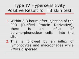
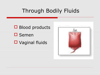
Microbiology
Hypersensitivity and Allergy
Presenter: Jaycris C. Agnes
3SED-SC
Hypersensitivity and Allergy
Hypersensitivity-An exaggerated
immune response that may cause
damage to the host. The trigger is often
an innocuous antigen
Allergy-A hypersensitive response to
an environmental antigen. Often
presents as “hay fever”, asthma,
dermatitis or anaphylaxis.
Four types of Hypersensitivity
Immediate-Type Hypersensitivity:
Type I (Anaphylactic/ Allergic Reactions)
 IgE-mediated
 e.g.most common allergies
Type II (Cytotoxic Reactions)
 IgG-mediated
 e.g.ABO transfusion reaction
Four types of Hypersensitivity
 Type III (Immune Complex Reactions)
 Immune-complex mediated
 e.g.serum sickness
Delayed-Type Hypersensitivity:
 Type IV (Cell Mediated Reaction)
 T cell-mediated; delayed type
 e.g.tuberculin reaction
Type I Hypersensitivity
 Allergens
 Proteins
 Examples: drugs, foods, house dust,
insect venom, latex, mold spores, &
pollens.
 Atopy-Predisposition to type I
hypersensitivity (atopic people)
 Higher levels of circulating IgE
 Greater numbers of eosinophils
Type I Hypersensitivity
 Factors in the Development of Type I
Hypersensitivity:
 Nature of Antigen
 Route of entry
 Amount of antigen
 Ability to produce IgE antibodies
 Frequency of exposure
 Length of exposure time
Table 1. Pharmacologic Mediators of Immediate
Hypersensitivity 
MEDIATOR
Preformed mediators in granules
histamine
bronchoconstriction, mucus secretion, vasodilatation,
vascular permeability
tryptase proteolysis
kininogenase
kinins and vasodilatation, vascular permeability,
edema
ECF-A
(tetrapeptides)
attract eosinophil and neutrophils
Newly formed mediators
leukotriene
B4
basophil attractant
leukotriene
C4, D4
same as histamine but 1000x more potent
prostagland
ins D2
edema and pain
PAF
platelet aggregation and heparin release:
microthrombi
Type I Hypersensitivity
 Localized Anaphylaxis
 Allergic reaction takes place in
specific part of the body.
 Systemic Anaphylaxis
 Allergic Reaction takes place in
different parts of the body.
Type I Hypersensitivity
 Clinical manifestations
 Allergic rhinitis
 Asthma
 Food allergies
 Systemic anaphylaxis
Type II Hypersensitivity
 Cell associated antigens
 Transfusion reactions
 Hemagglutinins
 Complement mediated
 Clinical symptoms include fever, chills,
nausea
Type II Hypersensitivity
 A typical Type Hypersensitivity reaction
might follow these sequence:
1. A particular drug binds to the surface of the
cell.
2. Anti-drug antibodies then bind to the drug.
3. This initiates complement activation on the
cell surface.
4. The complement cascade leads to the lysis
of the cell.
Type II Hypersensitivity
 Erythroblastosis fetalis
 Rh+ fetus born to Rh- mother
 First pregnancy sensitizes
 Subsequent pregnancies result in anti Rh
Ab
 Mild to severe anemia in fetus
 Rhogam
Type II Hypersensitivity
 Drug induced hemolytic anemia
 Some antibiotics can be antigenic
 Bind nonspecifically to RBC surface
proteins
 Ab fixes C and lyses RBCs
Type III Hypersensitivity
 Immune complexes consist of antigen
and antibodies bound together.
 Diseases involved are Systemic Lupus
Erythematosus and rheumatoid arthritis.
Type IV Hypersensitivity
 T cell mediated
 T helper 1 cells
 Effector response is through
macrophages not T cytotoxic cells
 Cytokine mediated
 IL3 Hematopoiesis
 Interferon, TNF, IL 1 Extravasation
 MCAF Attracts macrophages
 MIF Retains macrophages
   Delayed hypersensitivity reactions
Type
Reaction
time
Clinical
appearance
Histology Antigen and site
contact 48-72 hr eczema
lymphocytes, followed by
macrophages; edema of
epidermis
epidermal ( organic
chemicals, poison ivy,
heavy metals, etc.)
tuberculin 48-72 hr
local
induration
lymphocytes, monocytes,
macrophages
intradermal (tuberculin,
lepromin,etc.)
granuloma
21-28
days
hardening
macrophages, epitheloid
and giant cells, fibrosis
persistent antigen or
foreign body presence
(tuberculosis, leprosy, etc.)
Type IV Hypersensitivity
Positive Result for TB skin test
1. Within 2-3 hours after injection of the
PPD (Purified Protein Derivative),
there is an influx of
polymorphonuclear cells into the
site.
2. This is followed by an influx of
lymphocytes and macrophages while
PMN’s dispersed.
Type IV Hypersensitivity
3. Within 12-18 hours, the area become
red (erythematous) and swollen
(edematous).
4. The erythema (redness) and edema
(swell) reach maximum intensity bet.
24-48 hours.
5. With time, as the swelling and
redness disappear, the lymphocytes and
macrophages disperse.
Five possibilities why TB skin
test may be positive:
 A person has tuberculosis.
 A person has tuberculosis in the past.
 A person has been infected by M.
tuberculosis, but the organism has been
killed by that person’s host defence
mechanism.
 A person harvors live M. tuberculosis but
does not have TB.
 A person had received BCG (Bacille de
Calmette et Guérin) vaccine.
Table 5  -  Comparison of Different Types of hypersensitivity
Characteristics
type-I
(anaphylactic)
type-II
(cytotoxic)
type-III
(immune
complex)
type-IV
(delayed type)
antibody IgE IgG, IgM IgG, IgM None
antigen exogenous cell surface soluble
tissues &
organs
response time 15-30 minutes minutes-hours 3-8 hours 48-72 hours
appearance weal & flare lysis and necrosis
erythema and
edema,
necrosis
erythema and
induration
histology
basophils and
eosinophil
antibody and
complement
complement
and neutrophils
monocytes and
lymphocytes
transferred with antibody antibody antibody T-cells
examples
allergic
asthma, hay
fever
erythroblastosis
fetalis,
Goodpasture's
nephritis
SLE, farmer's
lung disease
tuberculin test,
poison ivy,
granuloma
Autoimmune Diseases
 Autoimmune Diseases result when a
person’s immune system can no
longer recognizes certain body tissues
as “self” and attempt to destroy those
tissues.
Immunosuppression
 Acquired Immunodeficiency maybe
caused by drugs, irradiation, or
certain infectious diseases.
 Inherited Immunodeficiency diseases
are inherited immune diseases.
H.I.V.
WHAT IS HIV??
 “Human Immunodeficiency Virus”
 A unique type of virus (a retrovirus)
 Invades the helper T cells (CD4 cells) in
the body of the host (defense
mechanism of a person)
 Threatening a global epidemic.
 Preventable, managable but not curable.
OTHER NAMES FOR HIV
 Former names of the virus include:
 Human T cell lymphotrophic virus (HTLV-
III)
 Lymphadenopathy associated virus (LAV)
 AIDS associated retrovirus (ARV)
WHAT IS AIDS ???
 “Acquired Immunodeficiency Syndrome”
 HIV is the virus that causes AIDS
 Disease limits the body’s ability to fight
infection due to markedly reduced helper T
cells.
 Patients have a very weak immune system
(defense mechanism)
 Patients predisposed to multiple
opportunistic infections leading to death.
AIDS (definition)
 Opportunistic infections and
malignancies that rarely occur in the
absence of severe immunodeficiency
(eg, Pneumocystis pneumonia, central
nervous system lymphoma).
 Persons with positive HIV serology who
have ever had a CD4 lymphocyte count
below 200 cells/mcL or a CD4
lymphocyte percentage below 14% are
considered to have AIDS.
Modes of HIV/AIDS
Transmission
Through Bodily Fluids
 Blood products
 Semen
 Vaginal fluids
IntraVenous Drug Abuse
 Sharing Needles
 Without sterilization Increases the
chances of contracting HIV
 Unsterilized blades
Through Sex
 Unprotected
Intercourse
 Oral
 Anal
Mother-to-Baby
 Before Birth
 During Birth
NATURAL COURSE OF HIV/AIDS
Stage 1 - Primary
 Short, flu-like
illness - occurs
one to six weeks
after infection
 Mild symptoms
 Infected person
can infect other
people
Stage 2 - Asymptomatic
 Lasts for an average of ten years
 This stage is free from symptoms
 There may be swollen glands
 The level of HIV in the blood drops
to low levels
 HIV antibodies are detectable in
the blood
Stage 3 - Symptomatic
 The immune system deteriorates
 Opportunistic infections and cancers
start to appear.
Stage 4 - HIV  AIDS
 The immune
system
weakens too
much as CD4
cells decrease in
number.
Opportunistic Infections associated
with AIDS
CD4<500
 Bacterial infections
 Tuberculosis (TB)
 Herpes Simplex
 Herpes Zoster
 Vaginal candidiasis
 Hairy leukoplakia
 Kaposi’s sarcoma
Opportunistic Infections associated
with AIDS
CD4<200
 Pneumocystic carinii
 Toxoplasmosis
 Cryptococcosis
 Coccidiodomycosis
 Cryptosporiosis
 Non hodgkin’s
lymphoma
CD4 <50
 Disseminated mycobacterium avium
complex (MAC) infection
 Histoplasmosis
 CMV retinitis
 CNS lymphoma
 Progressive multifocal leukoencephalopathy
 HIV dementia
Opportunistic Infections associated
with AIDS
Hypersensitivity with AIDS .

Hypersensitivity with AIDS .

  • 1.
  • 2.
    Hypersensitivity and Allergy Hypersensitivity-Anexaggerated immune response that may cause damage to the host. The trigger is often an innocuous antigen Allergy-A hypersensitive response to an environmental antigen. Often presents as “hay fever”, asthma, dermatitis or anaphylaxis.
  • 3.
    Four types ofHypersensitivity Immediate-Type Hypersensitivity: Type I (Anaphylactic/ Allergic Reactions)  IgE-mediated  e.g.most common allergies Type II (Cytotoxic Reactions)  IgG-mediated  e.g.ABO transfusion reaction
  • 4.
    Four types ofHypersensitivity  Type III (Immune Complex Reactions)  Immune-complex mediated  e.g.serum sickness Delayed-Type Hypersensitivity:  Type IV (Cell Mediated Reaction)  T cell-mediated; delayed type  e.g.tuberculin reaction
  • 6.
    Type I Hypersensitivity Allergens  Proteins  Examples: drugs, foods, house dust, insect venom, latex, mold spores, & pollens.  Atopy-Predisposition to type I hypersensitivity (atopic people)  Higher levels of circulating IgE  Greater numbers of eosinophils
  • 7.
    Type I Hypersensitivity Factors in the Development of Type I Hypersensitivity:  Nature of Antigen  Route of entry  Amount of antigen  Ability to produce IgE antibodies  Frequency of exposure  Length of exposure time
  • 9.
    Table 1. PharmacologicMediators of Immediate Hypersensitivity  MEDIATOR Preformed mediators in granules histamine bronchoconstriction, mucus secretion, vasodilatation, vascular permeability tryptase proteolysis kininogenase kinins and vasodilatation, vascular permeability, edema ECF-A (tetrapeptides) attract eosinophil and neutrophils
  • 10.
    Newly formed mediators leukotriene B4 basophilattractant leukotriene C4, D4 same as histamine but 1000x more potent prostagland ins D2 edema and pain PAF platelet aggregation and heparin release: microthrombi
  • 11.
    Type I Hypersensitivity Localized Anaphylaxis  Allergic reaction takes place in specific part of the body.  Systemic Anaphylaxis  Allergic Reaction takes place in different parts of the body.
  • 12.
  • 13.
    Type I Hypersensitivity Clinical manifestations  Allergic rhinitis  Asthma  Food allergies  Systemic anaphylaxis
  • 14.
    Type II Hypersensitivity Cell associated antigens  Transfusion reactions  Hemagglutinins  Complement mediated  Clinical symptoms include fever, chills, nausea
  • 15.
    Type II Hypersensitivity A typical Type Hypersensitivity reaction might follow these sequence: 1. A particular drug binds to the surface of the cell. 2. Anti-drug antibodies then bind to the drug. 3. This initiates complement activation on the cell surface. 4. The complement cascade leads to the lysis of the cell.
  • 17.
    Type II Hypersensitivity Erythroblastosis fetalis  Rh+ fetus born to Rh- mother  First pregnancy sensitizes  Subsequent pregnancies result in anti Rh Ab  Mild to severe anemia in fetus  Rhogam
  • 19.
    Type II Hypersensitivity Drug induced hemolytic anemia  Some antibiotics can be antigenic  Bind nonspecifically to RBC surface proteins  Ab fixes C and lyses RBCs
  • 20.
    Type III Hypersensitivity Immune complexes consist of antigen and antibodies bound together.  Diseases involved are Systemic Lupus Erythematosus and rheumatoid arthritis.
  • 21.
    Type IV Hypersensitivity T cell mediated  T helper 1 cells  Effector response is through macrophages not T cytotoxic cells  Cytokine mediated  IL3 Hematopoiesis  Interferon, TNF, IL 1 Extravasation  MCAF Attracts macrophages  MIF Retains macrophages
  • 22.
       Delayed hypersensitivityreactions Type Reaction time Clinical appearance Histology Antigen and site contact 48-72 hr eczema lymphocytes, followed by macrophages; edema of epidermis epidermal ( organic chemicals, poison ivy, heavy metals, etc.) tuberculin 48-72 hr local induration lymphocytes, monocytes, macrophages intradermal (tuberculin, lepromin,etc.) granuloma 21-28 days hardening macrophages, epitheloid and giant cells, fibrosis persistent antigen or foreign body presence (tuberculosis, leprosy, etc.)
  • 23.
    Type IV Hypersensitivity PositiveResult for TB skin test 1. Within 2-3 hours after injection of the PPD (Purified Protein Derivative), there is an influx of polymorphonuclear cells into the site. 2. This is followed by an influx of lymphocytes and macrophages while PMN’s dispersed.
  • 24.
    Type IV Hypersensitivity 3.Within 12-18 hours, the area become red (erythematous) and swollen (edematous). 4. The erythema (redness) and edema (swell) reach maximum intensity bet. 24-48 hours. 5. With time, as the swelling and redness disappear, the lymphocytes and macrophages disperse.
  • 25.
    Five possibilities whyTB skin test may be positive:  A person has tuberculosis.  A person has tuberculosis in the past.  A person has been infected by M. tuberculosis, but the organism has been killed by that person’s host defence mechanism.  A person harvors live M. tuberculosis but does not have TB.  A person had received BCG (Bacille de Calmette et Guérin) vaccine.
  • 26.
    Table 5  - Comparison of Different Types of hypersensitivity Characteristics type-I (anaphylactic) type-II (cytotoxic) type-III (immune complex) type-IV (delayed type) antibody IgE IgG, IgM IgG, IgM None antigen exogenous cell surface soluble tissues & organs response time 15-30 minutes minutes-hours 3-8 hours 48-72 hours appearance weal & flare lysis and necrosis erythema and edema, necrosis erythema and induration histology basophils and eosinophil antibody and complement complement and neutrophils monocytes and lymphocytes transferred with antibody antibody antibody T-cells examples allergic asthma, hay fever erythroblastosis fetalis, Goodpasture's nephritis SLE, farmer's lung disease tuberculin test, poison ivy, granuloma
  • 27.
    Autoimmune Diseases  AutoimmuneDiseases result when a person’s immune system can no longer recognizes certain body tissues as “self” and attempt to destroy those tissues.
  • 28.
    Immunosuppression  Acquired Immunodeficiencymaybe caused by drugs, irradiation, or certain infectious diseases.  Inherited Immunodeficiency diseases are inherited immune diseases.
  • 29.
  • 30.
    WHAT IS HIV?? “Human Immunodeficiency Virus”  A unique type of virus (a retrovirus)  Invades the helper T cells (CD4 cells) in the body of the host (defense mechanism of a person)  Threatening a global epidemic.  Preventable, managable but not curable.
  • 31.
    OTHER NAMES FORHIV  Former names of the virus include:  Human T cell lymphotrophic virus (HTLV- III)  Lymphadenopathy associated virus (LAV)  AIDS associated retrovirus (ARV)
  • 32.
    WHAT IS AIDS???  “Acquired Immunodeficiency Syndrome”  HIV is the virus that causes AIDS  Disease limits the body’s ability to fight infection due to markedly reduced helper T cells.  Patients have a very weak immune system (defense mechanism)  Patients predisposed to multiple opportunistic infections leading to death.
  • 33.
    AIDS (definition)  Opportunisticinfections and malignancies that rarely occur in the absence of severe immunodeficiency (eg, Pneumocystis pneumonia, central nervous system lymphoma).  Persons with positive HIV serology who have ever had a CD4 lymphocyte count below 200 cells/mcL or a CD4 lymphocyte percentage below 14% are considered to have AIDS.
  • 34.
  • 35.
    Through Bodily Fluids Blood products  Semen  Vaginal fluids
  • 36.
    IntraVenous Drug Abuse Sharing Needles  Without sterilization Increases the chances of contracting HIV  Unsterilized blades
  • 37.
  • 38.
  • 39.
  • 40.
    Stage 1 -Primary  Short, flu-like illness - occurs one to six weeks after infection  Mild symptoms  Infected person can infect other people
  • 41.
    Stage 2 -Asymptomatic  Lasts for an average of ten years  This stage is free from symptoms  There may be swollen glands  The level of HIV in the blood drops to low levels  HIV antibodies are detectable in the blood
  • 42.
    Stage 3 -Symptomatic  The immune system deteriorates  Opportunistic infections and cancers start to appear.
  • 43.
    Stage 4 -HIV  AIDS  The immune system weakens too much as CD4 cells decrease in number.
  • 44.
    Opportunistic Infections associated withAIDS CD4<500  Bacterial infections  Tuberculosis (TB)  Herpes Simplex  Herpes Zoster  Vaginal candidiasis  Hairy leukoplakia  Kaposi’s sarcoma
  • 45.
    Opportunistic Infections associated withAIDS CD4<200  Pneumocystic carinii  Toxoplasmosis  Cryptococcosis  Coccidiodomycosis  Cryptosporiosis  Non hodgkin’s lymphoma
  • 46.
    CD4 <50  Disseminatedmycobacterium avium complex (MAC) infection  Histoplasmosis  CMV retinitis  CNS lymphoma  Progressive multifocal leukoencephalopathy  HIV dementia Opportunistic Infections associated with AIDS