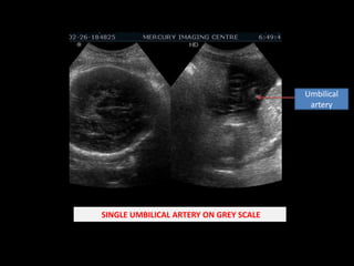
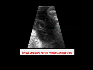
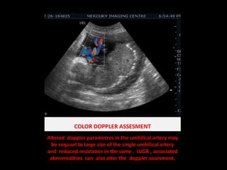
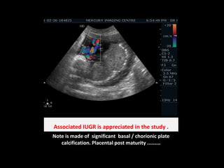
SINGLE UMBILICAL ARTERYSingle umbilical artery (SUA) has an incidence of 0.5%  witha higher prevalence in twin pregnancies. The aetiologyremains unknown but the most likely mechanism is atrophy of the second umbilical artery during development  PRESENT CASE :                                                24 yr Old Female with POG by USG  of 33 wks with IUGR. USG / Doppler assesment has e/o single umbilical artery . Previous pregnancy had e/o  Hydrocephalus with Meninigocele.MERCURY IMAGING INSTITUTE SCO 172-173 SEC 9C  CHANDIGARHMERCURY IMAGING CENTRE SCO 16-17 SEC 20D CHANDIGARH
Umbilical artery SINGLE UMBILICAL ARTERY ON GREY SCALE
SINGLE UMBILICAL ARTERY  WITH MAGNIFIED VIEW
COLOR DOPPLER ASSESMENTAltered  dopplerparametres in the umbilical artery may be sequael to large size of the single umbilical artery  and  reduced resistance in the same .  IUGR , associated abnormalities  can  also alter the  dopplerassesment.
All the fetalParametres for the growth of the are close to the lower limit of the  standard deviation suggestive of IUGR.
Associated IUGR is appreciated in the study .Note is made of  significant  basal / chorionic plate calcification. Placental post maturity .........
Normal umbilical CordThe normal umbilical cord consists of two umbilical arteriesand one umbilical vein surrounded by Wharton’s jelly.
A brief about single umbilical arterySingle umbilical artery is  the most  congenital abnormality of the umbilical cord.Associated with Hollow organ atresia . Gastrointestinal and urogenital abnormalities, musculoskeletal, cardiovascular and central nervous system malformations and limb reduction defects , Cystic renal dysplasia , meckels  syndrome , posterior uretheral valve , anterior uretheraldiverticulum , uretheral agenesis IUGR , IUD Premature labour .. EtiopathogenesisPrimary agenesisRegression of preformed artery  .Coagulopathy , Endothelial dysfunction , Ischemic sequale are possible mechanisms that can lead to mentioned vascular disruption.  Altered  blood supply of rest of the developing organs can also disturb  there genesis  and hence can lead to atresia.
A suggested protocol...........A detailed sonographic evaluation with fetal echocardiography.

Single umbilical artery

  • 1.
    SINGLE UMBILICAL ARTERYSingleumbilical artery (SUA) has an incidence of 0.5% witha higher prevalence in twin pregnancies. The aetiologyremains unknown but the most likely mechanism is atrophy of the second umbilical artery during development PRESENT CASE : 24 yr Old Female with POG by USG of 33 wks with IUGR. USG / Doppler assesment has e/o single umbilical artery . Previous pregnancy had e/o Hydrocephalus with Meninigocele.MERCURY IMAGING INSTITUTE SCO 172-173 SEC 9C CHANDIGARHMERCURY IMAGING CENTRE SCO 16-17 SEC 20D CHANDIGARH
  • 2.
    Umbilical artery SINGLEUMBILICAL ARTERY ON GREY SCALE
  • 3.
    SINGLE UMBILICAL ARTERY WITH MAGNIFIED VIEW
  • 4.
    COLOR DOPPLER ASSESMENTAltered dopplerparametres in the umbilical artery may be sequael to large size of the single umbilical artery and reduced resistance in the same . IUGR , associated abnormalities can also alter the dopplerassesment.
  • 5.
    All the fetalParametresfor the growth of the are close to the lower limit of the standard deviation suggestive of IUGR.
  • 6.
    Associated IUGR isappreciated in the study .Note is made of significant basal / chorionic plate calcification. Placental post maturity .........
  • 7.
    Normal umbilical CordThenormal umbilical cord consists of two umbilical arteriesand one umbilical vein surrounded by Wharton’s jelly.
  • 8.
    A brief aboutsingle umbilical arterySingle umbilical artery is the most congenital abnormality of the umbilical cord.Associated with Hollow organ atresia . Gastrointestinal and urogenital abnormalities, musculoskeletal, cardiovascular and central nervous system malformations and limb reduction defects , Cystic renal dysplasia , meckels syndrome , posterior uretheral valve , anterior uretheraldiverticulum , uretheral agenesis IUGR , IUD Premature labour .. EtiopathogenesisPrimary agenesisRegression of preformed artery .Coagulopathy , Endothelial dysfunction , Ischemic sequale are possible mechanisms that can lead to mentioned vascular disruption. Altered blood supply of rest of the developing organs can also disturb there genesis and hence can lead to atresia.
  • 9.
    A suggested protocol...........Adetailed sonographic evaluation with fetal echocardiography.