This guideline provides recommendations for investigating and managing small-for-gestational-age (SGA) fetuses. It discusses risk factors for SGA, screening and diagnostic methods, fetal monitoring options, and optimal timing of delivery. The guideline recommends assessing all women for SGA risk factors at their first prenatal visit. Women with major risk factors or three minor factors should undergo additional ultrasounds and Doppler studies for surveillance. Serial fundal height measurements and ultrasounds are also recommended for monitoring high-risk pregnancies. The guideline provides guidance on investigations, fetal testing, and deciding when delivery is appropriate for SGA fetuses.
![RCOG Green-top Guideline No. 31 2 of 34 © Royal College of Obstetricians and Gynaecologists
The Investigation and Management of the
Small–for–Gestational–Age Fetus
This is the second edition of this guideline. It replaces the first edition which was published in November
2002 under the same title.
Executive Summary of Recommendations
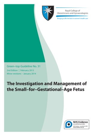
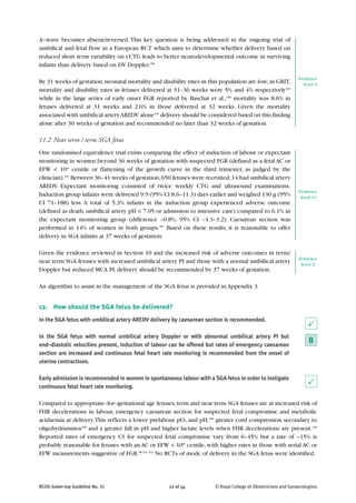
Risk factors for a SGA fetus/neonate
All women should be assessed at booking for risk factors for a SGA fetus/neonate to identify those who
require increased surveillance.
Women who have a major risk factor (Odds Ratio [OR] > 2.0) should be referred for serial ultrasound
measurement of fetal size and assessment of wellbeing with umbilical artery Doppler from 26–28
weeks of pregnancy (Appendix 1).
Women who have three or more minor risk factors should be referred for uterine artery Doppler at 20–24
weeks of gestation (Appendix 1).
Second trimester DS markers have limited predictive accuracy for delivery of a SGA neonate.
A low level (< 0.415 MoM) of the first trimester marker PAPP–A should be considered a major risk factor
for delivery of a SGA neonate.
In high risk populations uterine artery Doppler at 20–24 weeks of pregnancy has a moderate predictive
value for a severely SGA neonate.
In women with an abnormal uterine artery Doppler at 20–24 weeks of pregnancy, subsequent
normalisation of flow velocity indices is still associated with an increased risk of a SGA neonate.
Repeating uterine artery Doppler is therefore of limited value.
Women with an abnormal uterine artery Doppler at 20–24 weeks (defined as a pulsatility index [PI]
> 95th
centile) and/or notching should be referred for serial ultrasound measurement of fetal size and
assessment of wellbeing with umbilical artery Doppler commencing at 26–28 weeks of pregnancy.
Women with a normal uterine artery Doppler do not require serial measurement of fetal size and serial
assessment of wellbeing with umbilical artery Doppler unless they develop specific pregnancy
complications, for example antepartum haemorrhage or hypertension. However, they should be offered
a scan for fetal size and umbilical artery Doppler during the third trimester.
Serial ultrasound measurement of fetal size and assessment of wellbeing with umbilical artery Doppler
should be offered in cases of fetal echogenic bowel.
Abdominal palpation has limited accuracy for the prediction of a SGA neonate and thus should not be
routinely performed in this context.
Serial measurement of symphysis fundal height (SFH) is recommended at each antenatal appointment
from 24 weeks of pregnancy as this improves prediction of a SGA neonate.
B
P
P
P
P
B
A
C
P
C
C
B](https://image.slidesharecdn.com/iugrrcogguidelines-140928120309-phpapp01/85/Iugr-rcog-guidelines-2-320.jpg)
![RCOG Green-top Guideline No. 31 7 of 34 © Royal College of Obstetricians and Gynaecologists
methodology register, ACP journal club, DARE HTA, Maternity and Infant Care), EMBASE and TRIP were
searched for relevant randomised controlled trials (RCTs), systematic reviews, meta–analyses and cohort
studies.The search was restricted to articles published between 2002 and September 2011. Search words
included ‘fetal growth retardation’,‘fetal growth restriction’,‘infant, small for gestational age’, including all
relevant Medical Subject Heading (MeSH) terms.The search was limited to humans and the English language.
5. What are the risk factors for a SGA fetus/neonate? What is the optimum method of
screening for the SGA fetus/neonate and care of “at risk” pregnancies?
Methods employed in the first and second trimesters,to predict the likelihood of a SGA fetus/neonate include:
medical and obstetric history and examination, maternal serum screening and uterine artery Doppler.
Methods of screening for the SGA fetus/neonate in the second and third trimester are abdominal palpation
and measurement of symphysis fundal height (SFH) (including customised charts).
5.1 History
All women should be assessed at booking for risk factors for a SGA fetus/neonate to identify those who
require increased surveillance.
Women who have a major risk factor (Odds Ratio [OR] > 2.0) should be referred for serial ultrasound
measurement of fetal size and assessment of wellbeing with umbilical artery Doppler from 26–28
weeks of pregnancy (Appendix 1).
Women who have three or more minor risk factors should be referred for uterine artery Doppler at 20–24
weeks of gestation (Appendix 1).
A table of risk factors and associated odds ratios (ORs) for the birth of a SGA neonate, where evidence is
consistent and not affected by adjustment for confounders, is presented in Appendix 1. It is acknowledged
that other risk factors may need to be considered on an individual basis.
Women that have previously had a SGA neonate have at least a twofold increased risk of a subsequent SGA
neonate.6–8
The risk is increased further after two SGA births.7
Classification of prior infant birthweight is best
done using customised centiles.1–2
This can be done using computer software that can be downloaded from the
internet.9
Women with a prior history of other placenta–mediated diseases are also at increased risk of a
subsequent SGA neonate.This includes prior pre-eclampsia8
and prior stillbirth,7
and in particular those with a
history of previous preterm unexplained stillbirth, due to the association with FGR.10
While termination of
pregnancy is not a risk factor for a SGA infant,11
the evidence regarding recurrent miscarriage is inconsistent.12,13
Maternal medical conditions associated with an increased risk of a SGA neonate are diabetes with vascular
disease,14
moderate and severe renal impairment (especially when associated with hypertension),15
antiphospholipid syndrome16
and chronic hypertension.17
Systemic lupus erythematosus18
and certain types
of congenital heart disease, in particular cyanotic congenital heart disease, are associated with increased
likelihood of a SGA neonate but there are no papers reporting ORs.19
The risk will therefore need to be
assessed on an individual basis.The evidence for an association with asthma, thyroid disease, inflammatory
bowel disease and depression is less convincing. Studies report a weak or non–significant association with
LBW but do not differentiate between the effect on SGA and preterm birth, and with confidence intervals
[CIs] often crossing one.Therefore, if uncomplicated and adequately treated, these are not considered to be
risk factors for a SGA fetus.20,21
Maternal risk factors associated with an increased risk of a SGA neonate are maternal age ≥ 35 years, with a
further increase in those ≥ 40 years old,22
African American23
or Indian/Asian ethnicity,2,24
nulliparity,25
social
deprivation,26
unmarried status,27
body mass index (BMI) < 20,28–30
BMI > 25,28,29
maternal SGA,31
daily vigorous
P
P
B](https://image.slidesharecdn.com/iugrrcogguidelines-140928120309-phpapp01/85/Iugr-rcog-guidelines-7-320.jpg)
![exercise,32
a short (< 6 months) or long (> 60 months) inter–pregnancy interval33
and heavy vaginal bleeding
during the first trimester.34
The effect of some of these risk factors is reduced once adjusted for other
associated factors and thus they are not included in Appendix 1. Maternal exposure to domestic violence
during pregnancy has been shown in a systematic review to be associated with low birth weight (Adjusted
OR [AOR] 1.53,95% CI 1.28–1.82).35
Low maternal weight gain has been shown to be associated with a SGA
infant in a preterm population (OR 4.9,95% CI 1.9–12.6)13
but it is no longer recommended that women are
routinely weighed during pregnancy.36
Several maternal exposures have a seemingly causative relationship with a SGA infant, including moderate
alcohol intake,37
drug use (with cocaine use during pregnancy being the most significant)38
and cigarette
smoking.39
The effects of smoking are dose dependent.29
Other risk factors are maternal caffeine consumption ≥ 300 mg per day in the third trimester40
and a low fruit
intake pre–pregnancy,while a high green leafy vegetable intake pre–pregnancy has been reported to be protective
(AOR 0.44,95% CI 0.24–0.81).32
Singleton pregnancies following IVF are also a risk factor for a SGA fetus.41
Changing paternity has been associated with an increased risk of a SGA infant,42
although a recent
systematic review demonstrated inconclusive evidence.43
A paternal history of SGA birth is a risk
factor for a SGA fetus.44
There is insufficient evidence to determine how risk factors relate to each other in the individual woman and
consequently how these risk factors should be managed.This includes abnormal maternal Down syndrome
serum markers (see below). Further evidence may become available from the SCOPE study.45
This guideline
has therefore categorized risk factors into major and minor based on published ORs for the birth of a SGA
neonate. Major risk factors (OR > 2.0) should prompt referral for serial ultrasound measurement of fetal size
and assessment of wellbeing with umbilical artery Doppler.The presence of multiple minor risk factors is
likely to constitute a significant risk for the birth of a SGA neonate and there is a rationale for further screening
using uterine artery Doppler at 20 weeks (see below).
5.2 Biochemical markers used for Down Syndrome (DS) Screening
Second trimester DS markers have limited predictive accuracy for delivery of a SGA neonate.
A low level (< 0.415 MoM) of the first trimester marker PAPP–A should be considered a major risk factor
for delivery of a SGA neonate.
Due to their placental origin, several biochemical markers have been investigated as screening tests for a
SGA fetus.
Two systematic reviews found low predictive accuracy for alpha fetoprotein (AFP) (> 2.5 MoM or
< 0.25 MoM),elevated hCG (> 3.0 MoM) and inhibinA (≥ 2.0 MoM),low unconjugated estriol (< 0.5
MoM) and the combined triple test to predict a SGA fetus.46,47
One review found methodological and
reporting limitations in all studies, resulting in great heterogeneity, concluding that serum markers
were only useful as a means of contributing to the overall assessment of risk for a pregnancy.47
In women with elevatedAFP,there is no evidence that increased fetal surveillance has any benefit.48
Similarly, there is a lack of evidence for the use of aspirin in women with raised hCG.49
In a large series of 49801 women at 11+
0 to 13+6
weeks, low PAPP–A (but not beta HCG) was inversely
associated with risk of being SGA. Using a 5th
centile (0.415 MoM) cut off, ORs for A SGA infant (birthweight
< 10th
centile) and severe SGA (birthweight < 3rd
centile) were 2.7 and 3.66 respectively.50
A systematic review
8 of 34RCOG Green-top Guideline No. 31 © Royal College of Obstetricians and Gynaecologists
Evidence
level 3
P
B
Evidence
level 3
Evidence
level
1+/2+](https://image.slidesharecdn.com/iugrrcogguidelines-140928120309-phpapp01/85/Iugr-rcog-guidelines-8-320.jpg)
![RCOG Green-top Guideline No. 31 9 of 34 © Royal College of Obstetricians and Gynaecologists
found that an unexplained low first trimester PAPP–A (< 0.4 MoM) and/or a low hCG (< 0.5 MoM) were
associated with an increased frequency of adverse obstetrical outcome including a SGA infant.47
There is some evidence that addition of fetal size at 18–20 weeks of gestation or fetal growth
between 11–14 and 18–20 weeks of gestation to first trimester serum markers improves prediction
of a SGA infant.51,52
However,different ultrasound parameters have been used and it is unclear what
combination provides optimum prediction.
5.3 Uterine artery Doppler
In high risk populations uterine artery Doppler at 20–24 weeks of pregnancy has a moderate predictive
value for a severely SGA neonate.
In women with an abnormal uterine artery Doppler at 20–24 weeks of pregnancy, subsequent
normalisation of flow velocity indices is still associated with an increased risk of a SGA neonate.
Repeating uterine artery Doppler is therefore of limited value.
Women with an abnormal uterine artery Doppler at 20–24 weeks (defined as a pulsatility index [PI]
> 95th
centile) and/or notching should be referred for serial ultrasound measurement of fetal size and
assessment of wellbeing with umbilical artery Doppler commencing at 26–28 weeks of pregnancy.
Women with a normal uterine artery Doppler do not require serial measurement of fetal size and serial
assessment of wellbeing with umbilical artery Doppler unless they develop specific pregnancy
complications, for example antepartum haemorrhage or hypertension. However, they should be offered
a scan for fetal size and umbilical artery Doppler during the third trimester.
SGA birth, particularly when severe (birth weight < 3rd
centile) or necessitating delivery < 36 weeks of gestation,
is characterised by failure of trophoblast invasion of the myometrial uterine spiral arteries and reduced
uteroplacental blood flow. Non–pregnant and first trimester artery blood flow velocity waveforms are associated
with low end–diastolic velocities and an early diastolic notch.Persistent notching or abnormal flow velocity ratios
after 24 weeks of gestation are associated with inadequate trophoblast invasion of the myometrial spiral arteries.53
However reduced endovascular trophoblast invasion of decidual spiral arteries has been associated with the same
waveform abnormalities as early as 10–14 weeks of pregnancy.54
A systematic review and meta–analysis summarised the results from 61 studies testing 41131 pregnant women
with uterine artery Doppler (in both first and second trimesters) and assessed the value of different Doppler
flow velocity indices.55
SGA birth in low risk patients was best predicted by an increased pulsatility index (PI)
(defined as > 95th
centile) with diastolic notching (positive likelihood ratio [LR+] 9.1,95% CI 5.0–16.7;negative
likelihood ratio [LR–] 0.89, 95% CI 0.85–0.93). Severe SGA (birthweight < 5th
or < 3rd
centile) in low risk
populations was best predicted in the second trimester by an increased PI (LR+ 13.7, 95% CI 10.3–16.9; LR–
0.34, 95% CI 0.23–0.48) or an increased PI with notching (LR+ 14.6, 95% CI 7.8–26.3; LR– 0.78, 95% CI
0.68–0.87). Uterine artery Doppler to predict a SGA infant in high risk populations overall showed low
predictive characteristics;an increased PI or notching in the second trimester best predicted a SGA infant (LR+
3.6, 95% CI 2.0–5.1; LR– 0.40, 95% CI 0.14–0.65). Prediction of severe SGA showed moderate utility with the
best prediction by a resistance index (> 0.58 or > 90th
centile) and notching in the second trimester (LR+ 10.9,
95% CI 10.4–11.4;LR– 0.20,95% CI 0.14–0.26).Although first trimester uterine artery Doppler studies suggest
a high specificity (91–96%) and high negative predictive values (91–99%), the low sensitivity (12–25%) for a
SGA neonate suggest early screening cannot be recommended on current evidence.55
There were three studies included in this review that looked at prediction of early onset SGA, all
of which were in low risk/unselected populations.55
Increased PI in the second trimester has been
shown to be predictive of delivery of a SGA fetus < 34 weeks in two studies (LR+ 13.7, 95% CI
P
P
Evidence
level 2+
A
C
Evidence
level 1](https://image.slidesharecdn.com/iugrrcogguidelines-140928120309-phpapp01/85/Iugr-rcog-guidelines-9-320.jpg)
![has also been shown to improve the prediction of adverse prenatal outcome;90,91
OR of adverse
outcomes (stillbirths, neonatal deaths, referral to higher level or special care unit or Apgar score <
7 at 5 minutes) for SGA neonates versus those not SGA was 1.59 (95% CI 1.53–1.66) for the
non–customised fetal weight reference compared with 2.84 (95% CI 2.71–2.99) for the customised
reference.90
Prediction of perinatal mortality was also improved by the customised reference (OR
3.65, 95% CI 3.40–3.92 versus OR 1.77, 95% CI 1.65–1.89).91
A further study demonstrated that
individual growth trajectories of low risk fetuses with normal outcome were less likely to cross
below the 10th
centile for fetal weight when using customised reference standards than when
unadjusted standards were used.92
However, no trials were identified that compared customised
with non–customised EFW chartsp.
A meta–analysis, including eight trials comprising 27024 women, found no evidence that routine
fetal biometry (with or without assessment of amniotic fluid volume and placental grade) after
24 weeks of pregnancy improved perinatal outcome in a low risk population (SGA neonate relative
risk [RR] 0.98, 95% CI 0.74–1.28; perinatal mortality RR 0.94, 95% CI 0.55–1.61).93
The timing and
content of the ultrasound scan varied substantially between studies and the authors noted high
heterogeneity between studies in the reduction of the risk of a SGA neonate, mainly due to the
findings of one study in which routine estimation of fetal weight, amniotic fluid volume and
placental grading at 30–32 and 36–37 weeks of gestation was shown to result in the birth of fewer
SGA neonates (10.4% versus 6.9%, RR 0.67, 95% CI 0.50–0.89).94
The change in fetal size between two time points is a direct measure of fetal growth and hence
serial measurement of AC or EFW (growth velocities) should allow the diagnosis of FGR.However
the optimal method of using serial ultrasound measurements is not clear.Although ‘eyeballing’ a
chart of individual AC or EFW measurements may give an impression of FGR a more objective
definition requires establishment of growth rate standards from longitudinally collected data.
Several standards have been reported,95,96
including conditional centiles for fetal growth,97
although
none has been adopted in clinical practice. Reported mean growth rates for AC and EFW after
30 weeks of gestation are 10 mm/14 days and 200 g/14 days although greater variation exists in the
lower limits (reflecting the methods used to derive the standard deviation [SD]).98
However a
change in AC of < 5mm over 14 days is suggestive of FGR.95
In a high risk population, identified as
being SGA,Chang et al.99,100
showed that a change inAC or EFW (defined as a change in SD score of
≥ –1.5) were better predictors of wasting at birth (ponderal index, mid–arm circumference/head
circumference ratio or subscapular skinfold thickness < 2 SD below mean) and adverse perinatal
outcome than the final AC or EFW before delivery.
Mongelli et al.101
used a mathematical model to estimate the impact of time interval between
examinations on the false positive rates for FGR (defined as no apparent growth in fetalAC between
two consecutive examinations).When the initial scan was performed at 32 weeks of gestation,the
false positive rates were 30.8%,16.9%,8.1% and 3.2% for intervals of 1,2,3 and 4 weeks respectively.
False positive rates were higher when the first scan was performed at 36 weeks of gestation (34.4%,
22.1%,12.7%,6.9% respectively).These findings suggest that if two measurements are to be used to
estimate velocity, they should be a minimum of 3 weeks apart to minimise false–positive rates for
diagnosing FGR.This recommendation does not preclude more frequent ultrasound measurements
of AC/EFW to predict fetal size at birth but rather indicates which measurements should be used
to interpret growth.
6.2 Biophysical tests
Biophysical tests,including amniotic fluid volume,cardiotocography (CTG) and biophysical scoring are poor
at diagnosing a small or growth restricted fetus.102–104
A systematic review of the accuracy of umbilical artery
13 of 34RCOG Green-top Guideline No. 31 © Royal College of Obstetricians and Gynaecologists
Evidence
level 3
Evidence
level 1+
Evidence
level 2+
Evidence
level 3](https://image.slidesharecdn.com/iugrrcogguidelines-140928120309-phpapp01/85/Iugr-rcog-guidelines-13-320.jpg)


![8. Ananth CV,Peltier MR,Chavez MR,Kirby RS,Getahun D,
Vintzileos AM.Recurrence of ischemic placental disease.Obstet
Gynecol 2007;110:128–33.9.
9. Gestation Network Growth Charts.[https://www.gestation.net/
fetal_growth/download_grow.htm].
10. Gardosi J,Kady SM,McGeown P,FrancisA,TonksA.Classification
of stillbirth by relevant condition of death (ReCoDe):
population based cohort study.BMJ 2005;331:113–7.
11. Shah PS,Zao J.Induced termination of pregnancy and low
birthweight and preterm birth:a systematic review and
meta–analyses.BJOG 2009;116:1425–42.
12. Spinillo A,Capuzzo E,Piazzi G,Nicola S,Colonna L,Iasci A.
Maternal high–risk factors and severity of growth deficit in
small for gestational age infants.Early Hum Dev
1994;38:35–43.
13. Lang JM,Lieberman E,Cohen A.A comparison of risk–factors
for preterm labour and term small–for–gestational–age birth.
Epidemiology 1996;7:369–76.
14. Howarth C,Gazis A,James D.Associations of type 1 diabetes
mellitus,maternal vascular disease and complications of
pregnancy.Diabet Med 2007;24:1229–34.
15. Fink JC,Schwartz M,BenedettiTJ,Stehman–Breen CO.
Increased risk of adverse maternal and fetal outcomes among
women with renal disease.Paediatr Perinat Epidemiol
1998;12:277–87.
16. Yasuda M,Takakuwa K,Tokunaga A,Tanaka K.Prospective
studies of the association between anticardiolipin antibody and
outcome of pregnancy.Obstet Gynecol 1995;86:555–9.
17. Allen VM,Joseph KS,Murphy KE,Magee LA,Ohlsson A.The
effect of hypertensive disorders in pregnancy on small for
gestational age and stillbirth:a population based study.BMC
Pregnancy Childbirth 2004;4:17–25.
18. Yasmeen S,Wilkins EE,Field NT,Sheikh RA,Gilbert WM.
Pregnancy outcomes in women with systemic lupus
erythematosus.J Matern Fetal Med 2001;10:91–6.
19. Drenthen W,Pieper PG,Roos–Hesselink JW,van Lottum WA,
Voors AA,Mulder BJ,et al.Outcome of pregnancy in women
with congenital heart disease:a literature review.J Am Coll
Cardiol 2007;49:2303–11.
20. McCowan L,Horgan RP.Risk factors for small for gestational
age infants.Best Pract Res Clin Obstet Gynaecol
2009;23:779–93.
21. Grote NK,Bridge JA,Gavin AR,Melville JL,Iyengar S,Katon WJ.
A meta–analysis of depression during pregnancy and the risk
of preterm birth,low birth weight,and intrauterine growth
restriction.Arch Gen Psychiatry 2010;67:1012–24.
22. Odibo AO,Nelson D,Stamilio DM,Sehdev HM,Macones GA.
Advanced maternal age is an independent risk factor for
intrauterine growth restriction.Am J Perinatol 2006;23:325–8.
23. Kramer MS.Determinants of low birth weight:methodological
assessment and meta–analysis.BullWorld Health Organ
1987;65:663–737.
24. Alexander GR,Wingate MS,Mor J,Boulet S.Birth outcomes of
Asian–Indian–americans.Int J Gynaecol Obstet 2007;97:215–20.
25. Shah PS,Knowledge Synthesis Group on Determinants of
LBW/PT Births.Parity and low birth weight and pre–term
birth:a systematic review and meta–analyses.Acta Obstet
Gynecol Scand 2010;89:862–75.
26. Blumenshine P,Egarter S,Barclay CJ,Cubbin C,Braveman PA.
Socioeconomic disparities in adverse birth outcomes:a
systematic review.Am J Prev Med 2010;39:263–72.
27. Shah PS,Zao J,Ali S.Maternal marital status and birth
outcomes:a systematic review and meta–analyses.Matern
Child Health J 2011;15:1097–109.
28. Gardosi J,FrancisA.Adverse pregnancy outcome and association
with small for gestational age birthweight by customized and
popualtion–based centiles.Am J Obstet Gynecol 2009;201:1–8.
29. Kramer MS,Platt R,Yang H,McNamara H,Usher RH.Are all
growth restricted newborns created equal(ly)? Pediatrics
1999;103:599–602.
30. Han Z,Mulla S,Beyene J,Liao G,McDonald SD;Knowledge
Synthesis Group.Maternal underweight and the risk of
preterm birth and low birth weight:a systematic review and
meta–analyses.Int J Epidemiol 2011;40:65–101.
31. Shah PS,Shah V,Knowledge Synthesis Group on Determinants
of LBW/PT Births.Influence of maternal birth status on
offspring:a systematic review and meta–analysis.Acta Obstet
Gynecol Scand 2009;88:1307–18.
32. McCowan LM,Roberts CT,Dekker GA,Taylor RS,Chan EH,
Kenny LC,et al.Risk factors for small–for–gestational–age
infants by customised birthweight centiles:data from an
international prospective cohort study.BJOG
2010;117:1599–607.
33. Conde–Agudelo A,Rosas–Bermúdez A,Kafury–Goeta AC.Birth
spacing and risk of adverse perinatal outcomes:a
meta–analysis.JAMA 2006;295:1809–23.
34. Weiss JL,Malone FD,Vidaver J,Ball RH,Nyberg DA,Comstock
CH,et al.Threatened abortion:A risk factor for poor pregnancy
outcome,a population–based screening study.Am J Obstet
Gynecol 2004;190:745–50.
35. Shah JS,Shah J,Knowledge Synthesis Group on Determinants
of LBW/PT Births.Maternal exposure to domestic violence and
pregnancy and birth outcomes:a systematic review and
meta–analysis.JWomens Health 2010;19:2017–31.
36. National Institute for Health and Clinical Excellence (NICE).
Antenatal care.Routine care for the healthy pregnant
woman. London:NICE;2008.
37. Jaddoe VW,Bakker R,Hofman A,Mackenbach JP,Moll HA,
Steegers EA,et al.Moderate alcohol consumption during
pregnancy and the risk of low birth weight and preterm birth.
The generation R study.Ann Epidemiol 2007;17:834–40.
38. Gouin K,Murphy K,Shah PS,Knowledge Synthesis Group on
Determinants of LBW/PT Births.Effects of cocaine use during
pregnancy on low birthweight and preterm birth:systematic
review and metaanalyses.Am J Obstet Gynecol
2011;204:340:1–12.
39. McCowan LM,Dekker GA,Chan E,Stewart A,Chappell LC,
Hunter M,et al.Spontaneous preterm birth and small for
gestational age infants in women who stop smoking early in
pregnancy:prospective cohort study.BMJ 2009;338:b1081.
40. CARE Study group.Maternal caffeine intake during pregnancy
and risk of fetal growth restriction:a large prospective
observational study.BMJ 2008;337:a2332.
41. Jackson RA,Gibson KA,WuYW,Croughan MS.Perinatal
outcomes in singletons following in vitro fertilization:a meta-
analysis.Obstet Gynecol 2004;103:551–63.
42. Krulewitch CJ,HermanAA,Yu Kf,JohnsonYR.Does changing
paternity contribute to the risk of intrauterine growth
retardation? Paediatr Perinat Epidemiol 1997;11(Suppl 1):41–7.
43. Shah PS,Knowledge Synthesis Group on determinants of
LBW/PT births.Paternal factors and low birthweight,preterm
and small for gestational age births:a systematic review.Am J
Obstet Gynecol 2010;202:103–23.
44. Jaquet D,Swaminathan S,Alexander GR,Czernichow P,Collin
D,Salihu HM,et al.Significant paternal contribution to the risk
for small for gesational age.BJOG 2005;112:153–9.45.
45. The SCOPE Pregnancy Research Study.
[http://www.scopestudy.net/].
46. GagnonA,Wilson RD,Audibert F,AllenVM,Blight C,Brock JA,et
al.Obstetrical complications associated with abnormal maternal
serum markers analytes.J Obstet Gynaecol Can 2008;30:918–49.
47. Morris RK,Cnossen JS,Langejans M,Robson SC,Kleijnen J,Ter
Riet G,et al.Serum screening with Down's syndrome markers
to predict pre-eclampsia and small for gestational age:
systematic review and meta–analysis.BMC Pregnancy
Childbirth 2008;8:33.
48. Huerta–Enochian G,Katz V,Erfurth S.The association of
abnormal alpha–fetoprotein and adverse pregnancy outcome;
does increased fetal surveillance affect pregnancy outcome?
Am J Obstet Gynecol 2001;184:1549–53.
49. Wenstrom KD,Hauth JC,Goldenberg RL,DuBard MB,Lea C.
The effect of low–dose aspirin on pregnancies complicated by
elevated human chorionic gonadotrophin levels.Am J Obstet
Gynecol 1995;173:1292–6.
RCOG Green-top Guideline No. 31 24 of 34 © Royal College of Obstetricians and Gynaecologists](https://image.slidesharecdn.com/iugrrcogguidelines-140928120309-phpapp01/85/Iugr-rcog-guidelines-24-320.jpg)

![91. Mikolajczyk RT,Zhang J,Betran AP,Souza JP,Mori R,
Gülmezoglu AM,et al.A global reference for fetal–weight and
birthweight percentiles.Lancet 2011;377:1855–61.
92. Mongelli M,Gardosi J.Reduction of false–positive diagnosis of
fetal growth restriction by application of customized fetal
growth standards.Obstet Gynecol 1996;88:844–8.
93. Bricker L,Neilson JP,DowswellT.Routine ultrasound in late
pregnancy (after 24 weeks’gestation).Cochrane Database Syst
Rev 2008;(4):CD001451.
94. McKenna D,Tharmaratnam S,Mahsud S,Bailie C,Harper A,
Dornan J.A randomized trial using ultrasound to identify the
high–risk fetus in a low–risk population.Obstet Gynecol
2003;101:626–32.
95. Owen P,Donnet ML,Ogston SA,Christie AD,Howie PW,Patel
NB.Standards for ultrasound fetal growth velocity.BJOG
1996;103:60–9.
96. LarsenT,Petersen S,Greisen G,Larsen JF.Normal fetal growth
evaluated by longitudinal ultrasound examinations.Early Hum
Dev 1990;24:37–45.
97. Royston P.Calculation of unconditional and conditional
reference intervals for foetal size and growth from longitudinal
measurements.Stat Med 1995;14:1417–36.
98. Robson SC,ChangTC.Intrauterine growth retardation.In:Reed
G,Claireaux A,Cockburn F,editors.Diseases of the Fetus and
the Newborn. 2nd ed.London:Chapman and Hall;1994.
p.277–86.
99. ChangTC,Robson SC,Spencer JA,Gallivan S.Identification of
fetal growth retardation:comparison of Doppler waveform
indices and serial ultrasound measurements of abdominal
circumference and fetal weight.Obstet Gynecol 1993;82:230–6.
100. ChangTC,Tobson SC,Spencer JA,Gallivan S.Prediction of
perinatal morbidity at term in small fetuses:comparison of fetal
growth and Doppler ultrasound.BJOG 1994;101:422–7.
101. Mongelli M,Sverker EK,Tambyrajia R.Screening for fetal
growth restriction:a mathematical model of the effect of time
interval and ultrasound error.Obstet Gynecol 1998;92:908–12.
102. Chauhan SP,Magann EF,Dohrety DA,Ennen CS,Niederhauser
A,Morrison JC.Prediction of small for gestational age
newborns using ultrasound estimated and actual amniotic fluid
volume:published data revisited.ANZJOG 2008;48:160–4.
103. Owen P,Khan KS,Howie P.Single and serial estimates of
amniotic fluid volume and umbilical artery resistance in the
prediction of intrauterine growth restriction.Ultrasound
Obstet Gynecol 1999;13:415–9.
104. Niknafs P,Sibbald J.Accuracy of single ultrasound parameters in
detection of fetal growth restriction.Am J Perinatol
2001;18:325–34.
105. Morris RK,Malin G,Robson SC,Kleijnen J,Zamora J,Khan KS.
Fetal umbilical artery Doppler to predict compromise of
fetal/neonatal wellbeing in a high–risk population:systematic
review and bivariate meta–analysis.Ultrasound Obstet
Gynecol 2011;37:135–42.
106. Snijders RJ,Sherrod C,Gosden CM,Nicolaides KH.Fetal growth
retardation:associated malformations and chromosomal
abnormalities.Am J Obstet Gynecol 1993;168:547–55.
107. Anandakumar C,Chew S,WongYC,Malarvishy G,Po LU,
Ratnam SS.Early asymmetric IUGR and aneuploidy.J Obstet
Gynaecol Res 1996;22:365–70.
108. Hendrix N,Berghella V.Non–placental causes of intrauterine
growth restriction.Semin Perinatol 2008;32:161–5.
109. Freeman K,Oakley L,Pollak A,Buffolano W,Petersen E,
Semprini AE,et al.Association between congenital
toxoplasmosis and preterm birth,low birthweight and small
for gestational age birth.BJOG 2005;112:31–7.
110. Yakoob MY,Zakaria A,Waqar SN,Zafar S,Wahla AS,Zaidi SK,et
al.Does malaria during pregnancy affect the newborn? J Pak
Med Assoc 2005;55:543–6.
111. Severi FM,Bocchi C,Visentin A,Falco P,Cobellis L,Florio P,et al.
Uterine and fetal cerebral Doppler predict the outcome of
third–trimester small–for–gestational age fetuses with normal
umbilical artery Doppler.Ultrasound Obstet Gynecol
2002;19:225–8.
112. Vergani P,Roncaglia N,Ghidini A,Crippa I,Cameroni I,
Orsenigo F,et al.Can adverse neonatal outcome be predicted
in late preterm or term fetal growth restriction? Ultrasound
Obstet Gynecol 2010;36:166–70.
113. Oros D,Figueras F,Cruz–Martinez R,Meler E,Munmany M,
Gratacos E.Longitudinal changes in uterine,umbilical and fetal
cerebral Doppler indices in late–onset small–for–gestational
age fetuses.Ultrasound Obstet Gynecol 2011;37:191–5.
114. Duley L,Henderson–Smart DJ,Meher S,King JF.Antiplatelet
agents for preventing pre-eclampsia and its complications.
Cochrane Database Syst Rev 2007;(2):CD004659.
115.Askie LM,Duley L,Henderson–Smart DJ,Stewart LA,PARIS
Collaborative Group.Antiplatelet agents for prevention of
pre-eclampsia:a meta–analysis of individual patient data.
Lancet 2007;369:1791–8.
116. Bujold E,Roberge S,LacasseY,Bureau M,Audibert F,Marcoux S,
et al.Prevention of pre-eclampsia and intrauterine growth
restriction with aspirin started in early pregnancy.Obstet
Gynecol 2010;116:402–14.
117. Bujold E,Morency AM,Roberge S,LacasseY,Forest JC,Giguere
Y.Acetylsalicylic acid for the prevention of pre-eclampsia and
intra-uterine growth restriction in women with abnormal
uterine artery Doppler:a systematic review and meta–analysis.
J Obstet Gynecol Can 2009;31:818–26.
118. Kramer MS,Kakuma R.Energy and protein intake in
pregnancy.Cochrane Database Syst Rev 2010;(9):CD000032.
119. Haider BA,Bhutta ZA.Multiple–micronutrient supplementation
for women during pregnancy.Cochrane Database Syst Rev
2006;(4):CD004905.
120. Say L,Gulmezoglu AM,Hofmeyer JG.Maternal nutrient
supplementation for suspected impaired fetal growth.
Cochrane Summaries;2010 [http://summaries.cochrane.org/
CD000148/maternal–nutrient–supplementation–for–suspected
–impaired–fetal–growth].
121. Makrides M,Duley L,Olsen SF.Marine oil,and other
prostaglandin precursor,supplementation for pregnancy
complicated by pre-eclampsia or intrauterine growth
retardation.Cochrane Database Syst Rev 2006;(3):CD003402.
122. Meher S,Duley L.Progesterone for preventing pre-eclampsia
and its complications.Cochrane Database Syst Rev
2006;(4):CD006175.
123. Hofmeyr GJ,LawrieTA,Atallah AN,Duley L.Calcium
supplementation during pregnancy for preventing
hypertensive disorders and related problems.Cochrane
Database Syst Rev 2010;(8):CD001059.
124. Lumley J,Chamberlain C,DowswellT,Oliver S,Oakley L,
Watson L.Interventions to promote smoking cessation during
pregnancy.Cochrane Database Syst Rev 2009;(3):CD001055.
125. Dodd JM,McLeod A,Windrim R,Kingdom J.Anthithrombotic
therapy for improving maternal and infant health outcomes in
women considered at risk of placental dysfunction.Cochrane
Database Syst Rev 2010;(6):CD006780.
126. Abalos E,Duley L,Steyn WD,Henderson–Smart DJ.
Antihypertensive drug therapy for mild to moderate
hypertension during pregnancy.Cochrane Database Syst Rev
2007;(1):CD002252.
127. Magee L,Duley L.Oral beta–blockers for mild to moderate
hypertension during pregnancy.Cochrane Database Syst Rev
2003;(3):CD002863.
128. Nabhan AF,Elsedawy MM.Tight control of mild–moderate
pre–existing or non–proteinuric gestational hypertension.
Cochrane Database Syst Rev 2011;(7):CD006907.
129. Royal College of Obstetricians and Gynaecologists.Antenatal
Corticosteroids to Prevent Neonatal Morbidity and Mortality.
Green–top Guideline no.7.London:RCOG;2010.
130. Say L,Gulmezoglu MA,Hofmeyer JG.Bed rest in hospital for
suspected impaired fetal growth.Cochrane Database Syst Rev
1996;(1):CD000034.
131. Say L,Gulmezoglu MA,Hofmeyr GJ.Maternal oxygen
administration for suspected impaired fetal growth.Cochrane
Database Syst Rev 2003;(1):CD000137.
RCOG Green-top Guideline No. 31 26 of 34 © Royal College of Obstetricians and Gynaecologists](https://image.slidesharecdn.com/iugrrcogguidelines-140928120309-phpapp01/85/Iugr-rcog-guidelines-26-320.jpg)

![170. Baschat AA,Gembruch U,Harman CR.The sequence of
changes in Doppler and biophysical parameters as severe
fetal growth restriction worsens.Ultrasound Obstet Gynecol
2001;18:571–7.
171. Oros D,Figueras F,Cruz–Martinez R,Meler E,Munmany M,
Gratecos E.Longitudinal changes in uterine,umbilical and fetal
cerebral Doppler indices in late–onset small–for–gestational–age
fetuses.Ultrasound Obstet Gynecol 2011;37:191–5.
172. Hershkovitz R,Kingdom JC,Geary M,Rodeck CH.Fetal cerebral
blood flow redistribution in late gestation:identification of
compromise in small fetuses with normal umbilical artery
Doppler.Ultrasound Obstet Gynecol 2000;15:209–12.
173. Severi FM,Bocchi C,Visentin A,Falco P,Cobellis L,Florio P,et
al.Uterine and fetal cerebral Doppler predict the outcome of
third–trimester small–for–gestational age fetuses with normal
umbilical artery Doppler.Ultrsound Obstet Gynecol
2002;19:225–8.
174. Cruz–Martinez R,Figueras F,Hernandez–Andrade E,Oros D,
Gratecos E.Fetal brain Doppler to predict cesarean delivery
for nonreassuring fetal status in term small–for–gestational–age
fetuses.Obstet Gynecol 2011;117:618–26.
175. Yagel S,Kivilevitch Z,Cohen SM,Valsky DV,Messing B,Shen O,
et al.The fetal venous system,Part II:ultrasound evaluation of
the fetus with congenital venous system malformation or
developing circulatory compromise.Ultrasound Obstet
Gynecol 2010;36:93–111.
176. Morris RK,SelmanTJ,Verma M,Robson SC,Kleijnen J,Khan KS.
Systematic review and meta–analysis of the test accuracy of
ductus venosus Doppler to predict compromise of fetal/neonatal
wellbeing.Eur J Obstet Gynecol Reprod Biol 2010;152:3–12.
177. Baschat AA,Guclu S,Kush ML,Gembruch U,Weiner CP,Harman
CR.Venous Doppler in the prediction of acid–base status of
growth restricted fetuses with elevated placental blood flow
resistance.Am J Obstet Gynecol 2004;191:277–84.
178. Baschat AA.Doppler application in the delivery timing of the
preterm growth–restricted fetus;another step in the right
direction.Ultrasound Obstet Gynecol 2004;23:111–8.
179. ColeTJ,Hey E,Richmond S.The PREM score:a graphical tool
for predicting survival in very preterm births.Arch Dis Fetal
Neonat Ed 2010;95:14–9.
180. BaschatAA.Neurodevelopment following fetal growth restriction
and its relationship with antepartum parameters of placental
dysfunction.Ultrasound Obstet Gynecol 2011;37:501–14.
181. Ferrazzi E,Bozzo M,Rigano S,Bellotti M,Morabito A,Pardi G,et
al.Temporal sequence of abnormal Doppler changes in the
peripheral and central circulatory systems of the severely
growth–restricted fetus.Ultrasound Obstet Gynecol
2002;19:140–6.
182. Hecher K,Bilardo CM,Stigter RH,VilleY,Hackelöer BJ,Kok HJ,et
al.Monitoring of fetuses with intrauterine growth restriction:a
longitudinal study.Ultrasound Obstet Gynecol 2002;18:565–70.
183. GRIT Study Group.A randomised trial of timed delivery for the
compromised preterm fetus:short term outcomes and
Bayesian interpretation.BJOG 2003;110:27–32.
184. Thornton JG,Hornbuckle J,Vail A,Spiegelhalter DJ,Levene M;
GRIT study group.Infant wellbeing at 2 years of age in the
Growth Restriction InterventionTrial (GRIT):multicentred
randomised controlled trial.Lancet 2004;364:513–20.
185. Wilkinson AR,Ahluwalia J,Cole A,Crawford D,Fyle J,Gordon A,
et al.Management of babies born extremely preterm at less
than 26 weeks of gestation:a framework for clinical practice at
the time of birth.Arch Dis Child Fetal Neonatal Ed
2009;94:2–5.
186. Lees C.Protocol 02PRT/34Trial of umbilical and fetal flow in
Europe (TRUFFLE):a multicentre randomised study.[http://
www.thelancet.com/journals/lancet/misc/protocol/02PRT–34].
187. Boers KE,Vijgen SM,Bijlenga D,van der Post JA,Bekedam DJ,
Kwee A,et al.Induction versus expectant monitoring for
intrauterine growth restriction at term:randomised
equivalence trial (DIGITAT).BMJ 2010;341.
188. Nicolaides KH,Economides DL,Soothill PW.Blood gases,pH
and lactate in appropriate and small for gestational age fetuses.
Am J Obstet Gynecol 1989;161:996–1001.
189. Robson SC,Crawford RA,Spencer JA,Lee A.Intrapartum
amniotic fluid and its relationship to fetal distress.Am J Obstet
Gynecol 1992;166:78–82.
190. Lin CC,Moawad AH,Rosenow PJ,River P.Acid–base
characteristics of fetuses with intrauterine growth retardation
during labour and delivery.Am J Obstet Gynecol
1980;137:553–9.
191. Burke G,Stuart B,Crowley P,Ni Scanaill S,Drumm J.Is
intrauterine growth retardation with normal umbilical artery
blood flow a benign condition? BMJ 1990;300:1044–5.
192. Baschat AA,Weiner CP.Umbilical artery Doppler screening for
detection of the small fetus in need of antepartum
surveillance.Am J Obstet Gynecol 2000;182:154–8.
193. Karsdrop VH,van Vugt JM,van Geijn HP,Kostense PJ,Arduim D,
Montenegro N,et al.Clinical significance of absent or reversed
end diastolic waveforms in umbilical artery.Lancet
1994;344:1664–8.
194. Forouzan I.Absence of End–Diastolic Flow Velocity in the
Umbilical Artery:A Review.Obstet Gynecol Survey
1995;50:219–27.
195. Li H,Gudmundsson S,Olofsson P.Prospect of vaginal delivery
of growth restricted fetuses with abnormal umbilical artery
blood flow.Acta Obstet Gynecol Scand 2003;82:828–33.
196. Almström H,Axelsson O,Ekman G,Ingemarsson I,Maesel A,
Arström K,et al.Umbilical artery velocimetry may influence
clinical interpretation of intrapartum cardiotocograms.Acta
Obstet Gynecol Scand 1995;74:526–9.
197. National Institute for Health and Clinical Excellence (NICE).
Induction of labour. London;NICE:2008.
RCOG Green-top Guideline No. 31 28 of 34 © Royal College of Obstetricians and Gynaecologists](https://image.slidesharecdn.com/iugrrcogguidelines-140928120309-phpapp01/85/Iugr-rcog-guidelines-28-320.jpg)
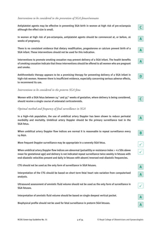
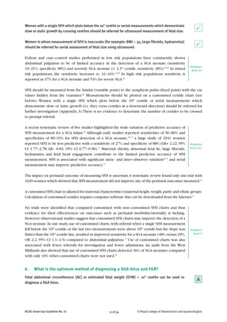
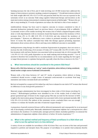
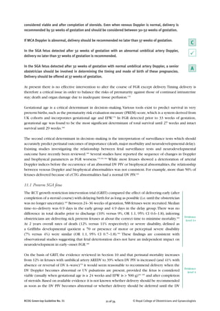
![31 of 34RCOG Green-top Guideline No. 31 © Royal College of Obstetricians and Gynaecologists
Referfor
fetalmedicine
specialist
opinion
SFH
Singlemeasurement<10th
customisedcentile
&/orserialmeasurementsindicativeofFGR
Delivery
Offerdeliveryby37weekswiththe
involvementofaseniorclinician
Recommenddeliveryby37weeksif
MCADopplerPI<5thcentile
Considerdelivery>34weeksifstaticgrowth
over3weeks
RecommendsteroidsifdeliveryisbyCS
(asperRCOGguidance)
Repeatultrasound
(Fortnightly)
Fetalbiometry
SingleACorEFW<10th
customisedcentile
SerialmeasurementsindicativeofFGR
Delivery
Recommenddeliveryby37weeks
ConsidersteroidsifdeliverybyCS
Considerdelivery>34weeksifstaticgrowth
over3weeks
RecommendsteroidsifdeliveryisbyCS
(asperRCOGguidance)
Repeatultrasound
Weekly
AC&EFW1,2
Twiceweekly
UADoppler
UADoppler
HighriskofSGAfetus/neonate
Basedonhistory,biochemistryoruterine
arteryDoppler
Delivery
Recommenddeliverybefore32weeksafter
steroidsif:
–abnormalDVDopplerand/orcCTG
provided≥24weeks&EFW>500g
Recommenddeliveryby32weeksaftersteroids
Considerdeliveryat30–32weeksevenwhen
DVDopplerisnormal
Repeatultrasound
Weekly
AC&EFW1,2
Daily
UADoppler
DVDoppler
[cCTG]3
1
Weeklymeasurementoffetalsizeisvaluableinpredictingbirthweightanddeterminingsize-for-gestationalage
2
IftwoAC/EFWmeasurementsareusedtoestimategrowth,theyshouldbeatleast3weeksapart
3
UsecCTGwhenDVDopplerisunavailableorresultsareinconsistent–recommenddeliveryifSTV<3ms
Abbreviations:AC,abdominalcircumference;EFW,estimatedfetalweight;PI,pulsatilityindex;RI,resistanceindex;UA,umbilicalartery;MCA,middlecerebralartery;DVductsvenosus;
SD,standarddeviation;AREDV.,Absent/reversedend–diastolicvelocities;cCTG,computerisedcardiotography;STV,shorttermvariation;SFH,symphysis–fundalheight;
FGR,fetalgrowthrestriction;EDV,end–diastolicvelocities.
PIorRI>2SDs,EDVpresentAREDVNormal
AC&EFW1,2
UADoppler
MCADopplerafter32weeks
APPENDIXIII:TheManagementoftheSmall–for–Gestational–Age(SGA)Fetus](https://image.slidesharecdn.com/iugrrcogguidelines-140928120309-phpapp01/85/Iugr-rcog-guidelines-31-320.jpg)


![CTEV [ clubfoot] DR ARUN LAL ,DR MOHAMED ASHRAF travancore medical college k...](https://cdn.slidesharecdn.com/ss_thumbnails/ctevclubfootdrarunlaldrmohamedashraftravancoremedicalcollegekollamkeralaindia-260208063247-18fc466c-thumbnail.jpg?width=640&height=640&fit=bounds)

![PERI-PROSTHETIC FRACTURE NAIL-PLATE CONSTRUCT [NPC].pptx](https://cdn.slidesharecdn.com/ss_thumbnails/drarunkumardrmohamedashrafperiprostheticfrasturenail-plateconstructnpc-260209164459-7e9d15a1-thumbnail.jpg?width=640&height=640&fit=bounds)


