Downloaded 553 times

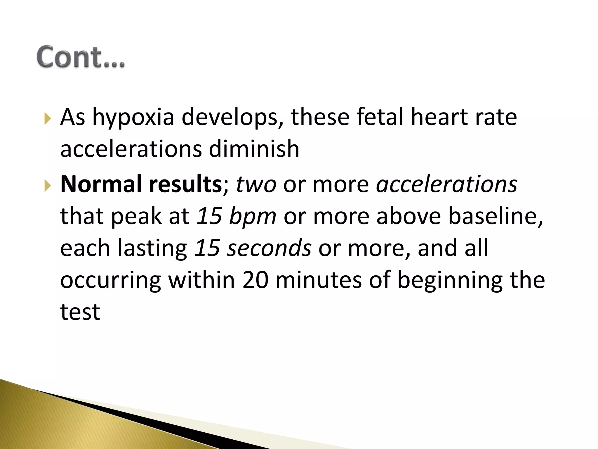


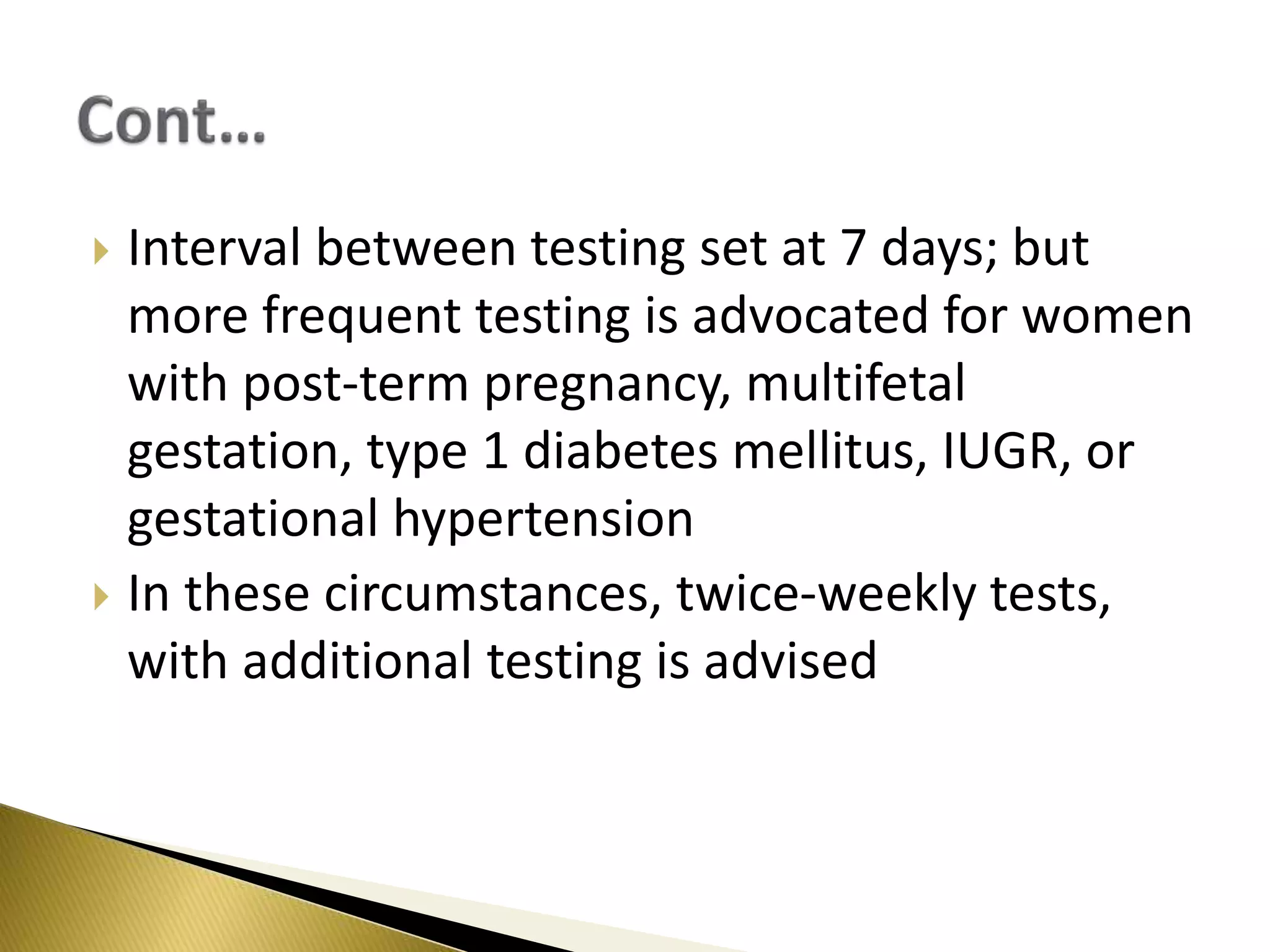


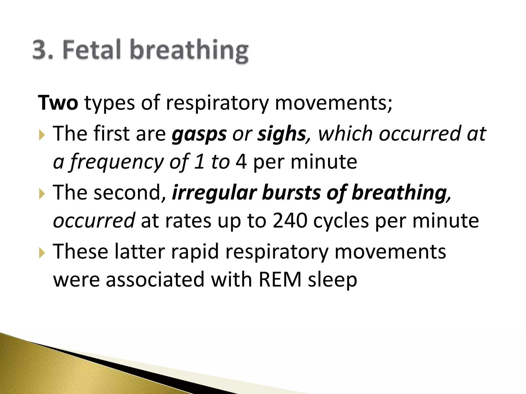


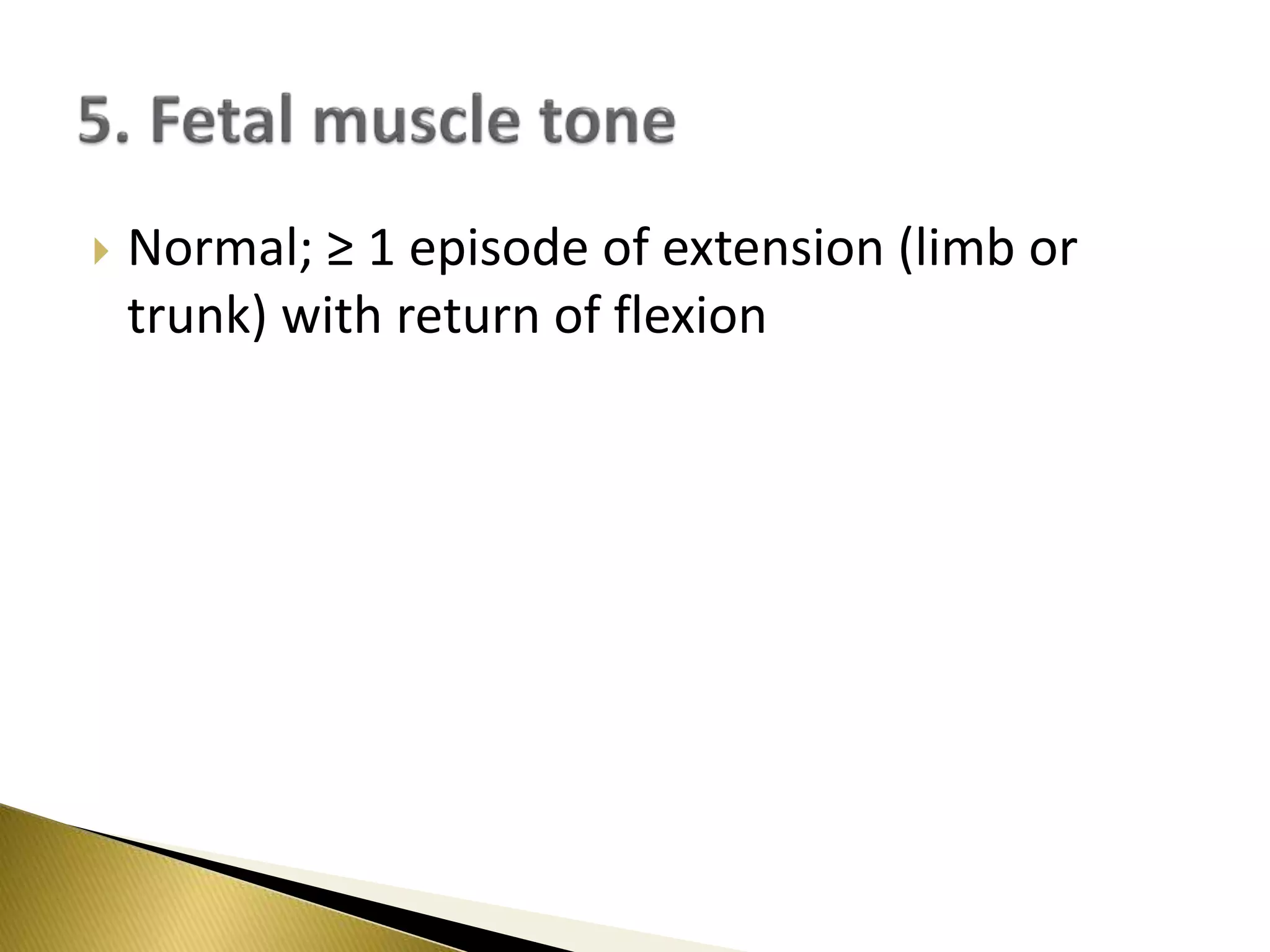


This document discusses the biophysical profile, a technique used to assess fetal well-being through 5 parameters: non-stress test (NST), fetal breathing, fetal movements, muscle tone, and amniotic fluid volume. It describes how each parameter is evaluated and provides details on interpreting results. Abnormal results in the biophysical profile are associated with conditions like IUGR and placental insufficiency and may indicate the need for delivery. The document also reviews other tests used to monitor fetal health like contraction stress tests, acoustic stimulation, and Doppler ultrasound assessments of fetal and placental blood flow.


