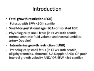
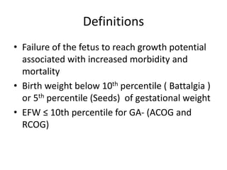
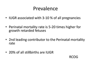
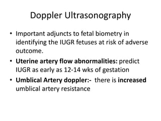
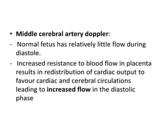
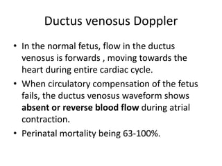
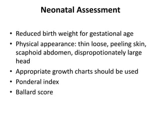
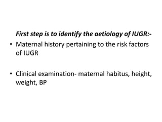
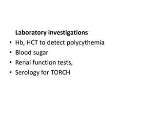
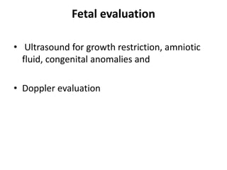
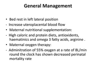
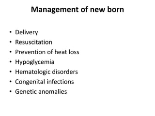
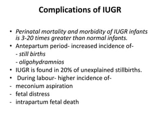
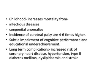
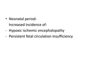
IUGR
Anupam Ghimire
GP/EM Resident
Content
• Introduction
• Prevalence
• Normal Fetal Growth
• Pathogenesis
• Diagnosis
• Management
Introduction
• Fetal growth restriction (FGR)
- Fetuses with EFW <10th centile
• Small-for-gestational age (SGA) or isolated FGR
- Physiologically small fetus (ie EFW<10th centile,
normal amniotic fluid volume and normal umbilical
artery Doppler)
• Intrauterine growth restriction (IUGR)
- Pathologically small fetus (ie EFW<10th centile,
oligohydramnios, abnormal UA Doppler AND/ OR poor
interval growth velocity AND/ OR EFW <3rd centile)
Definitions
• Failure of the fetus to reach growth potential
associated with increased morbidity and
mortality
• Birth weight below 10th percentile ( Battalgia )
or 5th percentile (Seeds) of gestational weight
• EFW ≤ 10th percentile for GA- (ACOG and
RCOG)
Prevalence
• IUGR associated with 3-10 % of all pregnancies
• Perinatal mortality rate is 5-20 times higher for
growth retarded fetuses
• 2nd leading contributor to the Perinatal mortality
rate
• 20% of all stillbirths are IUGR
RCOG
Normal Fetal Growth
• Normal fetal growth is characterized by
cellular hyperplasia followed by hyperplasia
and hypertrophy and lastly by hypertrophy
alone.
Normal Intrauterine Growth
Fetal weight gain
• Weight gain
• Fetal growth accelerates from about 5g per
day at 14 -15 wks of gestation to
- 10g per day at 20 wks
- Peaks at 30 -35g per day at 32-34wks
- After which growth rate decreases.
• Symphysiofundal height
- Increases by about 1cm per wk between 14
and 32 wks.
• Abdominal girth
- Increases by 1 inch per wk after 30 wks.
- About 30 inches at 30wks in an average built
woman.
Classification
1. Symmetrical IUGR
2. Asymmetrical IUGR
Symmetrical IUGR
• Head circumference,
length, and weight are all
proportionally reduced
for gestational age (below
10th percentile)
• Either decreased growth
potential of the fetus or
extrinsic conditions that
are active in pregnancy
Asymmetrical IUGR
• Fetal weight is reduced
out of proportion to
length and head
circumference
• Causes - uteroplacental
insufficiency, maternal
malnutrition, or extrinsic
conditions appearing late
in pregnancy
Etiology
• Fetal, maternal and placental disorders that affect
fetal growth.
A.Fetal Causes
1. Chromosomal Disorders
- usually result in early onset IUGR.
- Trisomies 13, 18, 21 contribute to 5% of IUGR
cases
- Sex chromosome disorders are frequently lethal,
fetuses that survive may have growth restriction
(Turner Syndrome)
2. Congenital Infections:
• Timing of infection is crucial:
- Effects depends on the phase of organogenesis.
• Viruses- rubella, CMV, varicella and HIV
- Rubella is the most embryotoxic virus
- CMV  cytolysis and localized necrosis in fetus.
- Protozoa- like malaria, toxoplasma, trypanosoma
3. Structural Anomalies-
• Defects of CNS,CVS,GIT, Genitourinary and
musculoskeletal system
• If growth restriction is associated with
polyhydramnios, the incidence of structural
anomaly is substantially increased.
4. Genetic Causes-
• Maternal genes have greater influence on
fetal growth.
• Inborn errors of metabolism like agenesis of
pancreas, congenital lipodystrophy,
galactosemia, phenylketonuria also result in
growth restriction of fetus.
B. Placental causes
• Channel for nutrition and oxygen supply to the
fetus
- Single umblical artery
- Abnormal placental implantation
- Velamentous umblical cord insertion
- Bilobed placenta
- Placental haemangiomas
C. Maternal causes
1. Maternal Characteristics:
Contributing to IUGR are-
- Extremes of maternal age
- Grandmultiparity
- History of IUGR in previous pregnancy
- Low maternal weight gain in pregnancy
4. Maternal diseases:
Uteroplacental insufficiency resulting from
medical complications like
- Hypertension
- Renal disease
- Autoimmune disease
- Hyperthyroidism
- Long term insulin dependent diabetes
• Smoking
- active or passive, especially during third trimester
• Alcohol and Drugs
- Crosses the placenta freely.
- Acts as a cellular toxin reducing fetal growth
potential.
• Cocaine and opiates are potent vasoconstrictors.
• Warfarin, anticonvulsants and antineoplastic
agents
• Thrombophilias
- Antiphospholipid antibody syndrome and
other thrombophilias  placental thrombosis
and impaired trophoblastic function
• Nutritional Deficiency
- leads to deficient substrate supply to the fetus
Diagnosis
Identifying mothers at risk:
• Poor maternal nutrition
• Poor BMI at conception
• Pre-eclampsia
• Renal disorders
• Diseases causing vascular insufficiency
• Infections (TORCH)
• Poor maternal weight gain during pregnancy
• Determination of gestational age is of utmost
importance-
– Can be calculated from the date of LMP- not
reliable
– Ultrasound dating before 21 wks of pregnancy
provides more accurate estimate
- CRL : until 13 +6
- HC : from 14 weeks onwards
Clinically
1. Serial measurement of fundal height and
abdominal girth.
• SFH - increases by 1cm per wk between 14
and 32 wks.
• A lag in fundal ht. of 4wks is suggestive of
moderate IUGR.
• A lag of >6 wks is suggestive of severe IUGR
USG evaluation
• Fetal biometry:
i. BPD(Biparietal Diameter)
- When growth rate of BPD is below 5th
percentile, 82% of births are below 10th
percentile
• AC and EFW are most accurate ultrasound
parameters for diagnosis of IUGR.
• AC < 5mm/wk reduction is suggestive of IUGR
iii. Measurement ratios-
- Age independent ratios to detect IUGR
• HC/AC: Persistence of a head to abdomen ratio
<1 late in gestation is predictive of asymmetric
IUGR.
• Femur length : serial measurements of femur
length are effective for detecting symmetric IUGR
• Placental Morphology: Acceleration of
placental maturation may occur with IUGR .
• Placental volume: helpful in predicting
subsequent fetal growth.
• Amniotic fluid volume:
- Amniotic fluid index(AFI) between 8 and 25 is
normal.
Doppler Ultrasonography
• Important adjuncts to fetal biometry in
identifying the IUGR fetuses at risk of adverse
outcome.
• Uterine artery flow abnormalities: predict
IUGR as early as 12-14 wks of gestation
• Umblical Artery doppler:- there is increased
umblical artery resistance
• Middle cerebral artery doppler:
- Normal fetus has relatively little flow during
diastole.
- Increased resistance to blood flow in placenta
results in redistribution of cardiac output to
favour cardiac and cerebral circulations
leading to increased flow in the diastolic
phase
Ductus venosus Doppler
• In the normal fetus, flow in the ductus
venosus is forwards , moving towards the
heart during entire cardiac cycle.
• When circulatory compensation of the fetus
fails, the ductus venosus waveform shows
absent or reverse blood flow during atrial
contraction.
• Perinatal mortality being 63-100%.
Neonatal Assessment
• Reduced birth weight for gestational age
• Physical appearance: thin loose, peeling skin,
scaphoid abdomen, dispropotionately large
head
• Appropriate growth charts should be used
• Ponderal index
• Ballard score
MANAGEMENT
Principles:
1. Identify the cause of growth restriction.
2. Treat the cause if found.
3. General management
First step is to identify the aetiology of IUGR:-
• Maternal history pertaining to the risk factors
of IUGR
• Clinical examination- maternal habitus, height,
weight, BP
Laboratory investigations
• Hb, HCT to detect polycythemia
• Blood sugar
• Renal function tests,
• Serology for TORCH
Fetal evaluation
• Ultrasound for growth restriction, amniotic
fluid, congenital anomalies and
• Doppler evaluation
Treatment of underlying cause
• Hypertension
• Cessation of smoking
• Protein energy supplementation in poorly
nourished and underweight women
General Management
• Bed rest in left lateral position
- Increase uteroplacental blood flow
• Maternal nutritional supplementation:
- High caloric and protein diets, antioxidents,
haematinics and omega 3 fatty acids, arginine .
• Maternal oxygen therapy:
- Adminitration of 55% oxygen at a rate of 8L/min
round the clock has shown decreased perinatal
mortality rate
Arginine
• Improbes GHRH secretion  increase plasma
GH  Somatic growth
• Precursor of NO
- NO helps in relaxation of smooth muscle 
improves placental blood supply
Pharmacological therapy
• Aspirin in low doses(1-2 mg/kg body wt.) have
been tried but all have failed to show any
significant difference in incidence of IUGR.
Role of steroids
• Antenatal glucocorticoid
- Reduces the incidence of respiratory distress
syndrome, intraventricular hemorrhage and
death
Timing of delivery
• The optimum timing of delivery is determined by
- Gestational age,
- Underlying etiology,
- Possibility of extrauterine survival and
- Fetal condition.
- Strict fetal surveillance is needed to monitor fetal
well being and to detect signs of fetal
compromise
• Recommend delivery from 34 weeks and
beyond
• With reassuring foetal surveillance, Vaginal
delivery may be attempted
• Expectant management can be guided using
Antepartum foetal surveillance
• Fetuses with significant IUGR
- Intrapartum continuous fetal heart monitoring
- Fetal blood sampling and
- Expert neonatal care.
Mode of Delivery
• Every case must be individualized
• Determinants: underlying aetiology, GA,
severity of IUGR, fetal surveillance
• Parameters, Facilities available
• Obstetric factors e.g. malpresentation
• CS better with severe growth restriction
• In cases of AEDF, delivery should be
considered no later than 34 weeks
• In cases of REDF, delivery should be
considered no later than 30 weeks gestation.
Earlier delivery may be indicated by a
deterioration of sonographic variables.
RCOG 2017
Management of new born
• Delivery
• Resuscitation
• Prevention of heat loss
• Hypoglycemia
• Hematologic disorders
• Congenital infections
• Genetic anomalies
Complications of IUGR
• Perinatal mortality and morbidity of IUGR infants
is 3-20 times greater than normal infants.
• Antepartum period- increased incidence of-
- still births
- oligohydramnios
• IUGR is found in 20% of unexplained stillbirths.
• During labour- higher incidence of-
- meconium aspiration
- fetal distress
- intrapartum fetal death
• Childhood- increases mortality from-
- infectious diseases
- congenital anomalies
• Incidence of cerebral palsy are 4-6 times higher.
• Subtle impairment of cognitive performance and
educational underachievement.
• Long term complications- increased risk of
coronary heart disease, hypertension, type II
diabetes mellitus, dyslipidaemia and stroke
• Neonatal period-
Increased incidence of-
- Hypoxic ischemic encephalopathy
- Persistent fetal circulation insufficiency
• Women who have delivered a growth
restricted infant <34 weeks gestation
- should be offered an appointment for
postnatal counselling
- review of placental histology and
investigation of underlying causes such as
thrombophilia screening
RCOG 2017
Take home message
• Early and proper identification and
management lowers this perinatal mortality
and morbidity
References
• RCOG Bulletein, 2017
• Arias, Practice guide for high risk pregnancy,
4th edition
• Essential of obstetrics, first edition
THANK YOU

Iugr

  • 1.
  • 2.
    Content • Introduction • Prevalence •Normal Fetal Growth • Pathogenesis • Diagnosis • Management
  • 3.
    Introduction • Fetal growthrestriction (FGR) - Fetuses with EFW <10th centile • Small-for-gestational age (SGA) or isolated FGR - Physiologically small fetus (ie EFW<10th centile, normal amniotic fluid volume and normal umbilical artery Doppler) • Intrauterine growth restriction (IUGR) - Pathologically small fetus (ie EFW<10th centile, oligohydramnios, abnormal UA Doppler AND/ OR poor interval growth velocity AND/ OR EFW <3rd centile)
  • 4.
    Definitions • Failure ofthe fetus to reach growth potential associated with increased morbidity and mortality • Birth weight below 10th percentile ( Battalgia ) or 5th percentile (Seeds) of gestational weight • EFW ≤ 10th percentile for GA- (ACOG and RCOG)
  • 5.
    Prevalence • IUGR associatedwith 3-10 % of all pregnancies • Perinatal mortality rate is 5-20 times higher for growth retarded fetuses • 2nd leading contributor to the Perinatal mortality rate • 20% of all stillbirths are IUGR RCOG
  • 6.
    Normal Fetal Growth •Normal fetal growth is characterized by cellular hyperplasia followed by hyperplasia and hypertrophy and lastly by hypertrophy alone.
  • 7.
  • 8.
    Fetal weight gain •Weight gain • Fetal growth accelerates from about 5g per day at 14 -15 wks of gestation to - 10g per day at 20 wks - Peaks at 30 -35g per day at 32-34wks - After which growth rate decreases.
  • 9.
    • Symphysiofundal height -Increases by about 1cm per wk between 14 and 32 wks. • Abdominal girth - Increases by 1 inch per wk after 30 wks. - About 30 inches at 30wks in an average built woman.
  • 10.
  • 11.
    Symmetrical IUGR • Headcircumference, length, and weight are all proportionally reduced for gestational age (below 10th percentile) • Either decreased growth potential of the fetus or extrinsic conditions that are active in pregnancy Asymmetrical IUGR • Fetal weight is reduced out of proportion to length and head circumference • Causes - uteroplacental insufficiency, maternal malnutrition, or extrinsic conditions appearing late in pregnancy
  • 12.
    Etiology • Fetal, maternaland placental disorders that affect fetal growth. A.Fetal Causes 1. Chromosomal Disorders - usually result in early onset IUGR. - Trisomies 13, 18, 21 contribute to 5% of IUGR cases - Sex chromosome disorders are frequently lethal, fetuses that survive may have growth restriction (Turner Syndrome)
  • 13.
    2. Congenital Infections: •Timing of infection is crucial: - Effects depends on the phase of organogenesis. • Viruses- rubella, CMV, varicella and HIV - Rubella is the most embryotoxic virus - CMV  cytolysis and localized necrosis in fetus. - Protozoa- like malaria, toxoplasma, trypanosoma
  • 14.
    3. Structural Anomalies- •Defects of CNS,CVS,GIT, Genitourinary and musculoskeletal system • If growth restriction is associated with polyhydramnios, the incidence of structural anomaly is substantially increased.
  • 15.
    4. Genetic Causes- •Maternal genes have greater influence on fetal growth. • Inborn errors of metabolism like agenesis of pancreas, congenital lipodystrophy, galactosemia, phenylketonuria also result in growth restriction of fetus.
  • 16.
    B. Placental causes •Channel for nutrition and oxygen supply to the fetus - Single umblical artery - Abnormal placental implantation - Velamentous umblical cord insertion - Bilobed placenta - Placental haemangiomas
  • 17.
    C. Maternal causes 1.Maternal Characteristics: Contributing to IUGR are- - Extremes of maternal age - Grandmultiparity - History of IUGR in previous pregnancy - Low maternal weight gain in pregnancy
  • 18.
    4. Maternal diseases: Uteroplacentalinsufficiency resulting from medical complications like - Hypertension - Renal disease - Autoimmune disease - Hyperthyroidism - Long term insulin dependent diabetes
  • 19.
    • Smoking - activeor passive, especially during third trimester • Alcohol and Drugs - Crosses the placenta freely. - Acts as a cellular toxin reducing fetal growth potential. • Cocaine and opiates are potent vasoconstrictors. • Warfarin, anticonvulsants and antineoplastic agents
  • 20.
    • Thrombophilias - Antiphospholipidantibody syndrome and other thrombophilias  placental thrombosis and impaired trophoblastic function • Nutritional Deficiency - leads to deficient substrate supply to the fetus
  • 21.
    Diagnosis Identifying mothers atrisk: • Poor maternal nutrition • Poor BMI at conception • Pre-eclampsia • Renal disorders • Diseases causing vascular insufficiency • Infections (TORCH) • Poor maternal weight gain during pregnancy
  • 22.
    • Determination ofgestational age is of utmost importance- – Can be calculated from the date of LMP- not reliable – Ultrasound dating before 21 wks of pregnancy provides more accurate estimate - CRL : until 13 +6 - HC : from 14 weeks onwards
  • 23.
    Clinically 1. Serial measurementof fundal height and abdominal girth. • SFH - increases by 1cm per wk between 14 and 32 wks. • A lag in fundal ht. of 4wks is suggestive of moderate IUGR. • A lag of >6 wks is suggestive of severe IUGR
  • 24.
    USG evaluation • Fetalbiometry: i. BPD(Biparietal Diameter) - When growth rate of BPD is below 5th percentile, 82% of births are below 10th percentile
  • 25.
    • AC andEFW are most accurate ultrasound parameters for diagnosis of IUGR. • AC < 5mm/wk reduction is suggestive of IUGR iii. Measurement ratios- - Age independent ratios to detect IUGR • HC/AC: Persistence of a head to abdomen ratio <1 late in gestation is predictive of asymmetric IUGR. • Femur length : serial measurements of femur length are effective for detecting symmetric IUGR
  • 26.
    • Placental Morphology:Acceleration of placental maturation may occur with IUGR . • Placental volume: helpful in predicting subsequent fetal growth. • Amniotic fluid volume: - Amniotic fluid index(AFI) between 8 and 25 is normal.
  • 27.
    Doppler Ultrasonography • Importantadjuncts to fetal biometry in identifying the IUGR fetuses at risk of adverse outcome. • Uterine artery flow abnormalities: predict IUGR as early as 12-14 wks of gestation • Umblical Artery doppler:- there is increased umblical artery resistance
  • 28.
    • Middle cerebralartery doppler: - Normal fetus has relatively little flow during diastole. - Increased resistance to blood flow in placenta results in redistribution of cardiac output to favour cardiac and cerebral circulations leading to increased flow in the diastolic phase
  • 29.
    Ductus venosus Doppler •In the normal fetus, flow in the ductus venosus is forwards , moving towards the heart during entire cardiac cycle. • When circulatory compensation of the fetus fails, the ductus venosus waveform shows absent or reverse blood flow during atrial contraction. • Perinatal mortality being 63-100%.
  • 30.
    Neonatal Assessment • Reducedbirth weight for gestational age • Physical appearance: thin loose, peeling skin, scaphoid abdomen, dispropotionately large head • Appropriate growth charts should be used • Ponderal index • Ballard score
  • 31.
    MANAGEMENT Principles: 1. Identify thecause of growth restriction. 2. Treat the cause if found. 3. General management
  • 32.
    First step isto identify the aetiology of IUGR:- • Maternal history pertaining to the risk factors of IUGR • Clinical examination- maternal habitus, height, weight, BP
  • 33.
    Laboratory investigations • Hb,HCT to detect polycythemia • Blood sugar • Renal function tests, • Serology for TORCH
  • 34.
    Fetal evaluation • Ultrasoundfor growth restriction, amniotic fluid, congenital anomalies and • Doppler evaluation
  • 35.
    Treatment of underlyingcause • Hypertension • Cessation of smoking • Protein energy supplementation in poorly nourished and underweight women
  • 36.
    General Management • Bedrest in left lateral position - Increase uteroplacental blood flow • Maternal nutritional supplementation: - High caloric and protein diets, antioxidents, haematinics and omega 3 fatty acids, arginine . • Maternal oxygen therapy: - Adminitration of 55% oxygen at a rate of 8L/min round the clock has shown decreased perinatal mortality rate
  • 37.
    Arginine • Improbes GHRHsecretion  increase plasma GH  Somatic growth • Precursor of NO - NO helps in relaxation of smooth muscle  improves placental blood supply
  • 38.
    Pharmacological therapy • Aspirinin low doses(1-2 mg/kg body wt.) have been tried but all have failed to show any significant difference in incidence of IUGR.
  • 39.
    Role of steroids •Antenatal glucocorticoid - Reduces the incidence of respiratory distress syndrome, intraventricular hemorrhage and death
  • 40.
    Timing of delivery •The optimum timing of delivery is determined by - Gestational age, - Underlying etiology, - Possibility of extrauterine survival and - Fetal condition. - Strict fetal surveillance is needed to monitor fetal well being and to detect signs of fetal compromise
  • 41.
    • Recommend deliveryfrom 34 weeks and beyond • With reassuring foetal surveillance, Vaginal delivery may be attempted • Expectant management can be guided using Antepartum foetal surveillance
  • 42.
    • Fetuses withsignificant IUGR - Intrapartum continuous fetal heart monitoring - Fetal blood sampling and - Expert neonatal care.
  • 43.
    Mode of Delivery •Every case must be individualized • Determinants: underlying aetiology, GA, severity of IUGR, fetal surveillance • Parameters, Facilities available • Obstetric factors e.g. malpresentation • CS better with severe growth restriction
  • 44.
    • In casesof AEDF, delivery should be considered no later than 34 weeks • In cases of REDF, delivery should be considered no later than 30 weeks gestation. Earlier delivery may be indicated by a deterioration of sonographic variables. RCOG 2017
  • 45.
    Management of newborn • Delivery • Resuscitation • Prevention of heat loss • Hypoglycemia • Hematologic disorders • Congenital infections • Genetic anomalies
  • 46.
    Complications of IUGR •Perinatal mortality and morbidity of IUGR infants is 3-20 times greater than normal infants. • Antepartum period- increased incidence of- - still births - oligohydramnios • IUGR is found in 20% of unexplained stillbirths. • During labour- higher incidence of- - meconium aspiration - fetal distress - intrapartum fetal death
  • 47.
    • Childhood- increasesmortality from- - infectious diseases - congenital anomalies • Incidence of cerebral palsy are 4-6 times higher. • Subtle impairment of cognitive performance and educational underachievement. • Long term complications- increased risk of coronary heart disease, hypertension, type II diabetes mellitus, dyslipidaemia and stroke
  • 48.
    • Neonatal period- Increasedincidence of- - Hypoxic ischemic encephalopathy - Persistent fetal circulation insufficiency
  • 49.
    • Women whohave delivered a growth restricted infant <34 weeks gestation - should be offered an appointment for postnatal counselling - review of placental histology and investigation of underlying causes such as thrombophilia screening RCOG 2017
  • 50.
    Take home message •Early and proper identification and management lowers this perinatal mortality and morbidity
  • 51.
    References • RCOG Bulletein,2017 • Arias, Practice guide for high risk pregnancy, 4th edition • Essential of obstetrics, first edition
  • 52.