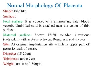
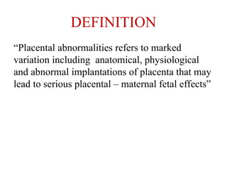
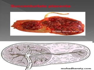
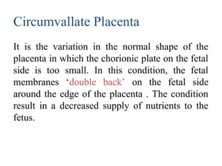
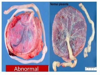
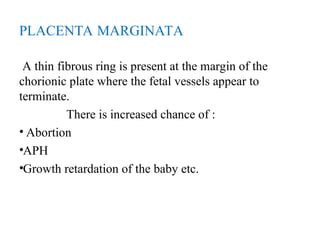
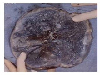
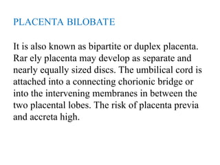
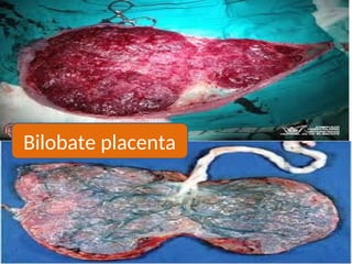
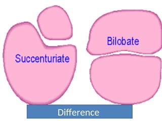
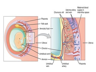
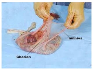
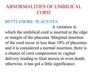
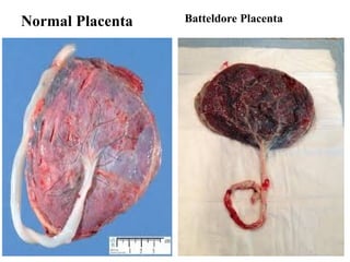
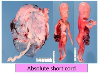
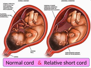
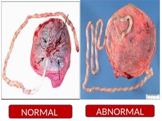
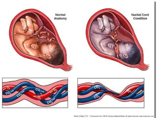
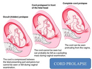
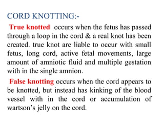
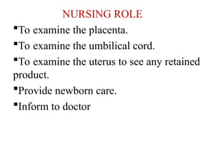
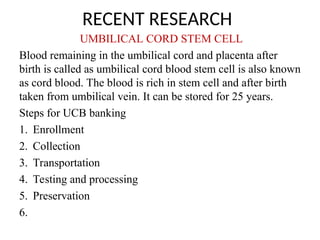
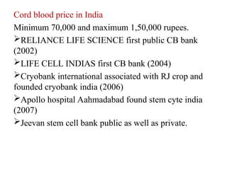
PRESENTATION ON
ABNORMALITIES OF PLACENTA
&
UMBLICAL CORD
PRESENTED BY
SAVITA SANJAY
HANAMSAGAR
Abnormalities of placenta
INTRODUCTION
The placenta is developed from two sources. The principal
component is fetal which develops from the chorion
frondosum and the maternal component consists of decidua
basalis.The placenta is the temporary organ that connects
the developing fetus via the umbilical cord to the uterine
wall. Which exchange material between fetal and maternal
blood. It includes
•Endocrine function
•Immunological function
•Respiratory function
•Excretory function
•Nutritive function
Normal Morphology Of Placenta
Shape: Disc like
Surface :
Fetal surface- It is covered with amnion and fetal blood
vessels. Umbilical cord is attached near the center of this
surface.
Maternal surface- Shows 15-20 rounded elevations
(cotyledon) with septa in between. Rough and red in color.
Site: At original implantation site which is upper part of
posterior wall of uterus.
Diameter :15-20cm
Thickness : about 3cm
Weight : about 450-500gm
DEFINITION
“Placental abnormalities refers to marked
variation including anatomical, physiological
and abnormal implantations of placenta that may
lead to serious placental – maternal fetal effects”
ABNORMALITIES OF PLACENTA
Placenta Succenturiata
One or more small lobes of placenta, size of
cotyledon, may be placed at varying distances
from the main placental margin. A leash of
vessels connecting the main to the small lobe
traverse through the membranes. The accessory
lobe is developed from the activated villi on the
chorionic laeve.In cases of absence of
communicating blood vessels, it is called
placenta spuria.The incidence of placenta
succenturiata is about 3% .
If the succenturiate lobe is retained, following
birth of the placenta, it may lead to –
Postpartum hemorrhage
Subinvolution
Uterine sepsis
Polyp formation
PLACENTA EXTRACHORIALS
Placenta extrachorialis are of two types-
•Circumvallate placenta
•Placenta marginata
Development- The placenta of such type is due
to the smaller chorionic plate than the basal
plate. The membranes ( amnion and chorion) are
folded, rollled back upon itself to form a ring
which reflected centrally .
Circumvallate Placenta
It is the variation in the normal shape of the
placenta in which the chorionic plate on the fetal
side is too small. In this condition, the fetal
membranes ‘double back’ on the fetal side
around the edge of the placenta . The condition
result in a decreased supply of nutrients to the
fetus.
Abnormal
PLACENTA MARGINATA
A thin fibrous ring is present at the margin of the
chorionic plate where the fetal vessels appear to
terminate.
There is increased chance of :
• Abortion
•APH
•Growth retardation of the baby etc.
PLACENTA BILOBATE
It is also known as bipartite or duplex placenta.
Rar ely placenta may develop as separate and
nearly equally sized discs. The umbilical cord is
attached into a connecting chorionic bridge or
into the intervening membranes in between the
two placental lobes. The risk of placenta previa
and accreta high.
Bilobate placenta
Difference
Abnormalities of
Umbilical cord
INTRODUCTION
The umbilical cord or funis extends from the
fetus to the placenta. It transmits the umbilical
blood vessels, which are two arteries and one
vein.
These are enclosed & Protected by a
gelantinous substance known as wharton’s jelly.
The two whole cord is covered in a layer of
amnion continues with that covering the placenta
It is the connection between placenta and fetus.
Lenght: 50-60cm
Diameter: 2cm
Shape : Tortous
Contents : One umbilical arteries, one umbilical
vein embedded in wharton’s jelly and
surrounded by amniotic membrane.
Attachments: It is attached to fetal surface of
placenta near its center.
Normal Morphology Of Umbilical Cord
Insertion of umbilical cord into
chorionic plate
Normally the umbilical cord inserts near the
center of the chorionic plate, which stabilizes
the fetal vessels as they leave the umbilical
cord. Like the roots of a tree, the fetal vessels
branch over the surface of the chorionic plate
and then dive into the placental parenchyma.
DEFINITION-:
“Cord abnormalities refers to marked variations
of umbilical cord including anatomical,
physiological alterations involving abnormal
insertion, length ,occlusion etc. that may result
in disturbance in feto-placental circulations.”
ABNORMALITIES OF UMBILICAL
CORD
BETTLEDORE PLACENTA
A variation in
which the umbilical cord is inserted at the edge
or margin of the placenta. Marginal insertion
of the cord occur in less than 10% of placentas
and it is considered a normal insertion, there is
a chance of cord compression in vaginal
delivery leading to fetal anoxia or even death;
otherwise, it has got a little significance.
Normal Placenta Batteldore Placenta
VELAMENTOUS PLACENTA
The cord is inserted into the membrane at
some distance from the edge of the placenta. The
umbilical vessel from the cord running through
the membrane for a variable distance before each
enters the placenta.
NORMAL
ABNORMAL
VASA PREVIA
Vasa Previa occurs when one or more blood vessels from the
umbilical cord or placenta cross the cervix underneath the
baby. The blood vessels, unprotected by the Wharton's jelly
in the umbilical cord or the tissue in the placenta, sometimes
tear when the cervix dilates or the membranes rupture. This
can result in life-threatening bleeding in the baby. Even if
the blood vessels do not tear, the baby may suffer from lack
of oxygen due to pressure on the blood vessels. Vasa Previa
occurs in 1 in 2,500 births
• when vasa Previa is diagnosed by ultrasound earlier in
pregnancy, fetal deaths generally can be prevented by
delivering the baby by cesarean section at about 35 weeks of
gestation.
Abnormal number of umbilical vessels in the
umbilical cord has high correlation with fetal
anomalies. About one third of babies born with
only one umbilical artery will have multiple and
severe malformation.
SINGLE UMBILICAL ARTERY-:
It is present in about 1-2% cases. It may be due
to failure of development of one artery or due to
its atrophy in later months. It is more common in
twins and in babies born of women with
eclampsia , diabetes, oligohydraminos and
antepartum hemorrage but exact cause is
unknown . It is frequently associated with
congenital malformation. There is increased
chance of abortion, fetal aneuploidy,
prematurity, IUGR and increased perinatal
mortality.
Microscopic structure of umbilical cord
Normal & Abnormal
ABNORMAL LENGTH
SHORT CORD:-
An absolute short cord is one that is short in
length.
A relative short cord is one of average length,
which has become short because of looping
around the body or neck of the fetus.
A short cord may be a causative factor in
failure of the fetus to descend. In such an event it
might additionally cause umbilical hernia, fetal
distress, rupture of the cord.
Absolute short cord
Normal cord & Relative short cord
EXCESSIVE LONG CORD:-
Long cord are more common than short
cords. Generally it has no significance.
However, a long cord becomes looped around
the fetal body or neck causing a relative short
cord. It can also become knotted or prolapsed in
front of the presenting part.
ABNORMAL
NORMAL
CORD PROLAPSE
CORD KNOTTING:-
True knotted occurs when the fetus has passed
through a loop in the cord & a real knot has been
created. true knot are liable to occur with small
fetus, long cord, active fetal movements, large
amount of amniotic fluid and multiple gestation
with in the single amnion.
False knotting occurs when the cord appears to
be knotted, but instead has kinking of the blood
vessel with in the cord or accumulation of
wartson’s jelly on the cord.
TRUE KNOT
FALSE
KNOT
NURSING ROLE
To examine the placenta.
To examine the umbilical cord.
To examine the uterus to see any retained
product.
Provide newborn care.
Inform to doctor
RECENT RESEARCH
UMBILICAL CORD STEM CELL
Blood remaining in the umbilical cord and placenta after
birth is called as umbilical cord blood stem cell is also known
as cord blood. The blood is rich in stem cell and after birth
taken from umbilical vein. It can be stored for 25 years.
Steps for UCB banking
1. Enrollment
2. Collection
3. Transportation
4. Testing and processing
5. Preservation
6.
Cord blood price in India
Minimum 70,000 and maximum 1,50,000 rupees.
RELIANCE LIFE SCIENCE first public CB bank
(2002)
LIFE CELL INDIAS first CB bank (2004)
Cryobank international associated with RJ crop and
founded cryobank india (2006)
Apollo hospital Aahmadabad found stem cyte india
(2007)
Jeevan stem cell bank public as well as private.
SUMMARY
I have discussed about the of abnormalities of
placenta and umbilical cord that includes–
introduction, morphology of placenta and
umbilical cord, abnormalities of placenta and
umbilical cord, recent research, bibliography.
BIBLIOGRAPHY
• D.C. DUTTA : A textbook of “Obstretics”
including perinatology & contraception, sixth
edition, published by New central book
agency, page no. 1-3,15.
• SUDIP CHAKRAVARTI & SHIRISH N DAFTORY :
“Mannual of obstretics”, 3rd
edition, published
by Elsevier, page no.2-6.
• JACOB ANNAMMA -: a textbook of midwifery
and gynecology nursing edition 4th
pg no. 200
• Anand & Verma : A textbook of “Human
Anatomy & physiology” for nursing, 2nd
Edition, published by Jaypee brothers Medical
publisher, page no.571-583.
• PR Ashalatha : A textbook of “Anatomy &
physiology” for nurses, 4th
edition, published
by Jaypee Brothers, Page no.470-481.
• https://www.slideshare.net.,shaliantony
• https://en.m.wikipedia.org.>wiki>female
1. The oxygen supply to the baby is at the rat of
A)8ml/kg/min B) 4ml/kg/min
C) 10ml/kg/min D) 6ml/kg/min
2. Umbilical cord blood can be stored for….
A) 25 year B) 30 year
C) 15 year D) 20 year
3. Normal diameter of placenta.
A)15-20cm B) 10-15cm
C) 20-25cm D) 5-10cm
4. Fetal aneuploidy is-
A)Presence of abnormal no. of chromosome
B) absence of chromosome
C) None of these
5. The cord can be seen protruding from vagina
is called –
A)Complete cord prolapse
B) Cord prolapse
C) Occult
D)None of these
THANK YOU
THANK YOU

430433181-Umblical-Cord-Abnormalities-ppt.ppt

  • 1.
    PRESENTATION ON ABNORMALITIES OFPLACENTA & UMBLICAL CORD PRESENTED BY SAVITA SANJAY HANAMSAGAR
  • 2.
  • 3.
    INTRODUCTION The placenta isdeveloped from two sources. The principal component is fetal which develops from the chorion frondosum and the maternal component consists of decidua basalis.The placenta is the temporary organ that connects the developing fetus via the umbilical cord to the uterine wall. Which exchange material between fetal and maternal blood. It includes •Endocrine function •Immunological function •Respiratory function •Excretory function •Nutritive function
  • 4.
    Normal Morphology OfPlacenta Shape: Disc like Surface : Fetal surface- It is covered with amnion and fetal blood vessels. Umbilical cord is attached near the center of this surface. Maternal surface- Shows 15-20 rounded elevations (cotyledon) with septa in between. Rough and red in color. Site: At original implantation site which is upper part of posterior wall of uterus. Diameter :15-20cm Thickness : about 3cm Weight : about 450-500gm
  • 5.
    DEFINITION “Placental abnormalities refersto marked variation including anatomical, physiological and abnormal implantations of placenta that may lead to serious placental – maternal fetal effects”
  • 6.
    ABNORMALITIES OF PLACENTA PlacentaSuccenturiata One or more small lobes of placenta, size of cotyledon, may be placed at varying distances from the main placental margin. A leash of vessels connecting the main to the small lobe traverse through the membranes. The accessory lobe is developed from the activated villi on the chorionic laeve.In cases of absence of communicating blood vessels, it is called placenta spuria.The incidence of placenta succenturiata is about 3% .
  • 8.
    If the succenturiatelobe is retained, following birth of the placenta, it may lead to – Postpartum hemorrhage Subinvolution Uterine sepsis Polyp formation
  • 9.
    PLACENTA EXTRACHORIALS Placenta extrachorialisare of two types- •Circumvallate placenta •Placenta marginata Development- The placenta of such type is due to the smaller chorionic plate than the basal plate. The membranes ( amnion and chorion) are folded, rollled back upon itself to form a ring which reflected centrally .
  • 10.
    Circumvallate Placenta It isthe variation in the normal shape of the placenta in which the chorionic plate on the fetal side is too small. In this condition, the fetal membranes ‘double back’ on the fetal side around the edge of the placenta . The condition result in a decreased supply of nutrients to the fetus.
  • 11.
  • 12.
    PLACENTA MARGINATA A thinfibrous ring is present at the margin of the chorionic plate where the fetal vessels appear to terminate. There is increased chance of : • Abortion •APH •Growth retardation of the baby etc.
  • 14.
    PLACENTA BILOBATE It isalso known as bipartite or duplex placenta. Rar ely placenta may develop as separate and nearly equally sized discs. The umbilical cord is attached into a connecting chorionic bridge or into the intervening membranes in between the two placental lobes. The risk of placenta previa and accreta high.
  • 15.
  • 16.
  • 17.
  • 18.
    INTRODUCTION The umbilical cordor funis extends from the fetus to the placenta. It transmits the umbilical blood vessels, which are two arteries and one vein. These are enclosed & Protected by a gelantinous substance known as wharton’s jelly. The two whole cord is covered in a layer of amnion continues with that covering the placenta
  • 19.
    It is theconnection between placenta and fetus. Lenght: 50-60cm Diameter: 2cm Shape : Tortous Contents : One umbilical arteries, one umbilical vein embedded in wharton’s jelly and surrounded by amniotic membrane. Attachments: It is attached to fetal surface of placenta near its center. Normal Morphology Of Umbilical Cord
  • 20.
    Insertion of umbilicalcord into chorionic plate Normally the umbilical cord inserts near the center of the chorionic plate, which stabilizes the fetal vessels as they leave the umbilical cord. Like the roots of a tree, the fetal vessels branch over the surface of the chorionic plate and then dive into the placental parenchyma.
  • 23.
    DEFINITION-: “Cord abnormalities refersto marked variations of umbilical cord including anatomical, physiological alterations involving abnormal insertion, length ,occlusion etc. that may result in disturbance in feto-placental circulations.”
  • 24.
    ABNORMALITIES OF UMBILICAL CORD BETTLEDOREPLACENTA A variation in which the umbilical cord is inserted at the edge or margin of the placenta. Marginal insertion of the cord occur in less than 10% of placentas and it is considered a normal insertion, there is a chance of cord compression in vaginal delivery leading to fetal anoxia or even death; otherwise, it has got a little significance.
  • 25.
  • 26.
    VELAMENTOUS PLACENTA The cordis inserted into the membrane at some distance from the edge of the placenta. The umbilical vessel from the cord running through the membrane for a variable distance before each enters the placenta.
  • 27.
  • 28.
    VASA PREVIA Vasa Previaoccurs when one or more blood vessels from the umbilical cord or placenta cross the cervix underneath the baby. The blood vessels, unprotected by the Wharton's jelly in the umbilical cord or the tissue in the placenta, sometimes tear when the cervix dilates or the membranes rupture. This can result in life-threatening bleeding in the baby. Even if the blood vessels do not tear, the baby may suffer from lack of oxygen due to pressure on the blood vessels. Vasa Previa occurs in 1 in 2,500 births • when vasa Previa is diagnosed by ultrasound earlier in pregnancy, fetal deaths generally can be prevented by delivering the baby by cesarean section at about 35 weeks of gestation.
  • 30.
    Abnormal number ofumbilical vessels in the umbilical cord has high correlation with fetal anomalies. About one third of babies born with only one umbilical artery will have multiple and severe malformation.
  • 31.
    SINGLE UMBILICAL ARTERY-: Itis present in about 1-2% cases. It may be due to failure of development of one artery or due to its atrophy in later months. It is more common in twins and in babies born of women with eclampsia , diabetes, oligohydraminos and antepartum hemorrage but exact cause is unknown . It is frequently associated with congenital malformation. There is increased chance of abortion, fetal aneuploidy, prematurity, IUGR and increased perinatal mortality.
  • 32.
  • 33.
  • 34.
    ABNORMAL LENGTH SHORT CORD:- Anabsolute short cord is one that is short in length. A relative short cord is one of average length, which has become short because of looping around the body or neck of the fetus. A short cord may be a causative factor in failure of the fetus to descend. In such an event it might additionally cause umbilical hernia, fetal distress, rupture of the cord.
  • 35.
  • 36.
    Normal cord &Relative short cord
  • 37.
    EXCESSIVE LONG CORD:- Longcord are more common than short cords. Generally it has no significance. However, a long cord becomes looped around the fetal body or neck causing a relative short cord. It can also become knotted or prolapsed in front of the presenting part.
  • 38.
  • 40.
  • 41.
    CORD KNOTTING:- True knottedoccurs when the fetus has passed through a loop in the cord & a real knot has been created. true knot are liable to occur with small fetus, long cord, active fetal movements, large amount of amniotic fluid and multiple gestation with in the single amnion. False knotting occurs when the cord appears to be knotted, but instead has kinking of the blood vessel with in the cord or accumulation of wartson’s jelly on the cord.
  • 42.
  • 43.
  • 44.
    NURSING ROLE To examinethe placenta. To examine the umbilical cord. To examine the uterus to see any retained product. Provide newborn care. Inform to doctor
  • 45.
    RECENT RESEARCH UMBILICAL CORDSTEM CELL Blood remaining in the umbilical cord and placenta after birth is called as umbilical cord blood stem cell is also known as cord blood. The blood is rich in stem cell and after birth taken from umbilical vein. It can be stored for 25 years. Steps for UCB banking 1. Enrollment 2. Collection 3. Transportation 4. Testing and processing 5. Preservation 6.
  • 46.
    Cord blood pricein India Minimum 70,000 and maximum 1,50,000 rupees. RELIANCE LIFE SCIENCE first public CB bank (2002) LIFE CELL INDIAS first CB bank (2004) Cryobank international associated with RJ crop and founded cryobank india (2006) Apollo hospital Aahmadabad found stem cyte india (2007) Jeevan stem cell bank public as well as private.
  • 47.
    SUMMARY I have discussedabout the of abnormalities of placenta and umbilical cord that includes– introduction, morphology of placenta and umbilical cord, abnormalities of placenta and umbilical cord, recent research, bibliography.
  • 48.
    BIBLIOGRAPHY • D.C. DUTTA: A textbook of “Obstretics” including perinatology & contraception, sixth edition, published by New central book agency, page no. 1-3,15. • SUDIP CHAKRAVARTI & SHIRISH N DAFTORY : “Mannual of obstretics”, 3rd edition, published by Elsevier, page no.2-6. • JACOB ANNAMMA -: a textbook of midwifery and gynecology nursing edition 4th pg no. 200
  • 49.
    • Anand &Verma : A textbook of “Human Anatomy & physiology” for nursing, 2nd Edition, published by Jaypee brothers Medical publisher, page no.571-583. • PR Ashalatha : A textbook of “Anatomy & physiology” for nurses, 4th edition, published by Jaypee Brothers, Page no.470-481. • https://www.slideshare.net.,shaliantony • https://en.m.wikipedia.org.>wiki>female
  • 50.
    1. The oxygensupply to the baby is at the rat of A)8ml/kg/min B) 4ml/kg/min C) 10ml/kg/min D) 6ml/kg/min 2. Umbilical cord blood can be stored for…. A) 25 year B) 30 year C) 15 year D) 20 year 3. Normal diameter of placenta. A)15-20cm B) 10-15cm C) 20-25cm D) 5-10cm
  • 51.
    4. Fetal aneuploidyis- A)Presence of abnormal no. of chromosome B) absence of chromosome C) None of these 5. The cord can be seen protruding from vagina is called – A)Complete cord prolapse B) Cord prolapse C) Occult D)None of these
  • 53.