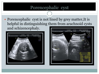
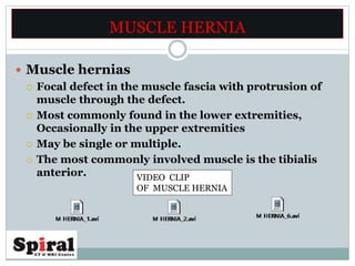
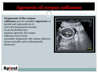
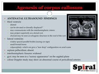
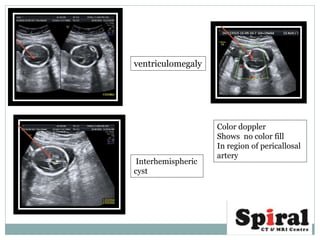
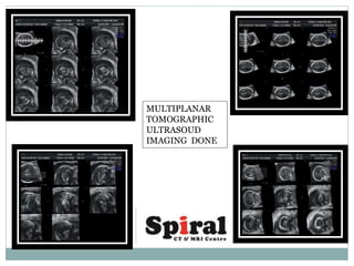
 Dr Arun Gupta
Director imaging
 Dr Rakhee gupta
Dr Vinayak Mittal
 Dr Gaurav
 Dr Kiran
Dr Ritesh Mahajan
•ADACTYLY IN FETUS
•PORENCEPHALIC CYST IN FETUS
•SEPTO-OPTIC DYSPLASIA IN
FETUS
•MUSCLE HERNIA IN ADULT
•FETAL REDUCTION
•AGENESIS OF CORPUS CALLOSUM
•FLAT FETAL FACIAL PROFILE
Porencephalic cyst
 Porencephaly is a rare congenital disorder that results in cystic
degeneration and encephalomalacia and the formation
of porencephalic cysts. Porencephalic cysts were originally described
by R Heschl in 1859
 Broadest definition being a cleft or cystic cavity within the brain .
 Narrow definition being a focal cystic area of encephalomalacia that
communicates with the ventricular system and/or the subarachnoid space.
The necessity of a cyst communicating with the ventricular system and
/ or subarachnoid space to earn the designation porencephaly is a point
of contention.
Porencephalic cysts can into
1. Internal (communicating with the ventricle)
2. External (communicating with the subarachnoid space).
 PRACTICALLY :
 porencephaly is taken to mean the following: a cystic lesion of
the brain due to an encephaloclastic insult (e.g. intrauterine
infections and ischaemia), lined by white matter, which
communicates with the ventricles and/or the subarachnoid
space.
Porencephalic cystPorencephalic cyst
 Aetiology
 Perinatal cerebral ischaemia
 Trauma
 Infection
 Antenatal intraparenchymal haemorrhage
 Familial porencephaly: mutations in the COL4A1 gene: fragile
blood vessels related to haemorrhagic strokes
Porencephalic cyst
 Porencephalic cyst is not lined by grey matter,It is
helpful in distinguishing them from arachnoid cysts
and schizencephaly.
Porencephalic cyst
MUSCLE HERNIA
 Muscle hernias
 Focal defect in the muscle fascia with protrusion of
muscle through the defect.
 Most commonly found in the lower extremities,
Occasionally in the upper extremities
 May be single or multiple.
 The most commonly involved muscle is the tibialis
anterior. VIDEO CLIP
OF MUSCLE HERNIA
 Muscle hernias are
 Typically asymptomic
 Can cause cramping sensations or pain with or after activity.
 May also present as a palpable mass and be referred for
imaging to evaluate for neoplasia.
 The mass may not be palpable when the patient is relaxed,
which is a clinical clue to the diagnosis.
MUSCLE HERNIA
 Ultrasound is the modality of choice in evaluation of suspected muscle
hernia, although MRI may be ordered if there is suspicion of neoplasia.
 Light pressure examination is preferable, so as not to reduce or efface
the herniation. Contraction of the involved muscle may reveal or
accentuate the lesion.
 The hernia is often hypoechoic to the surrounding muscle, and may
assume a mushroom shape as it protrudes through and over the fascial
defect (the defect is of the deep layer of the deep fascia.) It may thin or
elevate the superficial layer of the deep fascia that overlies the muscle.
MUSCLE HERNIA
SEPTO-OPTIC DYSPLASIA
 Septo-optic dysplasia (SOD) is a condition characterised by
 Optic nerve hypoplasia
 Absence of septum pellucidum
 Two thirds of patients hypothalamic-pituitary dysfunction.
 Being part of the holoprosencephaly spectrum.
ABSENT SEPTUM PELLUCIDUM
 Risk factors
 Maternal diabetes
 Medications
 quinidine ingestion
 antiepileptics
 Drug and alcohol abuse
 Cytomegalovirus infection
SEPTO-OPTIC DYSPLASIA
PERICALLOSAL ARTERY PRESENT
HENCE SURE ABOUT PRESENCE OF
CORPUS CALLOSUM .
SEPTO-OPTIC DYSPLASIA
CORONAL IMAGE WITH
E/O ABSENT SEPTUM PELLUCIDUM
SELLA TURCICA AND SELLA
PROPER APPRECIATED
ON ATTEMPTED SAGITTAL IMAGES
 Clinical presentation of septo-optic dysplasia is varied, and largely
dependent of whether or not it is associated with schizencephaly (~
50% of cases). This association is used to define two forms of
the condition .
 Not associated with schizencephaly
 visual apparatus more severely affected
 hypothalamic-pituitary dysfunction present in 60-80% of patients
 may present as hypoglycaemia in the neonatal period
 small pituitary gland with hypoplastic or absent infundibulum and ectopic
posterior pituitary seen as focus of T1 high signal intensity in median eminence
of hypothalamus
 olfactory bulbs may be absent (Kallmann syndrome)
 Associated with schizencephaly
 optic apparatus less severely affected
 cortical anomalies: polymicrogyria, cortical dysplasia
 may be etiologically different
 sometimes referred to as septo-optic dysplasia plus
SEPTO-OPTIC DYSPLASIA
FETAL REDUCTION
LUMBAR PUNCTURE
NEEDLE IN PLACE IN THE
AMNIOTIC CAVITY
VIDEO CLIP
OF FETAL REDUCTION
FETAL REDUCTION POST PROCEDURE
POST REDUCTION
NO COLOR FILL IN FETAL CARDIA
REST OF THE FETUS
SHOW NORMAL
COLOR FILL IN FETAL CARDIA
FLAT FETAL FACE
NORMAL FACE
PROFILE
•Forehead and chin on same plane .
•Nasal bone should be present
• Nose should project beyond plane of
forehead and chin.
•Top of ear at level of orbit
FLAT FACE
PROFILE
FLAT FETAL FACE
FETAL ADACTYLY
The detection of a fetal hand malformation
warrants a complete work-up, including
complete fetal and cardiac US
examinations, as well as genetic counseling
to determine whether familial inquiry and
karyotype analysis are necessary.
FOREARM BONES SEEN
ONLY THREE
RUDIMENTARY
PROXIMAL METACARPALS
APPRECIATED
FETAL LIMB ANOMALIES A BRIEF
 MALFORMATION
 A morphologic defect of an organ, part of an organ, or larger region of
the body resulting from an intrinsically abnormal developmental process
(eg, phocomelia, polydactyly).
 DEFORMATION
 An abnormal shape or position of part of the body caused by mechanical
forces (eg. clubfeet).
 DISRUPTION
 A morphologic defect of an organ, part of an organ, or a segment of the
body caused by an extrinsic factor interfering with an originally normal
developmental process (eg. amniotic band sequence).
 Higher in upper limb.
 More on Rt side.
 Usually unilateral than bilateral.
NORMAL
FETAL HAND
ADACTYLY
ABSENT FETAL HAND
TERMINOLOGY FOR QUICK REFERENCE
 Phocomelia
 Arms/forearms and thighs/calves are missing or foreshortened, the hands/feet may be normal or abnormal.
 Clinodactyly is a fixed deviation of the digitis.
 Asymmetrical hypoplasia of the mid-phalanx with the medial part being shorter than the lateral part, resulting in radial angulation of the distal phalanx.
 Clenched hand
 Second and fifth fingers overlap the third and fourth with an adducted thumb, it is important to evaluate, on ultrasound scan, if it is a persistent or a
temporary finding.
 Camptodactyly
 Flexion contracture of one of the interphalangeal joints.
 Abnormalities of length or width.
 Macrodactyly
 Rhizomelic (short femurs or humeri),
 Mesomelic (short forearms or calves)
 Acromelic (involving the hands or the feet).
 Polydactyly
 Presence of extra digit/s in the upper or lower extremities.
 Radial side (preaxial)
 Ulnar side (postaxial) polydactyly.
 ECTRODACTYLY:
 Split hand/foot deformity, also known as lobate claw hand/foot, results from the absence of the central digits/ toes with a deep V- or U-shaped central
cleft.
 Thumb abnormalities
 Thumb hypoplasia
 Triphalangeal thumb
 Broad thumb
 Hitchhiker thumb
 Abnormally abducted position of a more proximally inserted thumb .
Agenesis of corpus callosum
Dysgenesis of the corpus
callosum may be complete (agenesis) or
partial and represents an in
utero developmental anomaly.
It can be divided into:
primary agenesis: the corpus
callosum never forms
secondary dysgenesis: the corpus callosum
forms normally and is subsequently
destroyed
 ANTENATAL ULTRASOUND FINDINGS
 third ventricle
 dilated
 can be elevated or dorsally displaced 8
 may communicate with the interhemispheric cistern
 may project superiorly as a dorsal cyst
 choroid may be seen as echogenic structure in the roof of the cyst
 lateral ventricles
 widely spaced parallel bodies (racing car sign)
 small frontal horns
 colpocephaly: which can give a "tear drop" configuration on axial scans
 septum pellucidum: absent
 interhemispheric fissures: widened
 gyri: may be seen in a "sunray appearance" on the sagittal plane
 colour Doppler study may show an abnormal course of pericallosal arteries
Agenesis of corpus callosum
ventriculomegaly
Interhemispheric
cyst
Color doppler
Shows no color fill
In region of pericallosal
artery
MULTIPLANAR
TOMOGRAPHIC
ULTRASOUD
IMAGING DONE
REFERENCE
DIAGNOSTIC
ULTRASOUND
FOURTH EDITION
Carol M. Rumack, MD, FACR
J. William Charboneau, MD, FACR
Deborah Levine, MD, FACR

Ultrasound image gallery

  • 1.
     Dr ArunGupta Director imaging  Dr Rakhee gupta Dr Vinayak Mittal  Dr Gaurav  Dr Kiran Dr Ritesh Mahajan •ADACTYLY IN FETUS •PORENCEPHALIC CYST IN FETUS •SEPTO-OPTIC DYSPLASIA IN FETUS •MUSCLE HERNIA IN ADULT •FETAL REDUCTION •AGENESIS OF CORPUS CALLOSUM •FLAT FETAL FACIAL PROFILE
  • 2.
    Porencephalic cyst  Porencephalyis a rare congenital disorder that results in cystic degeneration and encephalomalacia and the formation of porencephalic cysts. Porencephalic cysts were originally described by R Heschl in 1859  Broadest definition being a cleft or cystic cavity within the brain .  Narrow definition being a focal cystic area of encephalomalacia that communicates with the ventricular system and/or the subarachnoid space.
  • 3.
    The necessity ofa cyst communicating with the ventricular system and / or subarachnoid space to earn the designation porencephaly is a point of contention. Porencephalic cysts can into 1. Internal (communicating with the ventricle) 2. External (communicating with the subarachnoid space).  PRACTICALLY :  porencephaly is taken to mean the following: a cystic lesion of the brain due to an encephaloclastic insult (e.g. intrauterine infections and ischaemia), lined by white matter, which communicates with the ventricles and/or the subarachnoid space. Porencephalic cystPorencephalic cyst
  • 4.
     Aetiology  Perinatalcerebral ischaemia  Trauma  Infection  Antenatal intraparenchymal haemorrhage  Familial porencephaly: mutations in the COL4A1 gene: fragile blood vessels related to haemorrhagic strokes Porencephalic cyst
  • 5.
     Porencephalic cystis not lined by grey matter,It is helpful in distinguishing them from arachnoid cysts and schizencephaly. Porencephalic cyst
  • 6.
    MUSCLE HERNIA  Musclehernias  Focal defect in the muscle fascia with protrusion of muscle through the defect.  Most commonly found in the lower extremities, Occasionally in the upper extremities  May be single or multiple.  The most commonly involved muscle is the tibialis anterior. VIDEO CLIP OF MUSCLE HERNIA
  • 7.
     Muscle herniasare  Typically asymptomic  Can cause cramping sensations or pain with or after activity.  May also present as a palpable mass and be referred for imaging to evaluate for neoplasia.  The mass may not be palpable when the patient is relaxed, which is a clinical clue to the diagnosis. MUSCLE HERNIA
  • 8.
     Ultrasound isthe modality of choice in evaluation of suspected muscle hernia, although MRI may be ordered if there is suspicion of neoplasia.  Light pressure examination is preferable, so as not to reduce or efface the herniation. Contraction of the involved muscle may reveal or accentuate the lesion.  The hernia is often hypoechoic to the surrounding muscle, and may assume a mushroom shape as it protrudes through and over the fascial defect (the defect is of the deep layer of the deep fascia.) It may thin or elevate the superficial layer of the deep fascia that overlies the muscle. MUSCLE HERNIA
  • 9.
    SEPTO-OPTIC DYSPLASIA  Septo-opticdysplasia (SOD) is a condition characterised by  Optic nerve hypoplasia  Absence of septum pellucidum  Two thirds of patients hypothalamic-pituitary dysfunction.  Being part of the holoprosencephaly spectrum. ABSENT SEPTUM PELLUCIDUM
  • 10.
     Risk factors Maternal diabetes  Medications  quinidine ingestion  antiepileptics  Drug and alcohol abuse  Cytomegalovirus infection SEPTO-OPTIC DYSPLASIA PERICALLOSAL ARTERY PRESENT HENCE SURE ABOUT PRESENCE OF CORPUS CALLOSUM .
  • 11.
    SEPTO-OPTIC DYSPLASIA CORONAL IMAGEWITH E/O ABSENT SEPTUM PELLUCIDUM SELLA TURCICA AND SELLA PROPER APPRECIATED ON ATTEMPTED SAGITTAL IMAGES
  • 12.
     Clinical presentationof septo-optic dysplasia is varied, and largely dependent of whether or not it is associated with schizencephaly (~ 50% of cases). This association is used to define two forms of the condition .  Not associated with schizencephaly  visual apparatus more severely affected  hypothalamic-pituitary dysfunction present in 60-80% of patients  may present as hypoglycaemia in the neonatal period  small pituitary gland with hypoplastic or absent infundibulum and ectopic posterior pituitary seen as focus of T1 high signal intensity in median eminence of hypothalamus  olfactory bulbs may be absent (Kallmann syndrome)  Associated with schizencephaly  optic apparatus less severely affected  cortical anomalies: polymicrogyria, cortical dysplasia  may be etiologically different  sometimes referred to as septo-optic dysplasia plus SEPTO-OPTIC DYSPLASIA
  • 13.
    FETAL REDUCTION LUMBAR PUNCTURE NEEDLEIN PLACE IN THE AMNIOTIC CAVITY VIDEO CLIP OF FETAL REDUCTION
  • 14.
    FETAL REDUCTION POSTPROCEDURE POST REDUCTION NO COLOR FILL IN FETAL CARDIA REST OF THE FETUS SHOW NORMAL COLOR FILL IN FETAL CARDIA
  • 15.
    FLAT FETAL FACE NORMALFACE PROFILE •Forehead and chin on same plane . •Nasal bone should be present • Nose should project beyond plane of forehead and chin. •Top of ear at level of orbit FLAT FACE PROFILE
  • 16.
  • 17.
    FETAL ADACTYLY The detectionof a fetal hand malformation warrants a complete work-up, including complete fetal and cardiac US examinations, as well as genetic counseling to determine whether familial inquiry and karyotype analysis are necessary. FOREARM BONES SEEN ONLY THREE RUDIMENTARY PROXIMAL METACARPALS APPRECIATED
  • 18.
    FETAL LIMB ANOMALIESA BRIEF  MALFORMATION  A morphologic defect of an organ, part of an organ, or larger region of the body resulting from an intrinsically abnormal developmental process (eg, phocomelia, polydactyly).  DEFORMATION  An abnormal shape or position of part of the body caused by mechanical forces (eg. clubfeet).  DISRUPTION  A morphologic defect of an organ, part of an organ, or a segment of the body caused by an extrinsic factor interfering with an originally normal developmental process (eg. amniotic band sequence).  Higher in upper limb.  More on Rt side.  Usually unilateral than bilateral.
  • 19.
  • 20.
    TERMINOLOGY FOR QUICKREFERENCE  Phocomelia  Arms/forearms and thighs/calves are missing or foreshortened, the hands/feet may be normal or abnormal.  Clinodactyly is a fixed deviation of the digitis.  Asymmetrical hypoplasia of the mid-phalanx with the medial part being shorter than the lateral part, resulting in radial angulation of the distal phalanx.  Clenched hand  Second and fifth fingers overlap the third and fourth with an adducted thumb, it is important to evaluate, on ultrasound scan, if it is a persistent or a temporary finding.  Camptodactyly  Flexion contracture of one of the interphalangeal joints.  Abnormalities of length or width.  Macrodactyly  Rhizomelic (short femurs or humeri),  Mesomelic (short forearms or calves)  Acromelic (involving the hands or the feet).  Polydactyly  Presence of extra digit/s in the upper or lower extremities.  Radial side (preaxial)  Ulnar side (postaxial) polydactyly.  ECTRODACTYLY:  Split hand/foot deformity, also known as lobate claw hand/foot, results from the absence of the central digits/ toes with a deep V- or U-shaped central cleft.  Thumb abnormalities  Thumb hypoplasia  Triphalangeal thumb  Broad thumb  Hitchhiker thumb  Abnormally abducted position of a more proximally inserted thumb .
  • 21.
    Agenesis of corpuscallosum Dysgenesis of the corpus callosum may be complete (agenesis) or partial and represents an in utero developmental anomaly. It can be divided into: primary agenesis: the corpus callosum never forms secondary dysgenesis: the corpus callosum forms normally and is subsequently destroyed
  • 22.
     ANTENATAL ULTRASOUNDFINDINGS  third ventricle  dilated  can be elevated or dorsally displaced 8  may communicate with the interhemispheric cistern  may project superiorly as a dorsal cyst  choroid may be seen as echogenic structure in the roof of the cyst  lateral ventricles  widely spaced parallel bodies (racing car sign)  small frontal horns  colpocephaly: which can give a "tear drop" configuration on axial scans  septum pellucidum: absent  interhemispheric fissures: widened  gyri: may be seen in a "sunray appearance" on the sagittal plane  colour Doppler study may show an abnormal course of pericallosal arteries Agenesis of corpus callosum
  • 23.
    ventriculomegaly Interhemispheric cyst Color doppler Shows nocolor fill In region of pericallosal artery
  • 24.
  • 25.
    REFERENCE DIAGNOSTIC ULTRASOUND FOURTH EDITION Carol M.Rumack, MD, FACR J. William Charboneau, MD, FACR Deborah Levine, MD, FACR