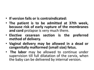
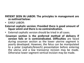
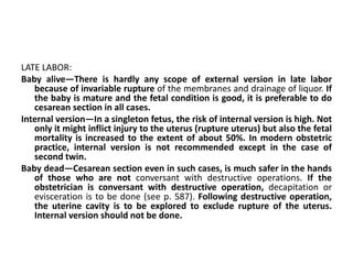
This document discusses transverse lie and cord prolapse during labor. It provides information on:
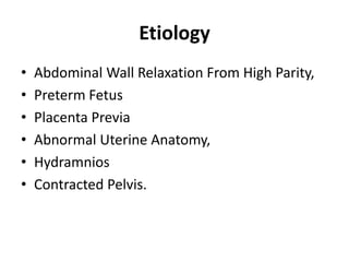
1) The definition and causes of transverse lie, where the fetus lies horizontally across the uterus with the shoulder over the pelvic inlet.
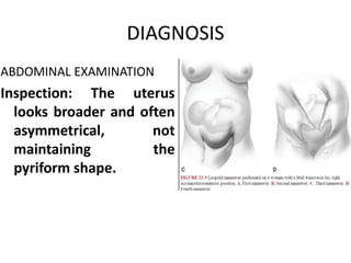
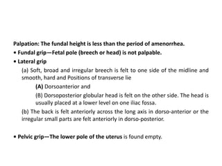
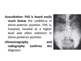
2) The diagnosis of transverse lie which involves abdominal and vaginal examinations to identify fetal parts and position. Ultrasound can also confirm the diagnosis.
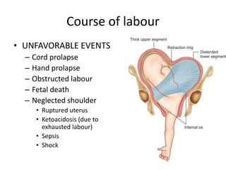
3) The risks of transverse lie including cord prolapse, obstructed labor, and fetal death. Management involves external cephalic version or cesarean section.
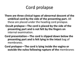
4) The definition, causes, diagnosis, and management of cord prolapse, which requires immediate delivery by cesarean section if the fetus is alive or
Anuman- An inference for helpful in diagnosis and treatment
Transverse lie and cord prolapse
1. TRANSVERSE LIE AND CORD
PROLAPSE
Dr. Hem Nath Subedi
RESIDENT
COMS-TH, BHARATPUR , NEPAL
2. TRANSVERSE LIE
• When the long axis forms an acute angle, an
oblique lie results. The latter is usually only
transitory, because either a longitudinal or
transverse lie commonly results when labor
supervenes.
• The position is determined by the direction of
the back, which is the denominator.
3. • In a transverse lie, the shoulder is usually
positioned over the pelvic inlet. The head
occupies one iliac fossa, and the breech the
other.
• This creates a shoulder presentation in which
the side of the mother on which the acromion
rests determines the designation of the lie as
right or left acromial.
4. POSITION
(1) Dorsoanterior, which is the commonest
(60%). The flexor surface of the fetus is better
adapted to the convexity of the maternal
spine.
(2) Dorsoposterior
(3) Dorsosuperior
(4) Dorso-inferior.
5.
6. • In dorsoposterior, chance of fetal extension is
common with increased risk of arm prolapse.
According to the position of the head, the
fetal position is termed right or left, the left
one being commoner than the right.
9. Palpation: The fundal height is less than the period of amenorrhea.
• Fundal grip—Fetal pole (breech or head) is not palpable.
• Lateral grip
(a) Soft, broad and irregular breech is felt to one side of the midline and
smooth, hard and Positions of transverse lie
(A) Dorsoanterior and
(B) Dorsoposterior globular head is felt on the other side. The head is
usually placed at a lower level on one iliac fossa.
(b) The back is felt anteriorly across the long axis in dorso-anterior or the
irregular small parts are felt anteriorly in dorso-posterior.
• Pelvic grip—The lower pole of the uterus is found empty.
10. Auscultation: FHS is heard easily
much below the umbilicus in
dorso-anterior position. FHS is,
however, located at a higher
level and often indistinct in
dorso-posterior position.
Ultrasonography and
radiography confirms the
diagnosis
11. VAGINAL EXAMINATION
During pregnancy, the presenting part is so high that it cannot be identified
properly but one can feel some soft parts.
During labor—Elongated bag of the membranes can be felt if it does not
rupture prematurely. The shoulder is identified by palpating the following
parts—acromion process, the scapula, the clavicle and axilla. The
characteristic landmarks are the feeling of the ribs and intercostal spaces
(grid iron feel).
On occasion, the arm is found prolapsed. It should be remembered that the
findings of a prolapsed arm is confined not only to transverse lie but it
may also be associated with compound presentation. Rarely, a leg may
be prolapsed. Frequently, a loop of cord may be found alongside the arm.
Determination of position: The thumb of the prolapsed hand, when
supinated, points toward the head, the palm corresponds to the ventral
aspect. The angle of the scapula, if felt, indicates the position of the back.
The side to which the prolapsed arm belongs, can be determined by
shaking hands with the fetus. If the right hand is required for this, the
prolapsed arm belongs to right side and vice-versa.
12.
13. Course of labour
• UNFAVORABLE EVENTS
– Cord prolapse
– Hand prolapse
– Obstructed labour
– Fetal death
– Neglected shoulder
• Ruptured uterus
• Ketoacidosis (due to
exhausted labour)
• Sepsis
• Shock
14. • FAVORABLE EVENTS
• The following are the favorable events that
may occur
– (1) spontaneous rectification or version
– (2) spontaneous evolution and
– (3) spontaneous expulsion (conduplicato
corpore).
These events are very rare and occur only when the
baby is premature or macerated.
15. Management of Labour
• External cephalic version should be done in
all cases beyond 35 weeks provided there is
no contraindication as mentioned in breech
presentation .
• If the lie fails to stabilize even at 36th week,
the case is to be managed as outlined in
unstable lie.
16. • If version fails or is contraindicated:
• The patient is to be admitted at 37th week,
because risk of early rupture of the membranes
and cord prolapse is very much there.
• Elective cesarean section is the preferred
method of delivery.
• Vaginal delivery may be allowed in a dead or
congenitally malformed (small size) fetus.
• The labor may be allowed to continue under
supervision till full dilatation of the cervix, when
the baby can be delivered by internal version.
17. PATIENT SEEN IN LABOR: The principles in management are
as outlined below:
• EARLY LABOR:
External cephalic version: Provided there is good amount of
liquor amnii and there is no contraindication.
• External cephalic version should be tried in all cases.
Cesarean section is the preferred method of delivery if
version fails or is contraindicated. Difficulties are faced
during cesarean section as the lower uterine segment is
poorly developed. However, the fetus can be manipulated
to a polar (cephalic/breech) presentation before entering
the uterus and a low transverse incision may be made.
Otherwise lower segment vertical incision may be made.
18. LATE LABOR:
Baby alive—There is hardly any scope of external version in late labor
because of invariable rupture of the membranes and drainage of liquor. If
the baby is mature and the fetal condition is good, it is preferable to do
cesarean section in all cases.
Internal version—In a singleton fetus, the risk of internal version is high. Not
only it might inflict injury to the uterus (rupture uterus) but also the fetal
mortality is increased to the extent of about 50%. In modern obstetric
practice, internal version is not recommended except in the case of
second twin.
Baby dead—Cesarean section even in such cases, is much safer in the hands
of those who are not conversant with destructive operations. If the
obstetrician is conversant with destructive operation, decapitation or
evisceration is to be done (see p. 587). Following destructive operation,
the uterine cavity is to be explored to exclude rupture of the uterus.
Internal version should not be done.
19. Cord prolapse
There are three clinical types of abnormal descent of the
umbilical cord by the side of the presenting part. All
these are placed under the heading cord prolapse.
Occult prolapse—The cord is placed by the side of the
presenting part and is not felt by the fingers on
internal examination.
Cord presentation—The cord is slipped down below the
presenting part and is felt lying in the intact bag of
membranes.
Cord prolapse—The cord is lying inside the vagina or
outside the vulva following rupture of the membranes
20. • INCIDENCE: The incidence of cord prolapse is
about 1 in 300 deliveries. It is mostly confined
to parous women.
• Incidence is reduced with the increased use
of elective CS in noncephalic presentations.
21.
22.
23. ETIOLOGY
The following are the associated factors:
– Malpresentations—the commonest being transverse (5–
10%) and breech (3%) especially with flexed legs or
footling and compound (10%) presentation
– Contracted pelvis,
– Prematurity
– Twins
– Hydramnios
– Placental factor—minor degree placenta praevia with
marginal insertion of the cord or long cord
– Iatrogenic—low rupture of the membranes, manual
rotation of the head, ECV, IPV
– Stabilizing induction
24.
25. DIAGNOSIS:
• Occult prolapse—is difficult to diagnose. The
possibility should be suspected if there is persistence
of variable deceleration of fetal heart rate pattern
detected on continuous electronic fetal monitoring.
• Cord presentation—The diagnosis is made by feeling
the pulsation of the cord through the intact
membranes.
• Cord prolapse—The cord is palpated directly by the
fingers and its pulsation can be felt if the fetus is alive.
Cord pulsation may cease during uterine contraction
which, however, returns after the contraction passes
off.
26. Management
Cord presentation: The aim is to preserve the membranes and to
expedite the delivery. Once the diagnosis is made, no attempt
should be made to replace the cord, as it is not only ineffective
but the membranes inevitably rupture leading to prolapse of the
cord.
If immediate vaginal delivery is not possible or contraindicated,
cesarean section is the best method of delivery. During the time
of preparing the patient for operative delivery, she is kept in
exaggerated Sims’ position to minimise cord compression.
A rare occasion is a multipara with longitudinal lie having good
uterine contractions with the cervix three-fourth (7–8 cm) dilated,
without any evidence of fetal distress. Watchful expectancy can be
adopted till full dilatation of the cervix, when the delivery can be
completed by forceps or breech extraction.
27. CORD PROLAPSE: Management protocol is to be guided by:
(1) Baby living or dead
(2) Maturity of the baby
(3) Degree of dilatation of the cervix.
BABY LIVING:
I. Definitive treatment:
• Cesarean section is the best treatment when the baby is sufficiently mature and is
alive. Just prior to making the abdominal incision, the fetal heart should be
auscultated once more to avoid unnecessary section on a dead baby. The
operation should be done quickly upto the delivery of the baby.
II. Immediate safe vaginal delivery is possible:
• If the head is engaged, delivery is to be completed by forceps. Ventouse may not be
ideal in such circumstances as it takes a longer time.
• If breech, the delivery is to be completed by breech extraction and in transverse lie,
it should be completed by internal version followed by breech extraction.
The same also applies in cases where the head is not engaged in second baby of
twins.
28. III. Immediate safe vaginal delivery is not possible: First aid management: The aim is
to minimize pressure on the cord till such time when the patient is prepared for
assisted delivery or is transferred to an equipped hospital. If an oxytocin infusion is
on, this should be stopped. At this time intravenous fluid and O2 by face mask is
given.
• Bladder filling has been done to raise the presenting part off the compressed cord
till such time that patient has delivered (either by CS or vaginally). Bladder is filled
with 400–750 mL of normal saline with a Foley’s catheter, the balloon is inflated
and the catheter is clamped. Bladder is emptied before cesarean delivery.
• To lift the presenting part off the cord, by Woman in labour wiht cord prolapse the
gloved fingers introduced into the vagina. The fingers should be placed inside the
vagina till definitive treatment is instituted.
• Postural treatment—exaggerated and elevated Sims’ position with a pillow or
wedge under the hip or thigh; Trendelenburg or knee-chest position has been
traditionally mentioned but may be tiring and irksome to the patient.
• To replace the cord into the vagina to minimize vasospasm due to irritation.
BABY DEAD: Labor is allowed to proceed awaiting spontaneous delivery.