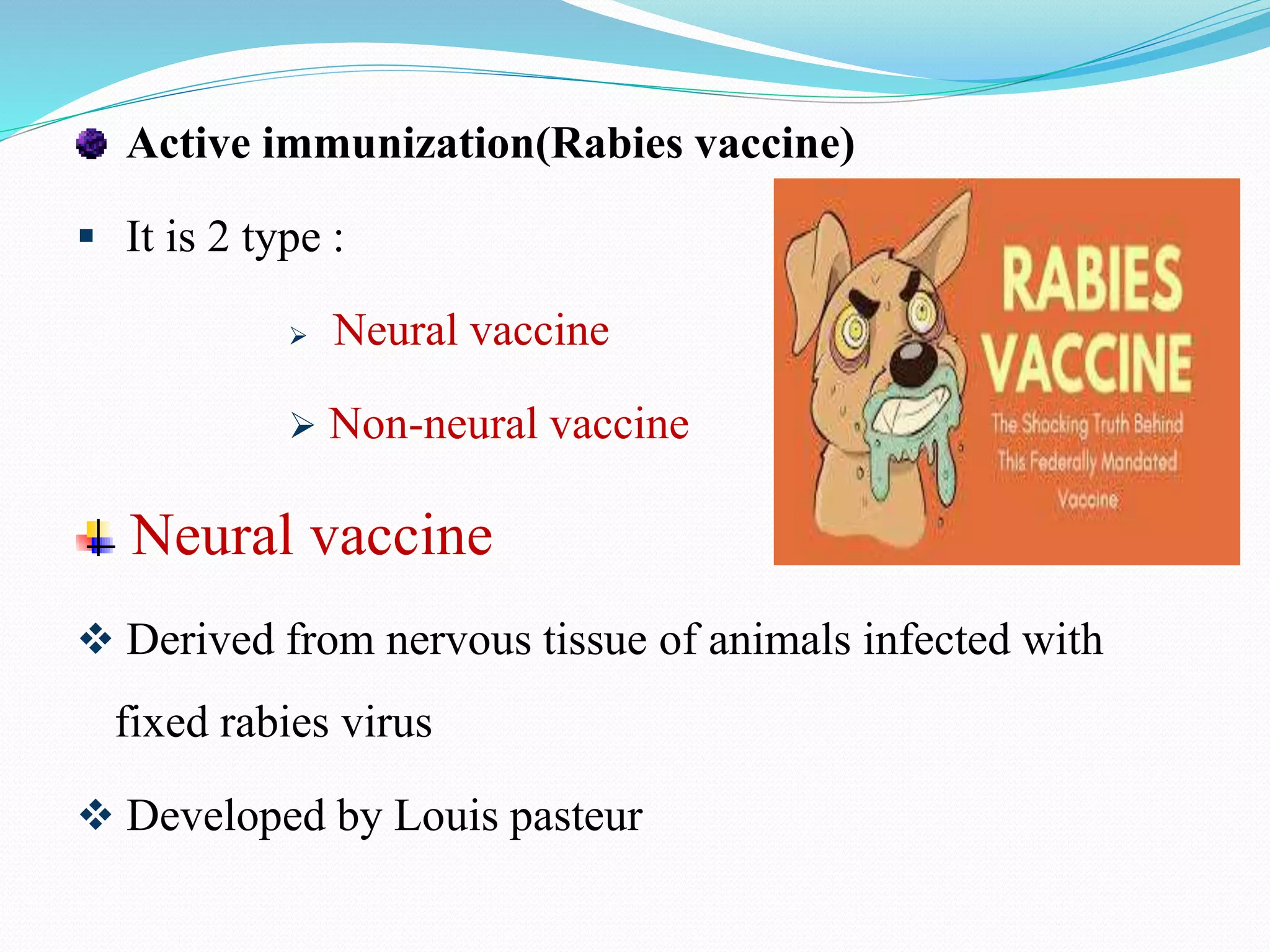
2. INTRODUCTION
The family Rhabdoviridae consists of more than 185 different
viruses.
They are enveloped, have helical nucleocapsid containing
negative sense ss RNA.
These are elongated, rod/bullet shaped particles , approx 100 -
430 nm long & 45 – 100 nm diameter .
Mature virions appear bullet shaped or bacilliform shaped.
3. There is 4 Genus,
Lyssavirus
Vesiculovirus
Ephemerovirus
Novirhabdovirus
Rhabdoviruses can replicate in plants, invertebrates or
vertebrates.
4. CLASSIFICATION
Family Rhabdoviridae, genus lyssavirus
Genotype Source Distribution
Rabies virus Dog, fox, raccoon, skunk,
bat
Widespread
Lagos bat virus Bats, cats Africa
Mokola Shrews, cats South Africa, Nigeria
Duvenhage Insectivorous bats South Africa
European bat lyssavirus 1a
& b
Bats Europe
European bat lyssavirus 2a
& b
Bats UK, Netherlands
Ausralian bat lyssavirus Flying foxes, Insectivorous
bats
Austalia
5. Oldest recognized infectious disease.
It causes- rapidly progressive acute infectious disease of
CNS .
Rabies is still considered as major public health problem
because it is almost always fatal.
6. MORPHOLOGY
Rhabdoviridae family has a unique morphology.
Bullet shaped
75 nm in width & 180 nm in length
Enveloped
They have lipid envelop with spikes.
Nucleocapsid
Helical symmetry & comprises a single stranded,
(-)sense RNA, nucleoprotein & polymerase protein
7.
8. ANTIGENIC STRUCTURE
Rabies virus has only one serotype.
There are 7 antigenic variants that are associated with
specific animal reservoirs and found in different
geographical areas.
Antigens are Glycoprotein
Nucleoprotein
Matrix protein
Envelop lipid Ag
9. Glycoprotein G
G protein is the major factor for neuroinvasiveness,
immunity, virulence & pathogenicity
There are spikes embedded in envelop
It is species specific
It has role in pathogenesis
It bind to acetyl choline receptor on neural tissue.
10. It possess haemagglutinine activity with mangoos
erythrocyte
It induces neutralising Antibody
Purified form of antigen is used to prepare subunit
vaccine.
Spikes are inactivated by heat ,ether, deoxycholate etc
11. Nucleoprotein N
These are capsid protein associated
with viral RNA
It is group specific
It cross react with rabies related virus
It does not have any role in pathogenesis
It induces complement fixing antibody
Other antigens are : M Ag (Matrix protein antigen)
L Ag (Envelop lipid Ag)
12. HOST RANGE
ANIMAL SUSCEPTIBILITY
Suscetibility varies among various animals
Very highly susceptible animals : Foxes
Wolves
Cotton rats
Highly susceptible animals : Rabbits
Cattle
Cats
Bats
13. Moderately susceptible animals : Dogs
Goats
Sheep
Horses
Low susceptible animal : Opossums
Based on the changes shown by serial passage in animals,
virus is classified into 2:
Street virus &
Fixed virus
14. Street Virus
These are freshly isolated strains in laboratory
It show variable I P ie. 1-3 months
It is pathogenic & affect salivary glands
It can produce intracytoplasmic inclusion bodies ie Negri
body.
Following inoculation by any route , street virus can cause
Fatal encephalitis in animal after long incubation period.
15. Fixed Virus
When street virus are undergone certain changes, they
lose their properties and become Fixed virus.
I P: 4- 6 days
It do not affect salivary gland
Negri bodies are absent
Non pathogenic
Used for vaccine preparation
17. Rapidly progressive acute infectious disease of CNS in
humans & animals.
Transmission
Bite
Transmitted to humans by the bite of infected animals
Dog bite is the most common mode
Other animal bites: Monkey, Sheep, Goat, Cat etc
18. Migrating fruit eating bats are transmit rabies in America
Human to human transmission is theoretically possible,
but it is rare
Non bite exposure
Rare Such as,
Lick on abrasion/mucosa
Inhalation of virus containing aerosols from infected bats
Corneal transplantation
19. Bite from rabid animal infection with rabies virus
Multiplication of rabies virus within new host
Spread of virus through nerve endings to CNS – virus
reaches the brain
Early symptoms include pain / paresthesia at site of
inoculation and other non specific flu like symptoms
(eg: malaise, fever, headache)
SPREAD OF VIRUS
20. Symptoms include cerebral dysfunction , anxiety, confusion,
agitation etc
Progression to delirium, abnormal behaviour, hallucinations,
insomnia, hydrophobia, aerophobia and photophobia
Coma
DEATH
21. Pathological changes
Histopathological changes produced in brain parenchyma
of rabies infected patients include,
Mononuclear cell infiltration
Perivascular cuffing of lymphocytes
Babes nodule consisting of glial cells
Negri bodies
22. Negri Bodies
Intracytoplasmic eosinophilic inclusions in brain neurons
composed of rabies virus protein and viral RNA
Contain basophilic inner granules
Commonly observed in Purkinje cells of cerebellum & in
pyramidal neurons of hippocampus
and less frequently seen in cortical
and brainstem neurons
23. CLINICAL MANIFESTATIONS
I P : Prolonged/ variable (20 – 90 days)
It is inversely related to the distance for the virus to travel
from the site of inoculation to CNS.
The I P will be shorter,
If the bite on head, neck
In the small children
In the low immunity people
The I P will be long,
If the bite on the leg and in adults
24. Clinical spectrum can be divided into 4 phases:
Short prodromal phase
Acute neurologic phase
Coma
Death
25. Short prodromal phase
It last for 2- 10 days with fever, malaise, anorexia,
nausea, vomiting, photophobia, sore throat etc
Acute neurologic phase
It is 2 type :
Encephalitic type
Paralytic type
26. Encephalitic rabies
Last for 2-7 days
Characterized by,
• Hyper excitability
• Lucid interval
• Autonomic dysfunction
• Hydrophobia/ aerophobia
27. Paralytic / Dumb rabies
It occurs in people who are partially vaccinated or
infected with bat rabies virus
Features are: Flaccid paralysis
Bitten limb . etc
Coma & Death
Following acute neurological phase, patient develops –
Coma and lead to Death within 14 days
28. LAB DIAGNOSIS
Specimen : Corneal smears , Skin biopsy from face or
neck , saliva ante-mortem, brain post-mortem
Rabies Ag detection
Direct immunofluorescence test
Also called as – Direct fluorescent Ab test
Performed to detect rabies nucleoprotein Ags in
specimens by using specific monoclonal Abs tagged with
fluorescent dye.
29. Gold standard method
Best specimen : Hair follicle of nape of neck
corneal impression smear is also be used.
Pos dFA Neg dFA
30. Immunohistochemistry
IHC based staining performed to detect rabies Ag
Peroxidase labelled specific Abs are used to detect viral
inclusions in formalin fixed tissues.
Sensitive & specific
31. Rabies Ab detection
Detection of CSF Abs is more significant than serum Abs
Serum Abs appear late & also prevent after vaccination
CSF Abs appear early and are produced only in rabies
infected individuals.
32. Other Ab detection tests are,
• Mouse neutralization test
• Rapid fluorescent focus inhibition test
• Fluorescent Ab virus neutralization test
• Indirect fluorescent assay
• Haemagglutination inhibition test
• Complete fixation test
• Immunoperoxidase inhibition assay
33. Viral isolation
Animal inoculation
Use animals such as mice, monkeys, guinea pigs, rabbits etc
Route of administration : Intraperitoneolly, indracerebrally,
intramuscularlly, intra subcutaneously & intranasally etc.
Intra cerebral inoculation into suckling mice can cause
encephalitis & death
34. Negri bodies & rabies Ag can be see in Brain biopsy of
inoculated animal.
35. Viral RNA detection
PCR is used to amplify
genes of rabies virus
Most sensitive & specific
Negri body detection
Useful for post-mortem diagnosis of rabies
It is an intracytoplasmic eosinophilic inclusion with
characteristic basophilic inner granules
36. Sharply demarcated spherical to oval about 2-10 µm size
Most common sites are, neurons of cerebellum &
hippocampus, less seen in cortical & brainstem neurons
Commonly used stains are histological stains such as
• H & E stain
• sellers stains
• Mann’s stain
• Giemsa stain
37.
38. PROPHYLAXIS
Mortality is 100%
Preventable by administration of past- exposure therapy
during the early incubation period.
PREVENTION OF HUMAN RABIES
Post exposure prophylaxis (PEP) includes
Local wound care
Active immunization
Passive immunization
39. Local Wound Care
Physical cleansing
Chemical inactivation
• Povidone iodine/alcohol
Biological neutralization
• Giving anti rabies immunoglobulin
Tetanus prophylaxis
Suturing contraindicated
Antibiotic treatment
40. Active immunization(Rabies vaccine)
It is 2 type :
Neural vaccine
Non-neural vaccine
Neural vaccine
Derived from nervous tissue of animals infected with
fixed rabies virus
Developed by Louis pasteur
42. Non neural vaccine
Egg derived vaccine
Recombinant glycoprotein vaccine
Cell line derived vaccine
Egg derived vaccine
Site : Allantoic cavity of embryonated egg
There is, - Purified Duck Embryo Vaccine (PDEV)
- Live Attenuated Chick Embryo Vaccine
43. Recombinant viral vaccine
Vaccinia virus carrying rabies surface glycoprotein gene
has been developed
Given orally
Cell culture derived vaccine
Highly immunogenic
Devoid of neurological complications
44. 3 vaccines are available
PCEC (Purified Chick Embryo Cell vaccine)
PVC ( Purified vero cell vaccine)
HDC ( Human diploid cell vaccine)
45. Passive immunization ( Rabies immunoglobulin)
Neutralization and infectivity of virus
Hence RIG are administrated locally at the site of
exposure
2 types RIG is available
Equine Rabies Immunoglobulin (ERIG)
• Dose of 40 IU/Kg
• Heterologous in origin
• Associated with serum sickness
46. Human Rabies Immunoglobulin (HRIG)
• Dose of 20 IU/ Kg
• No side effects
Max vol of RIG should be infiltrated into wound
remaining should be administrated by deep intramuscular
injection at the site.
47. National Guideline on Rabies prophylaxis
There is
Regimen for Post – exposure prophylaxis
IM regimen / Essen regimen (1-1-1-1-1)
5 doses of IM injection ; 1 dose given on 0,3,7,14,28.
ID regimen (Thai Red cross schedule (2-2-2-
0-2))
0.1 ml on 2 sites in ID site on 0,3,7,28.
Potency : 2.5 IU
48. SITE OF INJECTION
Deltoid region is ideal site
Gluteal region is not recommended
Antero lateral part of thigh – infants & young children
Regimen for Pre – exposure prophylaxis
Recommended for high risk groups like,
o Laboratory staff handling the virus
o Doctors /any other person attending to
rabies cases
49. o Veterinarians
o Animal handlers
3 doses are given on day – 0,7,21/28 days
Ab titre should be checked every 6 months for 2 years
Booster dose is given if titre is below 0.5 IU/ml
50. Regimen for Post – exposure prophylaxis in previously
vaccinated individuals.
This depends on Ab titre & severity of wound bite
In severe bite/ titre unknown, give 3 doses 0n 0,3,7 days
Less severe bite/ titre ˃0.5 IU/ml , give 2doses on 0& 3
days.
51. Type of contact, exposure and recommended post
exposure prophylaxis
Ca
teg
ory
Type of contact with a suspect or
non confirmed rabid domestic or
wild animal or animal un available
for testing
Type of
expos-
ure
Recommended post exposure
prophylaxis
1 Touching / feeding of animals
Licks on intact skin
None None, if reliable case history is
available
11 Nibbling of uncovered skin
Minor scratches or abrasions without
bleeding
Minor Administer vaccine immediately.
Stop treatment if animal remains
healthy throughout an observation
period of 10 days
111 Single /multiple transdermal bites or
scratches, licks on broken skin
Contamination of mucus membrane
with saliva
Exposures to bats
Severe Administer rabies immunoglobulin
and vaccine immediately.
Stop treatment if animal remains
healthy throughout an observation
period of 10 days
52. EPIDEMOLOGY
Rabies is an enzootic & epizootic disease of both wild and
domestic animals worldwide.
Source : Infected dog
Age : All ages are affected.
Rabies free countries are: Australia & Antartica
Britain
Iceland & Ireland
China
Japan
53.
54. REFERENCE
FIELDS VIROLOGY ; 6th edition. Vol.1
Ananthanarayan and Paniker’s Textbook of
MICROBIOLOGY. 10 th edition
Apurba Sankar Sastry & Sandhya Bhat K. Essentials of
MEDICAL MICROBIOLOGY textbook