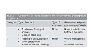
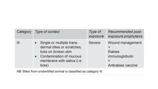
RABIES IN CHILDREN
DR JISHNU KR(PGT-2)
MODERATOR- DR MUKESH JAIN(ASSO PROFESSOR,DEPT OF PEDIATRICS
KIMS)
Introduction
• Bullet-shaped, negative-sense, single-stranded, enveloped RNA virus from the family
Rhabdoviridae, genus Lyssavirus.
• Seven Lyssavirus genotypes are associated with rabies in humans, although genotype 1 accounts
for the great majority of cases.
• Has a nonsegmented, negative-sense
genome that consists of 11,932 nucleotides
and encodes 5 proteins:
 Nucleocapsid protein,
 Phosphoprotein,
 Matrix protein,
 Glycoprotein
 Large polymerase protein.
Epidemiology
• Worldwide, transmission from dogs accounts for >90% of human cases.
• Approximately 50,000 cases of human rabies occur in Africa and Asia annually.
• In India, the most common transmitting animal is dog, accounting for more than 96% cases.
• As per the national multicentric rabies survey done in 2003,1 about 17 million animal bites occur
annually out of which 20,000 human rabies deaths occur in India.
• About 35% of these are in children.
• In Africa and Asia, other animals serve as prominent reservoirs, such as jackals, mongooses, and
raccoon dogs.
• In the United States, raccoons are the most commonly infected wild animal along the eastern
seaboard.
• Rabies is rare in small mammals, including mice, squirrels, and rabbits; to date, no animal-to-
human transmission from these animals has been documented
Transmission
• Found in large quantities in the saliva of infected animals.
• Transmission occurs almost exclusively through inoculation of the infected saliva through a bite or
scratch from a rabid mammal.
• The transmission rate is increased if the victim has suffered multiple bites and if the inoculation
occurs in highly innervated parts of the body such as the face and the hands
• Infection does not occur after exposure of intact skin to infected secretions, but virus may enter
the body through intact mucous membranes.
Pathogenesis
• After inoculation, rabies virus replicates slowly and at low levels in muscle or skin.
• This slow initial step likely accounts for the disease’s long incubation period.
• In muscles, the virus is known to bind to nicotinic acetylcholine receptors on postsynaptic
membranes at neuromuscular junctions, but the exact details of viral entry into the skin and
Subcutaneous tissues have not yet been clarified.
• Rabies virus spreads centripetally along peripheral nerves toward the spinal cord or brainstem via
retrograde fast axonal transport (rate, up to ~250 mm/d), with delays at intervals of ~12 h at each
synapse.
• Rapid dissemination occurs throughout the brain and spinal cord before symptoms appear.
• After CNS infection becomes established, there is centrifugal spread along sensory and
autonomic nerves to other tissues, including the salivary glands, heart, adrenal glands, and skin
• Infection of the dorsal root ganglia is apparently futile but causes characteristic radiculitis.
• Infection concentrates in the brainstem, accounting for autonomic dysfunction and relative
sparing of cognition.
Pathology
• Pathologic studies show mild inflammatory changes in the CNS in rabies,
 Mononuclear inflammatory infiltration in the leptomeninges,
Cuffing of perivascular regions
 Microglial nodules called Babes nodules
 Negri bodies are eosinophilic cytoplasmic inclusions in brain neurons that are composed of rabies
virus proteins and viral RNA.
• Commonly observed in Purkinje cells of the cerebellum and in pyramidal neurons of the
hippocampus, and are less frequently seen in cortical and brainstem neurons.
• Negri bodies are not observed in all cases of rabies
• Negri bodies can be absent in documented
rabies virus infection.
•
Clinical Features
• The incubation period for rabies is 1-3 months, but is variable.
• The clinical features of rabies begin with nononspecific prodromal manifestations, including fever,
malaise, headache, nausea, and vomiting.
• Anxiety or agitation may also occur.
• The earliest specific neurologic symptoms of rabies include paresthesias, pain, or pruritus near
the site of the exposure, one or more of which occur in 50–80% of patients and strongly suggest
rabies
• Two acute neurologic forms of rabies are seen in humans: the encephalitic (furious) form in 80%
and the paralytic form in 20%.
Encephalitic form
• Manifestations of encephalitic rabies include fever, confusion, hallucinations, combativeness, and
seizures.
• Autonomic dysfunction is common and may result in hypersalivation, gooseflesh, cardiac
arrhythmia, and priapism.
• Distinguished by early brainstem involvement, which results in the classic features :-
Hydrophobia (involuntary, painful contraction of the diaphragm and accessory respiratory,
laryngeal, and pharyngeal muscles in response to swallowing liquids)
Aerophobia(the same features caused by stimulation from a draft of air).
• These symptoms are probably due to dysfunction of infected brainstem neurons that normally
inhibit inspiratory neurons near the nucleus ambiguus, resulting in exaggerated defense reflexes
that protect the respiratory tract.
• Characteristically, patients with rabies encephalitis initially have periods of lucidity alternating
with periods of profound encephalopathy.
• The combination of hypersalivation and pharyngeal dysfunction is also responsible for the classic
appearance of “foaming at the mouth”
• Brainstem dysfunction progresses rapidly, and coma—followed within days by death—is the rule
unless the course is prolonged by supportive measures
• Late complications include
cardiac and/or respiratory failure,
disturbances of water balance (syndrome of inappropriate antidiuretic hormone secretion or
diabetes insipidus),
noncardiogenic pulmonary edema,
 gastrointestinal hemorrhage.
Paralytic Form
• About 20% of patients have paralytic rabies in which muscle weakness predominates and cardinal
features of encephalitic rabies (hyperexcitability, hydrophobia, and aerophobia) are lacking.
• Early and prominent flaccid muscle weakness,often beginning in the bitten extremity and
spreading to produce quadriparesis and facial weakness.
• Sphincter involvement is common, sensory involvement is usually mild, and these cases are
commonly misdiagnosed as Guillain-Barré syndrome.
Differential Diagnosis
• Severe cerebral infections,
• Tetanus,
• Autoimmune encephalitis,
• Psychiatric illness,
• Drug abuse,
• Conversion disorders.
• Paralytic rabies is most frequently confused with Guillain-Barré syndrome
Diagnosis
• CSF often reveals mild mononuclear-cell pleocytosis with a mildly elevated protein level.
• CT head scans are usually normal in rabies.
• MRI brain scans may show signal abnormalities in the brainstem or other gray-matter areas, but
these findings are variable and nonspecific.
• Electroencephalograms typically show only nonspecific abnormalities
• Diagnostically useful specimens include serum, CSF, fresh saliva, skin biopsy samples from the
neck, and brain tissue (rarely obtained before death).
• Because skin biopsy relies on the demonstration of rabies virus antigen cutaneous nerves at the
base of hair follicles, samples are usually taken from hairy skin at the nape of the neck
• Corneal impression smears are of low diagnostic yield and are generally not performed
• Detection of rabies virus RNA by RT-PCR is highly sensitive and specific. This technique can detect
virus in fresh saliva samples, skin biopsy specimens, CSF, and brain tissues.
• Direct fluorescent antibody (DFA) testing with rabies virus antibodies conjugated to fluorescent
dyes is highly sensitive and specific for the detection of rabies virus antigen in tissues; the test can
be performed quickly and applied to skin biopsy and brain tissue samples.
• Antirabies antibodies are present in the sera of patients who have received an incomplete course
of the rabies vaccine, precluding a meaningful interpretation in this setting
• Antibody in CSF is rarely detected after vaccination and is considered diagnostic of rabies
regardless of immunization status
Treatment
• Rabies is generally fatal(only 6 survivors worlwide)
• Neither rabies immunoglobulin (RIG) nor rabies vaccine provides benefit once symptoms have
appeared.
• Antiviral treatments have not been effective. Ribavirin delays the immune response and should
be avoided during early management.
Wound Category
Care of Wound
• The first step is thorough cleansing of the wound with soap and flushing under running water for
10 minutes.
• This should be followed by irrigation with a virucidal agent such as 70% alcohol or povidone
iodine.
• Antimicrobials and tetanus toxoid should be given if indicated.
• Rabies immunoglobulin (RIg) should be infiltrated in and around the wound in category III bites
• Any suturing of wound should be avoided.
• When suturing is unavoidable for purpose of hemostasis, it must be ensured that RIG has been
infiltrated in the wound prior to suturing.
Passive Immunisation
• Human Monoclonal Antibody:-
• Rabishield (rabies human monoclonal antibody), developed by an Indian firm Serum Institute of
India in technical collaboration with Mass Biologics, University of Massachusetts Medical School,
Boston, USA, is a safe alternative to RIg.
• It is a recombinant human immunoglobulin G1 (IgG1), antirabies monoclonal antibody (SII RMab)
superior alternate to serum-derived RIG
• This human IgG1 monoclonal antibody (Mab) binds to the ectodomain of G glycoprotein
• Rabies human monoclonal antibody (HuMAb) (Rabishield) neutralizes 25 different wild-type or
street RABV isolates.
• HuMAb 17C7 was the most promising antibody identified because it neutralized all RABV isolates
tested.
• Dose: 3.33 IU/kg, hence less quantity and less pain.
• A total of 50,000 vials used postmarketing have not reported any serious adverse events.
• RMab can be started till 7th day of first dose of vaccine.
• Presentation: Vial of 2.5 mL, 1 mL containing 40 IU, 100 IU per vial.
• Rabies Immunoglobulin
• It contains specific antirabies antibodies that neutralize the RABV and provide passive protection
till active immunity is generated.
• There are two types of RIg:
1. Human rabies immunoglobulin (HRIg—dose is 20 U/kg body weight, maximum dose 1,500 IU)
2. Equine rabies immunoglobulin (ERIg—dose is 40 U/kg, maximum dose 3,000 IU).
• Equine rabies immunoglobulin carry a small risk of anaphylaxis
• Administration: RIg is indicated in all cases of category III wounds where it should be infiltrated
thoroughly into and around the wound.
• The remaining part if any is to be injected intramuscularly (IM) into the deltoid region or
anterolateral aspect of thigh away from the site of vaccine administration to avoid vaccine
neutralization.
• If RIg could not be given when antirabies vaccination was began, it should be administered as
early as possible but no later than the 7th day after the first dose of vaccine was given.
• From the 8th day onward, RIG is not indicated since an antibody response to the vaccine is
presumed to have occurred.
• RIg is also not indicated in individuals who have received pre-exposure prophylaxis/PEP in the
past.
• Adverse reactions: Include tenderness/stiffness at the injection site, low-grade fever; sensitization
may occur after repeated injections.
Active Immunisation
• Rabies Vaccines:-
• The cell culture vaccines (CCVs) include purified chick embryo cell vaccine (PCECV), human
diploid cell vaccine (HDCV), purified Vero cell rabies vaccine (PVRV)
• Purified Duck Embryo vaccine (PDEV).
• All CCVs and PDEV should have potency (antigen content) greater than 2.5 IU per intramuscular
dose irrespective of whether it is 0.5 mL or 1.0 mL vaccine by volume.
• These vaccines induce protective antibodies in more than 99% of vaccinees following
preexposure prophylaxis/PEP
• The main adverse effects are local pain, swelling, and redness and less commonly fever,
headache, dizziness, and gastrointestinal side effects.
Post Exposure Prophylaxis
• Post-exposure prophylaxis is a medical urgency and is indicated following a significant contact
with any warm-blooded animal.
• Include dogs, cats, cows, buffaloes, sheep, goats, pigs, donkeys, horses, camels, foxes, jackals,
monkeys, mongoose, bears, and others.
• In case of bites by pet animals, PEP may be deferred only if the pet at the origin of exposure is
more than a year old and has a vaccination certificate indicating that it has received at least two
doses of a potent vaccine, the first not earlier than 3 months of age and the second within 6–12
months of the first dose and in the past 1 year.
• If vaccination is deferred, the pet should be observed for 10 days; if the dog shows any sign of
illness during the observation period, the patient should receive full rabies PEP urgently.
• Rabies due to rodent bites has not been reported in India till date and PEP is not normally
recommended for these bites.
• Because rabies is a lethal disease, there are no contraindications for PEP including infants, and
pregnant and lactating women.
Schedule Of Vaccination
• The standard schedule (Essen protocol) is five doses on days 0, 3, 7, 14, and 30, with day “0”
being the day of commencement of vaccination.
• Shortened Essen regimen, consisting of one dose on each of days 0, 3, 7, and 14, may be used as
an alternative for healthy, fully immune competent, exposed people provided that they receive
wound care plus rabies immunoglobulin in category III as well as in category II exposures.
• If a patient misses the dose scheduled for day 7 and presents for vaccination on day 10, the day 7
dose should be administered that day and the schedule resumed, maintaining the same interval
between doses. In this scenario, the remaining doses would be administered on days 17 and 31.
• The dose is same at all ages and is 1 mL IM for HDCV, PCEV, PDEV, and 0.5 mL for PVRV.
• As an alternative, the 2-1-1 regimen (Zagreb schedule) may be used.
• Two doses are given on day 0 in the deltoid muscle, right and left arm.
• In addition, one dose in the deltoid muscle on day 7 and one on day 21 are administered.
• This schedule is, however, not approved for use in India.
• Intradermal vaccination is a cost-effective alternative to intramuscular vaccination as the dose
required is only 0.1 mL.
• Only two of the three WHO-prequalified vaccines—purified Vero cell rabies vaccine and purified
chick embryo cell vaccine—have been shown to be safe and effective when administered
intradermally at a dose of 0.1 mL
• Drug Controller General of India (DCGI) has recently decided to allow ID route administration of
tissue culture based antirabies vaccine for PEP in a phased manner in certain government
antirabies centers.
• The schedules permitted in the first phase include the Thai Red Cross Regimen (2-2-2-0-1-1, two
intradermal doses on the deltoid on days 0, 3, and 7, and one dose on day 30 and 90) and the
Updated Thai Red Cross Regimen (2-2-2-0-2-0 and two doses on days 0, 3, 7, and 30)
• Postexposure prophylaxis in previously vaccinated children:
• Children who have received previously full rabies PEP or pre-exposure vaccination (either IM or
ID route) with CCV/PDEV should be given only two booster doses, either intramuscularly (0.5
mL/1 mL) or intradermally (0.1 mL at a single site only, using ID compliant vaccine)on days 0 and
3.
• In these situations, treatment with RIg is not necessary.
Pre-exposure Prophylaxis
• Eliminates the need for RIG (awareness, cost, and availability of RIg is a problem).
• Recommended for certain high-risk groups enumerated as follows:
1. Continuous exposure: Lab personnel involved with rabies research and production of
rabies biologics. Source and exposure may be unrecognized.
2. Frequent exposure: Veterinarians, laboratory personnel involved with rabies diagnosis,
medical, and paramedical staff treating rabies patients, dog catchers, zoo keepers, and
forest staff.
3. Infrequent exposure: – Postmen, policemen, and courier boys,Travelers to rabies
endemic countries particularly those who intend to backpack/trek.
• Three doses are given intramuscularly in deltoid/anterolateral thigh on days 0, 7, and 28 (day 21
may be used if time is limited but day 28 preferred).
• The intradermal schedule is 0.1 mL of any vaccine by the intradermal route on days 0, 7, and
21/28.
• Desirable to monitor antibody titers every 6 months in those with continuous exposure and every
year in those with frequent exposure
• Booster is recommended if antibody levels fall below 0.5 IU/mL
THANK YOU

Rabies in Children

  • 1.
    RABIES IN CHILDREN DRJISHNU KR(PGT-2) MODERATOR- DR MUKESH JAIN(ASSO PROFESSOR,DEPT OF PEDIATRICS KIMS)
  • 2.
    Introduction • Bullet-shaped, negative-sense,single-stranded, enveloped RNA virus from the family Rhabdoviridae, genus Lyssavirus. • Seven Lyssavirus genotypes are associated with rabies in humans, although genotype 1 accounts for the great majority of cases.
  • 3.
    • Has anonsegmented, negative-sense genome that consists of 11,932 nucleotides and encodes 5 proteins:  Nucleocapsid protein,  Phosphoprotein,  Matrix protein,  Glycoprotein  Large polymerase protein.
  • 4.
    Epidemiology • Worldwide, transmissionfrom dogs accounts for >90% of human cases. • Approximately 50,000 cases of human rabies occur in Africa and Asia annually. • In India, the most common transmitting animal is dog, accounting for more than 96% cases. • As per the national multicentric rabies survey done in 2003,1 about 17 million animal bites occur annually out of which 20,000 human rabies deaths occur in India. • About 35% of these are in children.
  • 7.
    • In Africaand Asia, other animals serve as prominent reservoirs, such as jackals, mongooses, and raccoon dogs. • In the United States, raccoons are the most commonly infected wild animal along the eastern seaboard. • Rabies is rare in small mammals, including mice, squirrels, and rabbits; to date, no animal-to- human transmission from these animals has been documented
  • 8.
    Transmission • Found inlarge quantities in the saliva of infected animals. • Transmission occurs almost exclusively through inoculation of the infected saliva through a bite or scratch from a rabid mammal. • The transmission rate is increased if the victim has suffered multiple bites and if the inoculation occurs in highly innervated parts of the body such as the face and the hands • Infection does not occur after exposure of intact skin to infected secretions, but virus may enter the body through intact mucous membranes.
  • 9.
    Pathogenesis • After inoculation,rabies virus replicates slowly and at low levels in muscle or skin. • This slow initial step likely accounts for the disease’s long incubation period. • In muscles, the virus is known to bind to nicotinic acetylcholine receptors on postsynaptic membranes at neuromuscular junctions, but the exact details of viral entry into the skin and Subcutaneous tissues have not yet been clarified. • Rabies virus spreads centripetally along peripheral nerves toward the spinal cord or brainstem via retrograde fast axonal transport (rate, up to ~250 mm/d), with delays at intervals of ~12 h at each synapse. • Rapid dissemination occurs throughout the brain and spinal cord before symptoms appear.
  • 11.
    • After CNSinfection becomes established, there is centrifugal spread along sensory and autonomic nerves to other tissues, including the salivary glands, heart, adrenal glands, and skin • Infection of the dorsal root ganglia is apparently futile but causes characteristic radiculitis. • Infection concentrates in the brainstem, accounting for autonomic dysfunction and relative sparing of cognition.
  • 12.
    Pathology • Pathologic studiesshow mild inflammatory changes in the CNS in rabies,  Mononuclear inflammatory infiltration in the leptomeninges, Cuffing of perivascular regions  Microglial nodules called Babes nodules  Negri bodies are eosinophilic cytoplasmic inclusions in brain neurons that are composed of rabies virus proteins and viral RNA. • Commonly observed in Purkinje cells of the cerebellum and in pyramidal neurons of the hippocampus, and are less frequently seen in cortical and brainstem neurons. • Negri bodies are not observed in all cases of rabies
  • 13.
    • Negri bodiescan be absent in documented rabies virus infection. •
  • 14.
    Clinical Features • Theincubation period for rabies is 1-3 months, but is variable. • The clinical features of rabies begin with nononspecific prodromal manifestations, including fever, malaise, headache, nausea, and vomiting. • Anxiety or agitation may also occur. • The earliest specific neurologic symptoms of rabies include paresthesias, pain, or pruritus near the site of the exposure, one or more of which occur in 50–80% of patients and strongly suggest rabies • Two acute neurologic forms of rabies are seen in humans: the encephalitic (furious) form in 80% and the paralytic form in 20%.
  • 15.
    Encephalitic form • Manifestationsof encephalitic rabies include fever, confusion, hallucinations, combativeness, and seizures. • Autonomic dysfunction is common and may result in hypersalivation, gooseflesh, cardiac arrhythmia, and priapism.
  • 16.
    • Distinguished byearly brainstem involvement, which results in the classic features :- Hydrophobia (involuntary, painful contraction of the diaphragm and accessory respiratory, laryngeal, and pharyngeal muscles in response to swallowing liquids) Aerophobia(the same features caused by stimulation from a draft of air). • These symptoms are probably due to dysfunction of infected brainstem neurons that normally inhibit inspiratory neurons near the nucleus ambiguus, resulting in exaggerated defense reflexes that protect the respiratory tract. • Characteristically, patients with rabies encephalitis initially have periods of lucidity alternating with periods of profound encephalopathy.
  • 17.
    • The combinationof hypersalivation and pharyngeal dysfunction is also responsible for the classic appearance of “foaming at the mouth” • Brainstem dysfunction progresses rapidly, and coma—followed within days by death—is the rule unless the course is prolonged by supportive measures • Late complications include cardiac and/or respiratory failure, disturbances of water balance (syndrome of inappropriate antidiuretic hormone secretion or diabetes insipidus), noncardiogenic pulmonary edema,  gastrointestinal hemorrhage.
  • 18.
    Paralytic Form • About20% of patients have paralytic rabies in which muscle weakness predominates and cardinal features of encephalitic rabies (hyperexcitability, hydrophobia, and aerophobia) are lacking. • Early and prominent flaccid muscle weakness,often beginning in the bitten extremity and spreading to produce quadriparesis and facial weakness. • Sphincter involvement is common, sensory involvement is usually mild, and these cases are commonly misdiagnosed as Guillain-Barré syndrome.
  • 19.
    Differential Diagnosis • Severecerebral infections, • Tetanus, • Autoimmune encephalitis, • Psychiatric illness, • Drug abuse, • Conversion disorders. • Paralytic rabies is most frequently confused with Guillain-Barré syndrome
  • 20.
    Diagnosis • CSF oftenreveals mild mononuclear-cell pleocytosis with a mildly elevated protein level. • CT head scans are usually normal in rabies. • MRI brain scans may show signal abnormalities in the brainstem or other gray-matter areas, but these findings are variable and nonspecific. • Electroencephalograms typically show only nonspecific abnormalities
  • 21.
    • Diagnostically usefulspecimens include serum, CSF, fresh saliva, skin biopsy samples from the neck, and brain tissue (rarely obtained before death). • Because skin biopsy relies on the demonstration of rabies virus antigen cutaneous nerves at the base of hair follicles, samples are usually taken from hairy skin at the nape of the neck • Corneal impression smears are of low diagnostic yield and are generally not performed • Detection of rabies virus RNA by RT-PCR is highly sensitive and specific. This technique can detect virus in fresh saliva samples, skin biopsy specimens, CSF, and brain tissues. • Direct fluorescent antibody (DFA) testing with rabies virus antibodies conjugated to fluorescent dyes is highly sensitive and specific for the detection of rabies virus antigen in tissues; the test can be performed quickly and applied to skin biopsy and brain tissue samples.
  • 22.
    • Antirabies antibodiesare present in the sera of patients who have received an incomplete course of the rabies vaccine, precluding a meaningful interpretation in this setting • Antibody in CSF is rarely detected after vaccination and is considered diagnostic of rabies regardless of immunization status
  • 23.
    Treatment • Rabies isgenerally fatal(only 6 survivors worlwide) • Neither rabies immunoglobulin (RIG) nor rabies vaccine provides benefit once symptoms have appeared. • Antiviral treatments have not been effective. Ribavirin delays the immune response and should be avoided during early management.
  • 24.
  • 25.
    Care of Wound •The first step is thorough cleansing of the wound with soap and flushing under running water for 10 minutes. • This should be followed by irrigation with a virucidal agent such as 70% alcohol or povidone iodine. • Antimicrobials and tetanus toxoid should be given if indicated. • Rabies immunoglobulin (RIg) should be infiltrated in and around the wound in category III bites • Any suturing of wound should be avoided. • When suturing is unavoidable for purpose of hemostasis, it must be ensured that RIG has been infiltrated in the wound prior to suturing.
  • 27.
    Passive Immunisation • HumanMonoclonal Antibody:- • Rabishield (rabies human monoclonal antibody), developed by an Indian firm Serum Institute of India in technical collaboration with Mass Biologics, University of Massachusetts Medical School, Boston, USA, is a safe alternative to RIg. • It is a recombinant human immunoglobulin G1 (IgG1), antirabies monoclonal antibody (SII RMab) superior alternate to serum-derived RIG • This human IgG1 monoclonal antibody (Mab) binds to the ectodomain of G glycoprotein
  • 29.
    • Rabies humanmonoclonal antibody (HuMAb) (Rabishield) neutralizes 25 different wild-type or street RABV isolates. • HuMAb 17C7 was the most promising antibody identified because it neutralized all RABV isolates tested. • Dose: 3.33 IU/kg, hence less quantity and less pain. • A total of 50,000 vials used postmarketing have not reported any serious adverse events. • RMab can be started till 7th day of first dose of vaccine. • Presentation: Vial of 2.5 mL, 1 mL containing 40 IU, 100 IU per vial.
  • 30.
    • Rabies Immunoglobulin •It contains specific antirabies antibodies that neutralize the RABV and provide passive protection till active immunity is generated. • There are two types of RIg: 1. Human rabies immunoglobulin (HRIg—dose is 20 U/kg body weight, maximum dose 1,500 IU) 2. Equine rabies immunoglobulin (ERIg—dose is 40 U/kg, maximum dose 3,000 IU). • Equine rabies immunoglobulin carry a small risk of anaphylaxis • Administration: RIg is indicated in all cases of category III wounds where it should be infiltrated thoroughly into and around the wound. • The remaining part if any is to be injected intramuscularly (IM) into the deltoid region or anterolateral aspect of thigh away from the site of vaccine administration to avoid vaccine neutralization.
  • 31.
    • If RIgcould not be given when antirabies vaccination was began, it should be administered as early as possible but no later than the 7th day after the first dose of vaccine was given. • From the 8th day onward, RIG is not indicated since an antibody response to the vaccine is presumed to have occurred. • RIg is also not indicated in individuals who have received pre-exposure prophylaxis/PEP in the past. • Adverse reactions: Include tenderness/stiffness at the injection site, low-grade fever; sensitization may occur after repeated injections.
  • 32.
    Active Immunisation • RabiesVaccines:- • The cell culture vaccines (CCVs) include purified chick embryo cell vaccine (PCECV), human diploid cell vaccine (HDCV), purified Vero cell rabies vaccine (PVRV) • Purified Duck Embryo vaccine (PDEV). • All CCVs and PDEV should have potency (antigen content) greater than 2.5 IU per intramuscular dose irrespective of whether it is 0.5 mL or 1.0 mL vaccine by volume.
  • 33.
    • These vaccinesinduce protective antibodies in more than 99% of vaccinees following preexposure prophylaxis/PEP • The main adverse effects are local pain, swelling, and redness and less commonly fever, headache, dizziness, and gastrointestinal side effects.
  • 34.
    Post Exposure Prophylaxis •Post-exposure prophylaxis is a medical urgency and is indicated following a significant contact with any warm-blooded animal. • Include dogs, cats, cows, buffaloes, sheep, goats, pigs, donkeys, horses, camels, foxes, jackals, monkeys, mongoose, bears, and others. • In case of bites by pet animals, PEP may be deferred only if the pet at the origin of exposure is more than a year old and has a vaccination certificate indicating that it has received at least two doses of a potent vaccine, the first not earlier than 3 months of age and the second within 6–12 months of the first dose and in the past 1 year.
  • 35.
    • If vaccinationis deferred, the pet should be observed for 10 days; if the dog shows any sign of illness during the observation period, the patient should receive full rabies PEP urgently. • Rabies due to rodent bites has not been reported in India till date and PEP is not normally recommended for these bites. • Because rabies is a lethal disease, there are no contraindications for PEP including infants, and pregnant and lactating women.
  • 38.
    Schedule Of Vaccination •The standard schedule (Essen protocol) is five doses on days 0, 3, 7, 14, and 30, with day “0” being the day of commencement of vaccination. • Shortened Essen regimen, consisting of one dose on each of days 0, 3, 7, and 14, may be used as an alternative for healthy, fully immune competent, exposed people provided that they receive wound care plus rabies immunoglobulin in category III as well as in category II exposures.
  • 39.
    • If apatient misses the dose scheduled for day 7 and presents for vaccination on day 10, the day 7 dose should be administered that day and the schedule resumed, maintaining the same interval between doses. In this scenario, the remaining doses would be administered on days 17 and 31. • The dose is same at all ages and is 1 mL IM for HDCV, PCEV, PDEV, and 0.5 mL for PVRV.
  • 40.
    • As analternative, the 2-1-1 regimen (Zagreb schedule) may be used. • Two doses are given on day 0 in the deltoid muscle, right and left arm. • In addition, one dose in the deltoid muscle on day 7 and one on day 21 are administered. • This schedule is, however, not approved for use in India.
  • 41.
    • Intradermal vaccinationis a cost-effective alternative to intramuscular vaccination as the dose required is only 0.1 mL. • Only two of the three WHO-prequalified vaccines—purified Vero cell rabies vaccine and purified chick embryo cell vaccine—have been shown to be safe and effective when administered intradermally at a dose of 0.1 mL
  • 42.
    • Drug ControllerGeneral of India (DCGI) has recently decided to allow ID route administration of tissue culture based antirabies vaccine for PEP in a phased manner in certain government antirabies centers. • The schedules permitted in the first phase include the Thai Red Cross Regimen (2-2-2-0-1-1, two intradermal doses on the deltoid on days 0, 3, and 7, and one dose on day 30 and 90) and the Updated Thai Red Cross Regimen (2-2-2-0-2-0 and two doses on days 0, 3, 7, and 30)
  • 43.
    • Postexposure prophylaxisin previously vaccinated children: • Children who have received previously full rabies PEP or pre-exposure vaccination (either IM or ID route) with CCV/PDEV should be given only two booster doses, either intramuscularly (0.5 mL/1 mL) or intradermally (0.1 mL at a single site only, using ID compliant vaccine)on days 0 and 3. • In these situations, treatment with RIg is not necessary.
  • 44.
    Pre-exposure Prophylaxis • Eliminatesthe need for RIG (awareness, cost, and availability of RIg is a problem). • Recommended for certain high-risk groups enumerated as follows: 1. Continuous exposure: Lab personnel involved with rabies research and production of rabies biologics. Source and exposure may be unrecognized. 2. Frequent exposure: Veterinarians, laboratory personnel involved with rabies diagnosis, medical, and paramedical staff treating rabies patients, dog catchers, zoo keepers, and forest staff. 3. Infrequent exposure: – Postmen, policemen, and courier boys,Travelers to rabies endemic countries particularly those who intend to backpack/trek.
  • 45.
    • Three dosesare given intramuscularly in deltoid/anterolateral thigh on days 0, 7, and 28 (day 21 may be used if time is limited but day 28 preferred). • The intradermal schedule is 0.1 mL of any vaccine by the intradermal route on days 0, 7, and 21/28. • Desirable to monitor antibody titers every 6 months in those with continuous exposure and every year in those with frequent exposure • Booster is recommended if antibody levels fall below 0.5 IU/mL
  • 46.