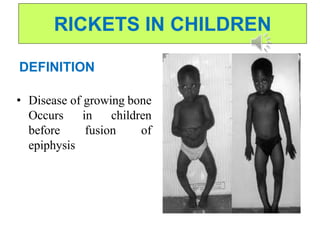
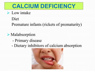
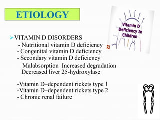
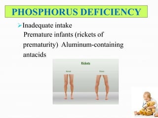
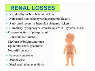
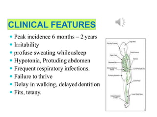
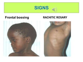
MRS.JEBAKUMARI SUTHA.A
ASSOCIATE PROFESSOR
HOD,PAEDIATRIC DEPARTMENT
GANGA COLLEGE OF NURSING
COIMBATORE-22
RICKETS IN CHILDREN
INTODUCTION
• Your child's body needs vitamin D to absorb
calcium and phosphorus from food. Rickets can
occur if your child's body doesn't get enough
vitamin D or if his or her body has problems
using vitamin D properly. Occasionally, not
getting enough calcium or lack of calcium and
vitamin D can cause rickets.
RICKETS IN CHILDREN
DEFINITION
• Disease of growing bone
Occurs in children
before fusion of
epiphysis
CALCIUM DEFICIENCY
 Low intake
Diet
Premature infants (rickets of prematurity)
 Malabsorption
- Primary disease
- Dietary inhibitors of calcium absorption
ETIOLOGY
VITAMIN D DISORDERS
- Nutritional vitamin D deficiency
- Congenital vitamin D deficiency
- Secondary vitamin D deficiency
Malabsorption Increased degradation
Decreased liver 25-hydroxylase
-Vitamin D–dependent rickets type 1
-Vitamin D–dependent rickets type 2
- Chronic renal failure
PHOSPHORUS DEFICIENCY
Inadequate intake
Premature infants (rickets of
prematurity) Aluminum-containing
antacids
RENAL LOSSES
 X-linked hypophosphatemic rickets
 Autosomal dominant hypophosphatemic rickets
 Autosomal recessive hypophosphatemic rickets
hypercalciuria Hereditary hypophosphatemic rickets with
Overproduction of phosphatonin
Tumor-induced rickets
McCune-Albright syndrome
Epidermal nevus syndrome
Neurofibromatosis
 Fanconi syndrome
 Dent disease
 Distal renal tubular acidosis
NUTRITIONAL RICKETS
Lack of vitamin D
 Commonestcause
 Most common in infancy
 Lack of exposure to U/ V sunlight
 Dark skin
 Covered body
 Kept in-door
 Exclusive breast feeding
 Limited intake of vitamin –D fortified milk and diaryproducts
 During rapid growth
 Infancy
 puberty
 Transplacental transport of vit D provide enough vit D
for first 1 to 2 months of life.
MALABSORPTION
Celiac disease
Pancreatic insufficiency
 Cystic fibrosis
Hepato-biliary disease
 BiliaryArtesia
 Cirrhosis
 Neonatal hepatitis
Drugs
▫ Anti-convulsants
 Phenobartbitone
 Phenytoin
Diet
▫ Excess of phytate in diet with impaired
calcium absorption (chapati flour)
PATHOPHYSIOLOGY.
• Rickets arises due to decreased availability of
phosphorus and calcium to mineralize the
skeletal matrix, leading to growth plate
disorganization and accumulation of
undermineralized osteoid.
• This results in growth plate expansion, bone
weakening, and skeletal deformities
CLINICAL FEATURES
 Peak incidence 6 months – 2 years
 Irritability
 profuse sweating whileasleep
 Hypotonia, Protuding abdomen
 Frequent respiratory infections.
 Failure to thrive
 Delay in walking, delayeddentition
 Fits, tetany.
SIGNS
Frontal bossing RACHITIC ROSARY
SIGNS
Harrison sulkus and
Pot belly Pigeon chest
SIGNS
Widening of wrists Widening of ankle joints
SIGNS
Bending of long bones Knock knee
SIGNS
Wind swept deformity Genu varum
SIGNS
Scoliosis Lordosis
CLINICAL EVALUATION
 Dietary history
 Maternal risk
 Medication
 Malabsorption
 Renal disease
 Family history
 Physical Examination
 Lab Test
DIAGNOSTIC EVALUATION
Serum Calcium low(normal 9-11mg/dl)
Serum phosphorus low (normal-5-
7mg/dl
Alkaline phosphatase israised.
• This is the most striking feature, shows
increased but ineffective activity ofosteoblasts.
 25-(OH) D levels less than 20mg/dl Confirms
of Vitamin Ddeficiency
LABORATORY FINDINGS
Elevated:
Alkaline phosphatase
Parathyroid hormone
Dihydroxyvitamin D
Decreased
Calcium
Phosphorus
Hydroxyvitamin D
RADIOLOGICAL FINDINGS
OF RICKETS
 Generalized osteopenia
 Widening of the unmineralised epiphyseal
growth plates
 Fraying of metaphysis of long bones
 Bowing of legs
 Pseudo-fractures (also called loozerzone)
 Transverse radio lucent band,usually
perpendicular to bonesurface
 Complete fractures
 Features of long standing secondary
hyperparathyroidism (Osteitis fibrosa cystica)
 Sub-periosteal resorption of phalanges
 Presence of bony cyst (brownTumor)
RADIOLOGY
Wrist x-rays in a
normal child (A) and
a child with
rickets (B). Child
with rickets has
metaphyseal fraying
and cupping of the
distal radius and
ulna.
TREATMENT
Stoss therapy – 300000 – 600000 IU Vitamin D
oral or IM, 2-4 doses over one day
Alternatively high dose vit D, 2000-5000 IU/day
over 4-6 wk
Followed by oral Vit D :
< 1 year of age - 400IU
> 1 years of age- 600IU
Symptomatic hypocalcemia –100 mg/kg
IV calcium gluconate followed by oral
calcium or calcitrol -0.05mcg/kg/day
1.Exposure to sunlight(ultraviolet light)
• Early morning and evening 30 minutes perday.
2.Foodfortified with Vit A and Vit D
specially butter,ghee and milk.
• Children under 5 should have 500ml of milk
daily or youghart or cheese daily.
 Daily intake of 400 i.u.vitamin D by
supplemention.
 Lactating mothers should receive
supplementation with milk or vitamin D to
ensure prevention of rickets in their babies.
 Sun exposure tomothers.
VITAMIN D SOURCE
 Sun light
 All Milk products (fortified)
 Cod liver oil
 Egg yolk
Vitamin D requirement:
• Infants- 200IU/day (5mcg) Children-
400IU/day (10mcg)
CONCLUSION
• Nutritional rickets is highly prevalent among
children in the State of Qatar. It can be considered
as a multifactorial condition, in which lack of
exposure to sunlight, calcium deficiency,
prolonged breast feeding without supplementation
and inadequate weaning practices are central.
Health education is important as it can influence
all of the above factors.
REFERENCES
• ACHAR TEXT BOOK OS PAEDIATRICS
• ESSENTIALS OF PAEDIATRICS,GUPTA.
• PRINCIPLES OF NEONATES AND PEDIATRIC
EMERGENCIES
• IAP TEXT BOOK OF PAEDIATRICS AND
NEONATAL EMERGENCIES
• NELSON ESSENTIALS OF PEDIATRIC
MEDICINE
• TEXT BOOK OF PAEDIATRICS ASUMA BEEVI
• GOOGLE REFERENCE
THANK
YOU
RICKETS IN CHILDREN

RICKETS IN CHILDREN

  • 1.
    MRS.JEBAKUMARI SUTHA.A ASSOCIATE PROFESSOR HOD,PAEDIATRICDEPARTMENT GANGA COLLEGE OF NURSING COIMBATORE-22
  • 2.
  • 3.
    INTODUCTION • Your child'sbody needs vitamin D to absorb calcium and phosphorus from food. Rickets can occur if your child's body doesn't get enough vitamin D or if his or her body has problems using vitamin D properly. Occasionally, not getting enough calcium or lack of calcium and vitamin D can cause rickets.
  • 4.
    RICKETS IN CHILDREN DEFINITION •Disease of growing bone Occurs in children before fusion of epiphysis
  • 5.
    CALCIUM DEFICIENCY  Lowintake Diet Premature infants (rickets of prematurity)  Malabsorption - Primary disease - Dietary inhibitors of calcium absorption
  • 6.
    ETIOLOGY VITAMIN D DISORDERS -Nutritional vitamin D deficiency - Congenital vitamin D deficiency - Secondary vitamin D deficiency Malabsorption Increased degradation Decreased liver 25-hydroxylase -Vitamin D–dependent rickets type 1 -Vitamin D–dependent rickets type 2 - Chronic renal failure
  • 7.
    PHOSPHORUS DEFICIENCY Inadequate intake Prematureinfants (rickets of prematurity) Aluminum-containing antacids
  • 8.
    RENAL LOSSES  X-linkedhypophosphatemic rickets  Autosomal dominant hypophosphatemic rickets  Autosomal recessive hypophosphatemic rickets hypercalciuria Hereditary hypophosphatemic rickets with Overproduction of phosphatonin Tumor-induced rickets McCune-Albright syndrome Epidermal nevus syndrome Neurofibromatosis  Fanconi syndrome  Dent disease  Distal renal tubular acidosis
  • 9.
    NUTRITIONAL RICKETS Lack ofvitamin D  Commonestcause  Most common in infancy  Lack of exposure to U/ V sunlight  Dark skin  Covered body  Kept in-door  Exclusive breast feeding  Limited intake of vitamin –D fortified milk and diaryproducts  During rapid growth  Infancy  puberty  Transplacental transport of vit D provide enough vit D for first 1 to 2 months of life.
  • 10.
    MALABSORPTION Celiac disease Pancreatic insufficiency Cystic fibrosis Hepato-biliary disease  BiliaryArtesia  Cirrhosis  Neonatal hepatitis Drugs ▫ Anti-convulsants  Phenobartbitone  Phenytoin Diet ▫ Excess of phytate in diet with impaired calcium absorption (chapati flour)
  • 11.
    PATHOPHYSIOLOGY. • Rickets arisesdue to decreased availability of phosphorus and calcium to mineralize the skeletal matrix, leading to growth plate disorganization and accumulation of undermineralized osteoid. • This results in growth plate expansion, bone weakening, and skeletal deformities
  • 12.
    CLINICAL FEATURES  Peakincidence 6 months – 2 years  Irritability  profuse sweating whileasleep  Hypotonia, Protuding abdomen  Frequent respiratory infections.  Failure to thrive  Delay in walking, delayeddentition  Fits, tetany.
  • 13.
  • 14.
  • 15.
    SIGNS Widening of wristsWidening of ankle joints
  • 16.
    SIGNS Bending of longbones Knock knee
  • 17.
  • 18.
  • 19.
    CLINICAL EVALUATION  Dietaryhistory  Maternal risk  Medication  Malabsorption  Renal disease  Family history  Physical Examination  Lab Test
  • 20.
    DIAGNOSTIC EVALUATION Serum Calciumlow(normal 9-11mg/dl) Serum phosphorus low (normal-5- 7mg/dl Alkaline phosphatase israised. • This is the most striking feature, shows increased but ineffective activity ofosteoblasts.  25-(OH) D levels less than 20mg/dl Confirms of Vitamin Ddeficiency
  • 21.
    LABORATORY FINDINGS Elevated: Alkaline phosphatase Parathyroidhormone Dihydroxyvitamin D Decreased Calcium Phosphorus Hydroxyvitamin D
  • 22.
    RADIOLOGICAL FINDINGS OF RICKETS Generalized osteopenia  Widening of the unmineralised epiphyseal growth plates  Fraying of metaphysis of long bones  Bowing of legs  Pseudo-fractures (also called loozerzone)  Transverse radio lucent band,usually perpendicular to bonesurface  Complete fractures  Features of long standing secondary hyperparathyroidism (Osteitis fibrosa cystica)  Sub-periosteal resorption of phalanges  Presence of bony cyst (brownTumor)
  • 23.
    RADIOLOGY Wrist x-rays ina normal child (A) and a child with rickets (B). Child with rickets has metaphyseal fraying and cupping of the distal radius and ulna.
  • 24.
    TREATMENT Stoss therapy –300000 – 600000 IU Vitamin D oral or IM, 2-4 doses over one day Alternatively high dose vit D, 2000-5000 IU/day over 4-6 wk Followed by oral Vit D : < 1 year of age - 400IU > 1 years of age- 600IU Symptomatic hypocalcemia –100 mg/kg IV calcium gluconate followed by oral calcium or calcitrol -0.05mcg/kg/day
  • 25.
    1.Exposure to sunlight(ultravioletlight) • Early morning and evening 30 minutes perday. 2.Foodfortified with Vit A and Vit D specially butter,ghee and milk. • Children under 5 should have 500ml of milk daily or youghart or cheese daily.
  • 26.
     Daily intakeof 400 i.u.vitamin D by supplemention.  Lactating mothers should receive supplementation with milk or vitamin D to ensure prevention of rickets in their babies.  Sun exposure tomothers.
  • 27.
    VITAMIN D SOURCE Sun light  All Milk products (fortified)  Cod liver oil  Egg yolk Vitamin D requirement: • Infants- 200IU/day (5mcg) Children- 400IU/day (10mcg)
  • 28.
    CONCLUSION • Nutritional ricketsis highly prevalent among children in the State of Qatar. It can be considered as a multifactorial condition, in which lack of exposure to sunlight, calcium deficiency, prolonged breast feeding without supplementation and inadequate weaning practices are central. Health education is important as it can influence all of the above factors.
  • 29.
    REFERENCES • ACHAR TEXTBOOK OS PAEDIATRICS • ESSENTIALS OF PAEDIATRICS,GUPTA. • PRINCIPLES OF NEONATES AND PEDIATRIC EMERGENCIES • IAP TEXT BOOK OF PAEDIATRICS AND NEONATAL EMERGENCIES • NELSON ESSENTIALS OF PEDIATRIC MEDICINE • TEXT BOOK OF PAEDIATRICS ASUMA BEEVI • GOOGLE REFERENCE
  • 30.