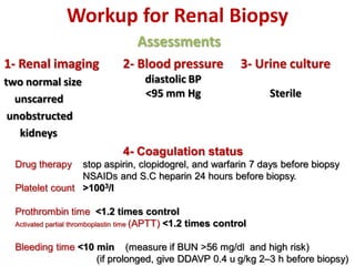
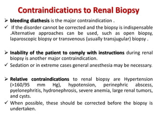
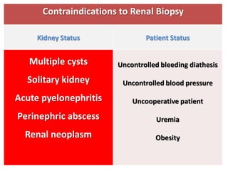
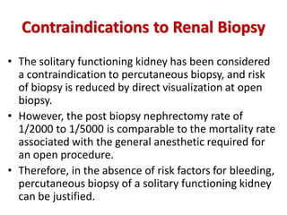
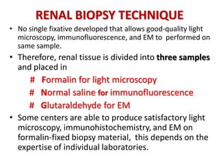
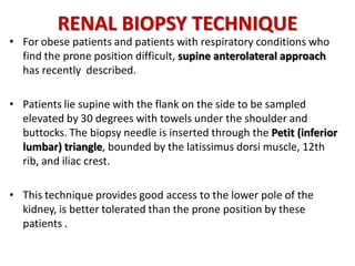
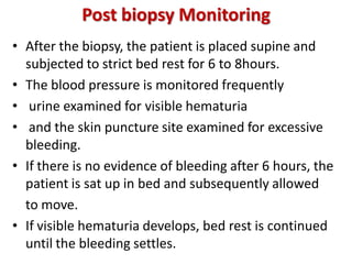
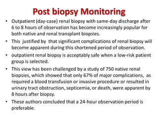
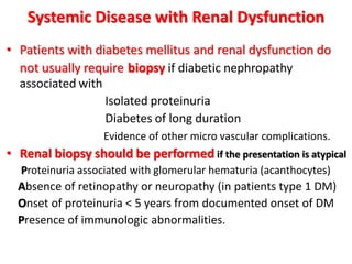
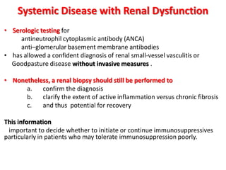
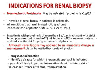
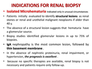
This document discusses renal biopsy procedures. It provides details on the history, technique, adequacy, contraindications, complications and indications for renal biopsy. Some key points include:
- Renal biopsy has evolved since the 1950s and can now provide a tissue diagnosis in over 95% of patients with a life-threatening complication rate of less than 0.1%.
- An adequate biopsy sample contains 10-15 glomeruli and provides samples for histology, immunofluorescence and electron microscopy.
- Contraindications include bleeding diathesis and inability to comply with instructions. Relative contraindications include hypertension and infection.
- Complications are rare but can include hematuria, pain, and rarely death from
random renal tru cut needle biopsy for histopathology
diffuse renal parenchymal disease and disorders
indications
technique
complications
contraindications
simple renal cyst lecture prepare by three medical student in Kerbala university / college of medicine department of surgery to presented as seminar
for download as ppt
https://drive.google.com/open?id=1bOP_UJuZl-dr6wJF6yv3reRw_uNqXGkt
Benign prostatic hyperplasia (BPH), also called prostate enlargement, is a noncancerous increase in size of the prostate gland. Symptoms may include frequent urination, trouble starting to urinate, weak stream, inability to urinate, or loss of bladder control.
This presentation comprises of congenital anomalies of kidney and urinary tract made concise and in depth for PG preparation. It contains all important topics of the regarding subject covered in detail.
How to Make Awesome SlideShares: Tips & TricksSlideShare
Turbocharge your online presence with SlideShare. We provide the best tips and tricks for succeeding on SlideShare. Get ideas for what to upload, tips for designing your deck and more.
Prolonged Pleural Effusion following Liver Biopsy in a 10-Year-Old Girlasclepiuspdfs
Pleural effusions in patients with liver disease are common. Bilious pleural effusion can occur following percutaneous biopsy if the pleura is traversed. We reported the case of a 10-year-old girl who had a liver biopsy. After this procedure, the girl had a pleural effusion during the 20-day period we were treated with the chest tube. After this period, the chest tube was removed and the patient continued conservative gastroenterological treatment for liver cirrhosis.
Prix Galien International 2024 Forum ProgramLevi Shapiro
June 20, 2024, Prix Galien International and Jerusalem Ethics Forum in ROME. Detailed agenda including panels:
- ADVANCES IN CARDIOLOGY: A NEW PARADIGM IS COMING
- WOMEN’S HEALTH: FERTILITY PRESERVATION
- WHAT’S NEW IN THE TREATMENT OF INFECTIOUS,
ONCOLOGICAL AND INFLAMMATORY SKIN DISEASES?
- ARTIFICIAL INTELLIGENCE AND ETHICS
- GENE THERAPY
- BEYOND BORDERS: GLOBAL INITIATIVES FOR DEMOCRATIZING LIFE SCIENCE TECHNOLOGIES AND PROMOTING ACCESS TO HEALTHCARE
- ETHICAL CHALLENGES IN LIFE SCIENCES
- Prix Galien International Awards Ceremony
Flu Vaccine Alert in Bangalore Karnatakaaddon Scans
As flu season approaches, health officials in Bangalore, Karnataka, are urging residents to get their flu vaccinations. The seasonal flu, while common, can lead to severe health complications, particularly for vulnerable populations such as young children, the elderly, and those with underlying health conditions.
Dr. Vidisha Kumari, a leading epidemiologist in Bangalore, emphasizes the importance of getting vaccinated. "The flu vaccine is our best defense against the influenza virus. It not only protects individuals but also helps prevent the spread of the virus in our communities," he says.
This year, the flu season is expected to coincide with a potential increase in other respiratory illnesses. The Karnataka Health Department has launched an awareness campaign highlighting the significance of flu vaccinations. They have set up multiple vaccination centers across Bangalore, making it convenient for residents to receive their shots.
To encourage widespread vaccination, the government is also collaborating with local schools, workplaces, and community centers to facilitate vaccination drives. Special attention is being given to ensuring that the vaccine is accessible to all, including marginalized communities who may have limited access to healthcare.
Residents are reminded that the flu vaccine is safe and effective. Common side effects are mild and may include soreness at the injection site, mild fever, or muscle aches. These side effects are generally short-lived and far less severe than the flu itself.
Healthcare providers are also stressing the importance of continuing COVID-19 precautions. Wearing masks, practicing good hand hygiene, and maintaining social distancing are still crucial, especially in crowded places.
Protect yourself and your loved ones by getting vaccinated. Together, we can help keep Bangalore healthy and safe this flu season. For more information on vaccination centers and schedules, residents can visit the Karnataka Health Department’s official website or follow their social media pages.
Stay informed, stay safe, and get your flu shot today!
Title: Sense of Taste
Presenter: Dr. Faiza, Assistant Professor of Physiology
Qualifications:
MBBS (Best Graduate, AIMC Lahore)
FCPS Physiology
ICMT, CHPE, DHPE (STMU)
MPH (GC University, Faisalabad)
MBA (Virtual University of Pakistan)
Learning Objectives:
Describe the structure and function of taste buds.
Describe the relationship between the taste threshold and taste index of common substances.
Explain the chemical basis and signal transduction of taste perception for each type of primary taste sensation.
Recognize different abnormalities of taste perception and their causes.
Key Topics:
Significance of Taste Sensation:
Differentiation between pleasant and harmful food
Influence on behavior
Selection of food based on metabolic needs
Receptors of Taste:
Taste buds on the tongue
Influence of sense of smell, texture of food, and pain stimulation (e.g., by pepper)
Primary and Secondary Taste Sensations:
Primary taste sensations: Sweet, Sour, Salty, Bitter, Umami
Chemical basis and signal transduction mechanisms for each taste
Taste Threshold and Index:
Taste threshold values for Sweet (sucrose), Salty (NaCl), Sour (HCl), and Bitter (Quinine)
Taste index relationship: Inversely proportional to taste threshold
Taste Blindness:
Inability to taste certain substances, particularly thiourea compounds
Example: Phenylthiocarbamide
Structure and Function of Taste Buds:
Composition: Epithelial cells, Sustentacular/Supporting cells, Taste cells, Basal cells
Features: Taste pores, Taste hairs/microvilli, and Taste nerve fibers
Location of Taste Buds:
Found in papillae of the tongue (Fungiform, Circumvallate, Foliate)
Also present on the palate, tonsillar pillars, epiglottis, and proximal esophagus
Mechanism of Taste Stimulation:
Interaction of taste substances with receptors on microvilli
Signal transduction pathways for Umami, Sweet, Bitter, Sour, and Salty tastes
Taste Sensitivity and Adaptation:
Decrease in sensitivity with age
Rapid adaptation of taste sensation
Role of Saliva in Taste:
Dissolution of tastants to reach receptors
Washing away the stimulus
Taste Preferences and Aversions:
Mechanisms behind taste preference and aversion
Influence of receptors and neural pathways
Impact of Sensory Nerve Damage:
Degeneration of taste buds if the sensory nerve fiber is cut
Abnormalities of Taste Detection:
Conditions: Ageusia, Hypogeusia, Dysgeusia (parageusia)
Causes: Nerve damage, neurological disorders, infections, poor oral hygiene, adverse drug effects, deficiencies, aging, tobacco use, altered neurotransmitter levels
Neurotransmitters and Taste Threshold:
Effects of serotonin (5-HT) and norepinephrine (NE) on taste sensitivity
Supertasters:
25% of the population with heightened sensitivity to taste, especially bitterness
Increased number of fungiform papillae
- Video recording of this lecture in English language: https://youtu.be/lK81BzxMqdo
- Video recording of this lecture in Arabic language: https://youtu.be/Ve4P0COk9OI
- Link to download the book free: https://nephrotube.blogspot.com/p/nephrotube-nephrology-books.html
- Link to NephroTube website: www.NephroTube.com
- Link to NephroTube social media accounts: https://nephrotube.blogspot.com/p/join-nephrotube-on-social-media.html
micro teaching on communication m.sc nursing.pdfAnurag Sharma
Microteaching is a unique model of practice teaching. It is a viable instrument for the. desired change in the teaching behavior or the behavior potential which, in specified types of real. classroom situations, tends to facilitate the achievement of specified types of objectives.
Report Back from SGO 2024: What’s the Latest in Cervical Cancer?bkling
Are you curious about what’s new in cervical cancer research or unsure what the findings mean? Join Dr. Emily Ko, a gynecologic oncologist at Penn Medicine, to learn about the latest updates from the Society of Gynecologic Oncology (SGO) 2024 Annual Meeting on Women’s Cancer. Dr. Ko will discuss what the research presented at the conference means for you and answer your questions about the new developments.
HOT NEW PRODUCT! BIG SALES FAST SHIPPING NOW FROM CHINA!! EU KU DB BK substit...GL Anaacs
Contact us if you are interested:
Email / Skype : kefaya1771@gmail.com
Threema: PXHY5PDH
New BATCH Ku !!! MUCH IN DEMAND FAST SALE EVERY BATCH HAPPY GOOD EFFECT BIG BATCH !
Contact me on Threema or skype to start big business!!
Hot-sale products:
NEW HOT EUTYLONE WHITE CRYSTAL!!
5cl-adba precursor (semi finished )
5cl-adba raw materials
ADBB precursor (semi finished )
ADBB raw materials
APVP powder
5fadb/4f-adb
Jwh018 / Jwh210
Eutylone crystal
Protonitazene (hydrochloride) CAS: 119276-01-6
Flubrotizolam CAS: 57801-95-3
Metonitazene CAS: 14680-51-4
Payment terms: Western Union,MoneyGram,Bitcoin or USDT.
Deliver Time: Usually 7-15days
Shipping method: FedEx, TNT, DHL,UPS etc.Our deliveries are 100% safe, fast, reliable and discreet.
Samples will be sent for your evaluation!If you are interested in, please contact me, let's talk details.
We specializes in exporting high quality Research chemical, medical intermediate, Pharmaceutical chemicals and so on. Products are exported to USA, Canada, France, Korea, Japan,Russia, Southeast Asia and other countries.
Explore natural remedies for syphilis treatment in Singapore. Discover alternative therapies, herbal remedies, and lifestyle changes that may complement conventional treatments. Learn about holistic approaches to managing syphilis symptoms and supporting overall health.
Ozempic: Preoperative Management of Patients on GLP-1 Receptor Agonists Saeid Safari
Preoperative Management of Patients on GLP-1 Receptor Agonists like Ozempic and Semiglutide
ASA GUIDELINE
NYSORA Guideline
2 Case Reports of Gastric Ultrasound
Couples presenting to the infertility clinic- Do they really have infertility...Sujoy Dasgupta
Dr Sujoy Dasgupta presented the study on "Couples presenting to the infertility clinic- Do they really have infertility? – The unexplored stories of non-consummation" in the 13th Congress of the Asia Pacific Initiative on Reproduction (ASPIRE 2024) at Manila on 24 May, 2024.
2. Renal Biopsy
Introduction
Is Renal Biopsy A Necessary Investigation?
Biopsy adequacy
Workup For Renal Biopsy
Contraindications To Renal Biopsy
Renal Biopsy Technique
Post Biopsy Monitoring
Complications Of Renal Biopsy
Indications For Renal Biopsy
3. INTRODUCTION
• Percutaneous renal biopsy was first described in the early 1950s .
• These early biopsies were performed with the patient in sitting position by
use of a suction needle and intravenous urography for guidance.
• An adequate tissue diagnosis was achieved in less than 40% of these early cases.
• In 1954, Kark described a modified technique using the Franklin modified Vim-
Silverman needle, with the patient in a prone position and an exploring needle
used to localize the kidney before insertion of the biopsy needle.
• These modifications yielded a tissue diagnosis in 96% of cases, and no major
complications were reported.
4. INTRODUCTION
• Since then, the basic renal biopsy procedure has remained largely
unchanged, although the use of real-time ultrasound and refinement of
biopsy needle design have offered significant improvements.
• Renal biopsy is now able to provide a tissue diagnosis in more than 95% of
patients, with a life-threatening complication rate of less than 0.1%.
5. Is Renal Biopsy a Necessary Investigation?
• Early studies suggested that renal biopsy provided
diagnostic clarity in majority of patients , but this
information did not alter management, with the exception
of those with heavy proteinuria or systemic disease.
• More recent prospective studies suggest that :
Renal biopsy identifies a diagnosis different from that
predicted on clinical grounds in 50% to 60% of patients
and leads to a treatment change in 20% to 50%.
• This is apparent in patients with heavy proteinuria or AKI,
more than 80% of whom have biopsy findings that alter
their management.
6. Biopsy Adequacy
• The number of glomeruli in the sample is the
major determinant of whether the biopsy will be
diagnostically informative.
• A typical diagnostically useful biopsy sample will
contain 10 to 15 glomeruli .
• Because of sampling issue, a biopsy sample of
this size will be unable to diagnose focal diseases
and at best will provide imprecise guidance on
the extent of glomerular involvement.
7. Biopsy Adequacy
• An adequate biopsy should provide samples for :
immunohistology and electron microscopy (EM).
• Immunohistology is provided by either immunofluorescence on
frozen material or immunoperoxidase on fixed tissue, according to
local protocols .
• It is helpful for the biopsy cores to be viewed immediately after
being taken under microscope to ensure that they contain cortex
and when cores are divided, immunohistology and EM samples
both contain glomeruli.
8. Biopsy Adequacy
• If the material obtained for a pathologic evaluation is
insufficient, a discussion with pathologist should address
how best to proceed before the tissue is placed in fixative .
• So that provide maximum information for specific clinical
scenario.
• For example, if patient has heavy proteinuria, most information
will be gained from EM because it is able to demonstrate
Podocyte foot process effacement
Focal sclerosis
Electron-dense deposits of immune complexes.
. Organized deposits of amyloid.
9. Workup for Renal Biopsy
Assessments
1- Renal imaging
two normal size
unscarred
unobstructed
kidneys
2- Blood pressure
diastolic BP
<95 mm Hg
3- Urine culture
Sterile
4- Coagulation status
Drug therapy stop aspirin, clopidogrel, and warfarin 7 days before biopsy
NSAIDs and S.C heparin 24 hours before biopsy.
Platelet count >1003/l
Prothrombin time <1.2 times control
Activated partial thromboplastin time (APTT) <1.2 times control
Bleeding time <10 min (measure if BUN >56 mg/dl and high risk)
(if prolonged, give DDAVP 0.4 u g/kg 2–3 h before biopsy)
10. Contraindications to Renal Biopsy
bleeding diathesis is the major contraindication .
If the disorder cannot be corrected and the biopsy is indispensable
.Alternative approaches can be used, such as open biopsy,
laparoscopic biopsy or transvenous (usually transjugular) biopsy .
Inability of the patient to comply with instructions during renal
biopsy is another major contraindication.
Sedation or in extreme cases general anesthesia may be necessary.
Relative contraindications to renal biopsy are Hypertension
(>160/95 mm Hg), hypotension, perinephric abscess,
pyelonephritis, hydronephrosis, severe anemia, large renal tumors,
and cysts.
When possible, these should be corrected before the biopsy is
undertaken.
11. Contraindications to Renal Biopsy
Kidney Status Patient Status
Multiple cysts
Solitary kidney
Acute pyelonephritis
Perinephric abscess
Renal neoplasm
Uncontrolled bleeding diathesis
Uncontrolled blood pressure
Uncooperative patient
Uremia
Obesity
12. Contraindications to Renal Biopsy
• The solitary functioning kidney has been considered
a contraindication to percutaneous biopsy, and risk
of biopsy is reduced by direct visualization at open
biopsy.
• However, the post biopsy nephrectomy rate of
1/2000 to 1/5000 is comparable to the mortality rate
associated with the general anesthetic required for
an open procedure.
• Therefore, in the absence of risk factors for bleeding,
percutaneous biopsy of a solitary functioning kidney
can be justified.
13. RENAL BIOPSY TECHNIQUE
Percutaneous native Renal Biopsy
• Biopsy is performed by nephrologists with
continuous (real-time) ultrasound guidance and
disposable automated biopsy needles.
• We use 16-gauge needles and the trend toward
fewer bleeding complications of smaller needles.
• For most patients, premedication or sedation is not
required.
• The patient is prone, and a pillow is placed under
the abdomen at the level of the umbilicus to
straighten the lumbar spine and to splint the
kidneys.
14. RENAL BIOPSY TECHNIQUE
• Ultrasound is used to localize the lower pole of
the kidney where the biopsy will be performed
(usually the left kidney).
• A pen mark is used to indicate the point of entry
of the biopsy needle.
• The skin is sterilized with povidone-iodine
(Betadine) . A sterile fenestrated sheet is placed
over the area to maintain a sterile field.
• Local anesthetic (2% lidocaine ) is infiltrated into
the skin at the point previously marked.
15. Renal biopsy procedure
• The biopsy needle is introduced at an angle of
approximately 70 degrees to the skin and is guided by
continuous ultrasound.
• The operator is shown wearing a surgical gown.
16. RENAL BIOPSY TECHNIQUE
• While the anesthetic takes effect, the ultrasound probe is covered
in a sterile sheath. Sterile ultrasound jelly is applied to the skin
• Under ultrasound guidance, a 10-cm, needle is guided to the renal
capsule.
• A stab incision is made through the dermis to ease passage of
the biopsy needle. This is passed under ultrasound guidance to the
kidney capsule .
• As the needle approaches the capsule, the patient is instructed to take a
breath until the kidney is moved to a position such that the lower pole
rests just under the biopsy needle, and then to stop breathing.
• The biopsy needle tip is advanced to the renal capsule, and the trigger
mechanism is released, firing the needle into the kidney .
• The needle is immediately withdrawn, the patient is asked to resume
breathing, and the contents of the needle are examined .
17. Renal biopsy imaging. Ultrasound scan
shows the needle
entering the lower pole of the left
kidney. Arrows indicate the needle track,
which appears as a fuzzy white line.
Renal biopsy imaging
CT left kidney
The angle of approach of
needle is demonstrated.
Note adjacency to
the lower pole of the
kidney
18. RENAL BIOPSY TECHNIQUE
• We examined the tissue core under an operating
microscope to ensure that renal cortex has been
obtained .
• A second pass of the needle is usually necessary to
obtain additional tissue for immunohistology and EM.
• If insufficient tissue is obtained, further passes of the
needle are made.
• However, passing the needle more than four times is
associated with a modest increase in the post biopsy
. complication rate.
• Once sufficient renal tissue has been obtained, the
skin incision is dressed and the patient rolled directly
into bed for observation.
19. • A core of renal tissue is demonstrated
in the sampling notch of the biopsy needle
20. Renal biopsy micrographs
• Appearance of renal biopsy material under the operating
microscope.
A Low-power view shows two good-sized cores.
B Higher-magnification view shows typical appearance
of glomeruli (arrows).
21. RENAL BIOPSY TECHNIQUE
• No single fixative developed that allows good-quality light
microscopy, immunofluorescence, and EM to performed on
same sample.
• Therefore, renal tissue is divided into three samples
and placed in
# Formalin for light microscopy
# Normal saline for immunofluorescence
# Glutaraldehyde for EM
• Some centers are able to produce satisfactory light
microscopy, immunohistochemistry, and EM on
formalin-fixed biopsy material, this depends on the
expertise of individual laboratories.
22. RENAL BIOPSY TECHNIQUE
• For obese patients and patients with respiratory conditions who
find the prone position difficult, supine anterolateral approach
has recently described.
• Patients lie supine with the flank on the side to be sampled
elevated by 30 degrees with towels under the shoulder and
buttocks. The biopsy needle is inserted through the Petit (inferior
lumbar) triangle, bounded by the latissimus dorsi muscle, 12th
rib, and iliac crest.
• This technique provides good access to the lower pole of the
kidney, is better tolerated than the prone position by these
patients .
23. RENAL BIOPSY TECHNIQUE
Renal Transplant Biopsy
• Biopsy of the transplant kidney is facilitated by the proximity of the
kidney to the anterior abdominal wall and the lack of movement on
respiration.
• It is performed under real-time ultrasound guidance with use of an
automated biopsy needle.
In most patients, renal transplant biopsy is performed to identify
cause of acute allograft dysfunction (acute rejection), therefore
diagnosis can be made on a formalin fixed sample alone for light
microscopy.
If vascular rejection is suspected, a snap-frozen sample for C4d
immunostaining should also be obtained (although some laboratories
are able to detect C4d onformalin-fixed material).
If recurrent or de novo GN is suspected in patients with chronic
allograft dysfunction, additional samples for EM and immunohistology
should be collected.
24. Post biopsy Monitoring
• After the biopsy, the patient is placed supine and
subjected to strict bed rest for 6 to 8hours.
• The blood pressure is monitored frequently
• urine examined for visible hematuria
• and the skin puncture site examined for excessive
bleeding.
• If there is no evidence of bleeding after 6 hours, the
patient is sat up in bed and subsequently allowed
to move.
• If visible hematuria develops, bed rest is continued
until the bleeding settles.
25. Post biopsy Monitoring
• Outpatient (day-case) renal biopsy with same-day discharge after
6 to 8 hours of observation has become increasingly popular for
both native and renal transplant biopsies.
• This justified by that significant complications of renal biopsy will
become apparent during this shortened period of observation.
• outpatient renal biopsy is acceptably safe when a low-risk patient
group is selected.
• This view has been challenged by a study of 750 native renal
biopsies, which showed that only 67% of major complications, as
required a blood transfusion or invasive procedure or resulted in
urinary tract obstruction, septicemia, or death, were apparent by
8 hours after biopsy.
• These authors concluded that a 24-hour observation period is
preferable.
26. Complications of Renal Biopsy
Complication Percentage
Visible hematuria 3.5%
Need for blood transfusion 0.9%
Need for intervention to control
bleeding
0.7% 0.6%angiographic
0.1%surgical
Death 0.02%
27. Complications of Renal Biopsy
• Dull ache Pain around the needle entry site when the local anesthetic
wears off after renal biopsy.
Simple analgesia with paracetamol usually suffices.
• More severe pain in the loin or abdomen on the side of the biopsy
suggests significant perirenal hemorrhage.
• The mean decrease in hemoglobin after a biopsy is approximately 1 g/dl.
• Significant perirenal hematomas are almost associated with severe loin
pain.
• Both visible hematuria and painful hematoma are seen in 3% to 4% of
patients after biopsy.
The initial management is strict bed rest and maintenance of normal
coagulation indices.
• If bleeding is brisk and associated with hypotension or prolonged and
fails to settle with bed rest, renal angiography should performed to
identify source of bleeding. Coil embolization can performed, and this
eliminate need for open surgical intervention and nephrectomy.
28. Complications of Renal Biopsy
• Most postbiopsy arteriovenous fistulas detected by Doppler
• Ultrasound or contrast-enhanced C T , can be found as many as
18% of patients.
• Because most are clinically silent and more than 95% resolve
spontaneously within 2 year .
• In a small minority of patients, arteriovenous fistulas can lead to
visible hematuria (typically recurrent, dark red, and often with
blood clots), hypertension, and renal impairment, which requires
embolization.
• Death resulting directly from renal biopsy become much less
common according to recent biopsy series compared with earlier
reports.
• Most deaths result from uncontrolled hemorrhage in
high-risk patients, particularly those with severe renal impairment.
29. INDICATIONS FOR RENAL BIOPSY
• Ideally, analysis of a renal biopsy sample
should identify :
a specific diagnosis .
reflect the level of disease activity .
provide information to allow decisions,
. planned treatment .
• Although renal biopsy not always able to fulfill
these criteria .
• It remains a valuable clinical tool and of
particular benefit in the clinical situations .
31. INDICATIONS FOR RENAL BIOPSY
Nephrotic Syndrome
1- Routinely indicated in adults .
2- In prepubertal children
only if clinical features atypical of .
. minimal change disease
• Nephrotic children with atypical features :
Microscopic hematuria
Reduced serum complement levels
Renal impairment
Failure to respond to corticosteroids.
32. INDICATIONS FOR RENAL BIOPSY
Acute Kidney Injury
Obstruction
Reduced renal perfusion
Acute tubular necrosis have been ruled out
• In a minority of patients, a confident diagnosis
cannot be made .
• Renal biopsy should be performed on an urgent
basis so that appropriate treatment started before
irreversible renal injury develops.
• This is particularly true in patients with AKI
accompanied by active urine sediment .
33. INDICATIONS FOR RENAL BIOPSY
Systemic Disease with Renal Dysfunction
• In patients with
1 Small-vessel vasculitis
2 Anti–glomerular basement membrane disease
3 Systemic lupus
• In patients with diabetes only if atypical features
present
34. Systemic Disease with Renal Dysfunction
• Patients with diabetes mellitus and renal dysfunction do
not usually require biopsy if diabetic nephropathy
associated with
Isolated proteinuria
Diabetes of long duration
Evidence of other micro vascular complications.
• Renal biopsy should be performed if the presentation is atypical
Proteinuria associated with glomerular hematuria (acanthocytes)
Absence of retinopathy or neuropathy (in patients type 1 DM)
Onset of proteinuria < 5 years from documented onset of DM
Presence of immunologic abnormalities.
35. Systemic Disease with Renal Dysfunction
• Serologic testing for
antineutrophil cytoplasmic antibody (ANCA)
anti–glomerular basement membrane antibodies
• has allowed a confident diagnosis of renal small-vessel vasculitis or
Goodpasture disease without invasive measures .
• Nonetheless, a renal biopsy should still be performed to
a. confirm the diagnosis
b. clarify the extent of active inflammation versus chronic fibrosis
c. and thus potential for recovery
This information
important to decide whether to initiate or continue immunosuppressives
particularly in patients who may tolerate immunosuppression poorly.
36. INDICATIONS FOR RENAL BIOPSY
• Non-nephrotic Proteinuria May be indicated if proteinuria >1 g/24 h
• The value of renal biopsy in patients is debatable.
• All conditions that result in nephrotic syndrome
can cause non-nephrotic proteinuria, except MCD.
• In patients with proteinuria of more than 1 g/day, treatment with strict
blood pressure control and (ACE) inhibitors or (ARBs) reduces proteinuria
and reduces the risk for progressive renal dysfunction .
• Although renal biopsy may not lead to an immediate change in
management : it can be justified because it will provide
- prognostic information
- identify a disease for which therapeutic approach is indicated
- provide clinically important information about the future risk of .
.disease recurrence after renal transplantation.
37. INDICATIONS FOR RENAL BIOPSY
• Isolated Microhematuria Indicated only in unusual circumstances
• Patients initially evaluated to identify structural lesions as renal
stones or renal and urothelial malignant neoplasms if older than
40 y.
• The absence of a structural lesion suggests that hematuria have
a glomerular source.
• Biopsy studies identified glomerular lesions in up to 75% of
biopsies.
• IgA nephropathy is the most common lesion, followed by
thin basement membrane .
• In the absence of nephrotic proteinuria, renal impairment, or
hypertension, the prognosis is excellent .
• because no specific therapies are available, renal biopsy is not
necessary and patients require only follow-up.
38. INDICATIONS FOR RENAL BIOPSY
• Biopsy should be performed only :
if the result would provide reassurance to a patient .
avoid repeated urologic investigations .
or provide specific information :
i. in evaluation of potential living kidney donors .
ii. in familial hematuria .
iii. or for life insurance and employment purposes .
39. INDICATIONS FOR RENAL BIOPSY
• Unexplained Chronic Kidney Disease
• Renal biopsy can be informative in the patient with
unexplained CKD and normal-sized kidneys .
• Studies shown that in these patients with CKD, the biopsy will
demonstrate disease that was not predicted in almost half.
• However, if both kidneys are small (<9 cm on ultrasound), the
risks of biopsy are increased, and the diagnostic information.
limited by extensive glomerulosclerosis and tubulointerstitial
fibrosis.
• However, immunofluorescence studies may still be informative :
For example, glomerular IgA deposition may be identified . .
. despite advanced structural damage.
40. INDICATIONS FOR RENAL BIOPSY
• Familial Renal Disease
• A renal biopsy performed in one affected family member
may secure the diagnosis for the whole family and avoid
the need for repeat investigation.
• Conversely, a renal biopsy may unexpectedly
identify disease that has an inherited basis,
thereby stimulating evaluation of other family
members.
41. INDICATIONS FOR RENAL BIOPSY
• Renal Transplant Dysfunction
• Renal allograft dysfunction in the absence of ureteral obstruction, urinary
sepsis, renal artery stenosis, or toxic levels of calcineurin inhibitors requires
a renal biopsy to determine the cause.
In the early post-transplantation period, this is most useful in differentiat
acute rejection from ATN and increasingly prevalent BK virus nephropathy.
Later , renal biopsy can differentiate late acute rejection from chronic allograft
nephropathy , recurrent or de novo glomerulonephritis (GN) , and calcineurin
inhibitor toxicity.
• The accessible location of the renal transplant in the iliac fossa facilitates
biopsy of the allograft and allows repeated biopsies when indicated.
• This encouraged many units to adopt a policy of protocol biopsies to detect
subclinical acute rejection and renal scarring and to guide the choice of
immunosuppressive therapy .
42. Role of Repeat Renal Biopsy
• In some patients, a repeat biopsy may be indicated.
• The pathologic changes in lupus nephritis may evolve,
necessitating treatment adjustment.
• Corticosteroid-resistant/dependent MCD or frequently
relapsing MCD may actually represent a missed diagnosis
of focal segmental glomerulosclerosis (FSGS), which may
be detected on repeat biopsy.