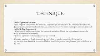
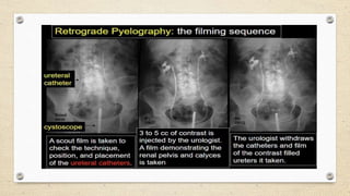
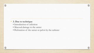
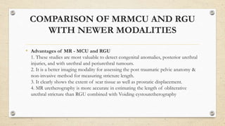
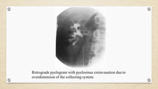
SlideShare a Scribd company logo
RETROGRADE
PYELOURETEROGRAPHY
PRESENTED BY:
DR. DILIP SHAH
INTRODUCTION
• It is the roentgenographic demonstration of the renal pelvis and
ureter by the retrograde injection of radio-opaque material through
the ureters.
INDICATIONS
• 1. Absent or unsatisfactory visualisation of the collecting system on IVU.
2. Unexplained hematuria, when the ureters have not been completely
visualised by IVU.
3. Evaluating persistent intraureteral or intrapelvic filling defects on
IVU.
4. Demonstrating the exact site of ureteral fistula.
5. Brushing and/or biopsy of suspected lesions.
6. Evaluating the collecting system in patients who cannot receive
intravenous contrast medium.
CONTRAINDICATION
• Acute urinary tract infection
• Recent instrumentation like cystoscopy or other transurethral endoscopy- the
small mucosal laceration may lead to contrast extravasation into the corpus
spongiosum thus leading to septic infection.
• Pregnancy
CONTRAST MEDIA
• Ionic contrast media can be used safely, however if there is any
specific contraindication like known hypersensitivity etc., Non ionic
contrast media may be used.
• The Ionic contrast media is preferred
due to its low cost.
• The strength of contrast media should be
150-200 mg I/ml.
• Contrast media should not be too dense as it will obscure small
lesions in the ureters and the pelvis.
PROCEDURE
• Patient Preparation
• Bowel preparation with cathartics is not routinely performed.
Preliminary Film
• Full length supine AP abdomen before the examination is started.
Anaesthesia
• May be performed under local anaesthesia although general
anesthesia is often required. Sterile precautions are mandatory
TECHNIQUE
• In the Operation theatre:
• The surgeon catheterizes the ureter via a cystoscope and advances the ureteric catheter to the
desired level. Contrast medium is injected under fluoroscopic control and spot films are exposed.
In the X-Ray Department
• With ureteric catheter(s) in situ, the patient is transferred from the operation theatre to the
X-ray department if necessary.
• Urine is aspirated and under fluoroscopic control.
• Contrast medium is slowly injected. About 3-5 ml is usually enough to fill the pelvis
but the injection should be terminated before this if the patient complains of pain or fullness in
the loin.
FILMS
• Using the undercouch tube
(a) Supine PA film of the kidney
(b) Both 35° anterior obliques of the kidneys. Low kVp (65-75 kVp) technique is
used to visualise calculi and contrast medium.
(c) If there is pelvi-ureteric junction obstruction, the contrast medium in the pelvis
is aspirated. The films are examined and if satisfactory, the catheter is withdrawn,
first to 10 cm below the renal pelvis and then to lie above the ureteric orifice. About
2ml of contrast medium is injected at each of these levels and films taken.
• AFTER CARE
1. Post anesthetic observation.
2. Prophylactic antibiotics may be used
COMPLICATIONS
• 1. Due to anaesthesia
• Complications of general anaesthesia.
2. Due to the contrast medium
• Contrast medium can be absorbed from the renal pelvis, giving rise to adverse
reactions. However, the risks are much less than with excretory urography
• Chemical pyelitis-if there is stasis of contrast medium.
• Extravasation due to overdistension of the pelvis.
• 3. Due to technique
• Introduction of infection
• Mucosal damage to the ureter
• Perforation of the ureter or pelvis by the catheter
COMPARISON OF MRMCU AND RGU
WITH NEWER MODALITIES
• Advantages of MR - MCU and RGU
1. These studies are most valuable to detect congenital anomalies, posterior urethral
injuries, and with urethral and periurethral tumours.
2. It is a better imaging modality for assessing the post traumatic pelvic anatomy &
non-invasive method for measuring stricture length.
3. It clearly shows the extent of scar tissue as well as prostatic displacement.
4. MR uretherography is more accurate in estimating the length of obliterative
urethral stricture than RGU combined with Voiding cystouretherography
Limitations of conventional RGU combined with
voiding cystouretherography:
• 1. It does not provide accurate length of the defect because of poor
prostatic urethral filling.
2. It does not provide information regarding extent of fbrosis of
corpora spongiosa or prostatic displacement.
3. The stricture length is overestimated if bladder neck does not
relax.
Advantages of CT urethrography
• 1. C.T. voiding uretherography is more comfortable to the patient
because it requires adaptation only in one position.
2. Less time consuming; takes only few seconds
3. Comparison of lual size & stricture length for follow up is
possible.
4. Extraluinal pathology can be detected
5. Good patient compliance
6. Ability to survey whole urinary tract from kidney to urethra.
Retrograde pyelogram
demonstrating
the pelvicalyceal system and ureter
down to the vesicoureteric
junction.
URETERAL
STENOSIS
Two views of the retrograde pyelography
showing stenosis of the lower portion of the
right ureter at the level of the iliac vessels.
There is severe dilatation of the medial and
proximal portions of the right ureter and
hydronephrosis.
URETERAL
STRICTURE
A case ofTCC
Filling defect is seen in the
right ureter –
RAT BITE SIGN
Goblet sign or
Champagne glass sign:
•irregular ureteric
narrowing with proximal
shouldering termed the
goblet sign
•Focally dilated
intraluminal mass
Causes of goblet cells
• TCC of ureter
•Metastatic disease in to
ureter
•Endometriosis involving
ureter
Retrograde pyelogram shows the area of narrowing at the site
of ureteral implantation into the bladder (arrow).
Retrograde pyelogram shows rounded filling defects in many
calyces representing papillary necrosis.
Retrograde pyelogram with pyelosinus extravasation due to
overdistension of the collecting system.
THANK YOU!

More Related Content

What's hot

IVU- Intravenous urography
IVU- Intravenous urographyIVU- Intravenous urography
IVU- Intravenous urography
Ashim Budhathoki
 
Ptc )percutaneous transhepatic cholangiography
Ptc )percutaneous transhepatic cholangiographyPtc )percutaneous transhepatic cholangiography
Ptc )percutaneous transhepatic cholangiography
Yashawant Yadav
 
Enteroclysis
EnteroclysisEnteroclysis
Anterograde/Retrograde urethrography (RGU/MCU)
Anterograde/Retrograde urethrography (RGU/MCU)Anterograde/Retrograde urethrography (RGU/MCU)
Anterograde/Retrograde urethrography (RGU/MCU)
Shubham Singhal
 
Hsg ppt
Hsg ppt Hsg ppt
Intravenous urography (IVU)- Avinesh Shrestha
Intravenous urography (IVU)- Avinesh ShresthaIntravenous urography (IVU)- Avinesh Shrestha
Intravenous urography (IVU)- Avinesh Shrestha
Avinesh Shrestha
 
Percutaneous transhepatic choledochography
Percutaneous transhepatic choledochographyPercutaneous transhepatic choledochography
Percutaneous transhepatic choledochography
Maajid Mohi ud din
 
Enteroclysis( small bowel enema)
Enteroclysis( small bowel enema)Enteroclysis( small bowel enema)
Enteroclysis( small bowel enema)
Ankit Mishra
 
Barium meal follow through
Barium meal follow throughBarium meal follow through
Barium meal follow through
Shiva Prakash
 
Barium Swallow Presentation
Barium Swallow  PresentationBarium Swallow  Presentation
Barium Swallow Presentationdrshaik
 
Hypotonic duodenography
Hypotonic duodenographyHypotonic duodenography
Hypotonic duodenography
Maajid Mohi ud din
 
Venography
VenographyVenography
Venography
SUJAN KARKI
 
Imaging of biliary tract
Imaging of biliary tractImaging of biliary tract
Imaging of biliary tract
Maajid Mohi ud din
 
Contrast media 4
Contrast media 4 Contrast media 4
Contrast media 4
Behzad Ommani
 
Loopogram
LoopogramLoopogram
Loopogram
Sam Shaikh
 
Intravenous Urography
Intravenous UrographyIntravenous Urography
Intravenous Urography
Youttam Laudari
 
CT Urography
CT UrographyCT Urography
IVP Best presnetation
IVP Best presnetationIVP Best presnetation
IVP Best presnetation
DR SACHIN SURA
 
Intravenous urography
Intravenous urographyIntravenous urography
Intravenous urography
RamanGhimire3
 
Venography/ Phlebography- Avinesh Shrestha
Venography/ Phlebography- Avinesh ShresthaVenography/ Phlebography- Avinesh Shrestha
Venography/ Phlebography- Avinesh Shrestha
Avinesh Shrestha
 

What's hot (20)

IVU- Intravenous urography
IVU- Intravenous urographyIVU- Intravenous urography
IVU- Intravenous urography
 
Ptc )percutaneous transhepatic cholangiography
Ptc )percutaneous transhepatic cholangiographyPtc )percutaneous transhepatic cholangiography
Ptc )percutaneous transhepatic cholangiography
 
Enteroclysis
EnteroclysisEnteroclysis
Enteroclysis
 
Anterograde/Retrograde urethrography (RGU/MCU)
Anterograde/Retrograde urethrography (RGU/MCU)Anterograde/Retrograde urethrography (RGU/MCU)
Anterograde/Retrograde urethrography (RGU/MCU)
 
Hsg ppt
Hsg ppt Hsg ppt
Hsg ppt
 
Intravenous urography (IVU)- Avinesh Shrestha
Intravenous urography (IVU)- Avinesh ShresthaIntravenous urography (IVU)- Avinesh Shrestha
Intravenous urography (IVU)- Avinesh Shrestha
 
Percutaneous transhepatic choledochography
Percutaneous transhepatic choledochographyPercutaneous transhepatic choledochography
Percutaneous transhepatic choledochography
 
Enteroclysis( small bowel enema)
Enteroclysis( small bowel enema)Enteroclysis( small bowel enema)
Enteroclysis( small bowel enema)
 
Barium meal follow through
Barium meal follow throughBarium meal follow through
Barium meal follow through
 
Barium Swallow Presentation
Barium Swallow  PresentationBarium Swallow  Presentation
Barium Swallow Presentation
 
Hypotonic duodenography
Hypotonic duodenographyHypotonic duodenography
Hypotonic duodenography
 
Venography
VenographyVenography
Venography
 
Imaging of biliary tract
Imaging of biliary tractImaging of biliary tract
Imaging of biliary tract
 
Contrast media 4
Contrast media 4 Contrast media 4
Contrast media 4
 
Loopogram
LoopogramLoopogram
Loopogram
 
Intravenous Urography
Intravenous UrographyIntravenous Urography
Intravenous Urography
 
CT Urography
CT UrographyCT Urography
CT Urography
 
IVP Best presnetation
IVP Best presnetationIVP Best presnetation
IVP Best presnetation
 
Intravenous urography
Intravenous urographyIntravenous urography
Intravenous urography
 
Venography/ Phlebography- Avinesh Shrestha
Venography/ Phlebography- Avinesh ShresthaVenography/ Phlebography- Avinesh Shrestha
Venography/ Phlebography- Avinesh Shrestha
 

Similar to Retrograde pyeloureterography

RGU and MCU by capt alauddin, MD phase A.pptx
RGU and MCU by capt alauddin, MD phase A.pptxRGU and MCU by capt alauddin, MD phase A.pptx
RGU and MCU by capt alauddin, MD phase A.pptx
Alauddin Md
 
MCU AND RGU
MCU AND RGUMCU AND RGU
MCU AND RGU
dypradio
 
Renal biopsy
Renal biopsyRenal biopsy
Renal biopsy
FarragBahbah
 
Renal biopsy
Renal biopsyRenal biopsy
Renal biopsy
FarragBahbah
 
Renal biopsy
Renal biopsyRenal biopsy
Renal biopsy
FarragBahbah
 
Spc ppt final
Spc ppt finalSpc ppt final
Spc ppt final
Shashank Dubey
 
CT urography
CT urography CT urography
CT urography
SabitaMandal1
 
PERCITANEOUS NEPHROSTOMY and HYSTEROSALPIONGOGRAPHY
PERCITANEOUS NEPHROSTOMY and HYSTEROSALPIONGOGRAPHYPERCITANEOUS NEPHROSTOMY and HYSTEROSALPIONGOGRAPHY
PERCITANEOUS NEPHROSTOMY and HYSTEROSALPIONGOGRAPHY
SharmaRajan4
 
Radiographic procedure PTC & PTB..D.pptx
Radiographic procedure PTC & PTB..D.pptxRadiographic procedure PTC & PTB..D.pptx
Radiographic procedure PTC & PTB..D.pptx
justinfan550
 
Intravenous urography
Intravenous urographyIntravenous urography
Intravenous urography
rajen ray
 
Urological investigations
Urological investigationsUrological investigations
Urological investigations
Khalid Mahmood Al-tameemi
 
VOIDING CYSTO URETHROGRAM
VOIDING CYSTO URETHROGRAMVOIDING CYSTO URETHROGRAM
VOIDING CYSTO URETHROGRAM
GovtRoyapettahHospit
 
INTRAVENOUS UROGRAPHY INDICATIONS CONTRAINDICATIONS
INTRAVENOUS UROGRAPHY INDICATIONS CONTRAINDICATIONSINTRAVENOUS UROGRAPHY INDICATIONS CONTRAINDICATIONS
INTRAVENOUS UROGRAPHY INDICATIONS CONTRAINDICATIONS
obscmch
 
Radiological importance of intravenous pyelography
Radiological importance of intravenous pyelographyRadiological importance of intravenous pyelography
Radiological importance of intravenous pyelography
Praful9764
 
Obstructive Uropathy of Urology
Obstructive Uropathy of UrologyObstructive Uropathy of Urology
Obstructive Uropathy of Urology
Muhammad Eimaduddin
 
Endoscopy in surgery
Endoscopy in surgery Endoscopy in surgery
Endoscopy in surgery
Nur Izzatul Najwa
 
Renal biopsy
Renal biopsyRenal biopsy
Renal biopsy
Ankit Data
 
URINARY SYSTEM - Copy.pptx
URINARY SYSTEM - Copy.pptxURINARY SYSTEM - Copy.pptx
URINARY SYSTEM - Copy.pptx
GokulnathMbbs
 
Obstructive uropathy
Obstructive uropathyObstructive uropathy
Obstructive uropathy
Pius Musau
 

Similar to Retrograde pyeloureterography (20)

RGU and MCU by capt alauddin, MD phase A.pptx
RGU and MCU by capt alauddin, MD phase A.pptxRGU and MCU by capt alauddin, MD phase A.pptx
RGU and MCU by capt alauddin, MD phase A.pptx
 
MCU AND RGU
MCU AND RGUMCU AND RGU
MCU AND RGU
 
Renal biopsy
Renal biopsyRenal biopsy
Renal biopsy
 
Renal biopsy
Renal biopsyRenal biopsy
Renal biopsy
 
Renal biopsy
Renal biopsyRenal biopsy
Renal biopsy
 
Spc ppt final
Spc ppt finalSpc ppt final
Spc ppt final
 
CT urography
CT urography CT urography
CT urography
 
PERCITANEOUS NEPHROSTOMY and HYSTEROSALPIONGOGRAPHY
PERCITANEOUS NEPHROSTOMY and HYSTEROSALPIONGOGRAPHYPERCITANEOUS NEPHROSTOMY and HYSTEROSALPIONGOGRAPHY
PERCITANEOUS NEPHROSTOMY and HYSTEROSALPIONGOGRAPHY
 
Ercp
ErcpErcp
Ercp
 
Radiographic procedure PTC & PTB..D.pptx
Radiographic procedure PTC & PTB..D.pptxRadiographic procedure PTC & PTB..D.pptx
Radiographic procedure PTC & PTB..D.pptx
 
Intravenous urography
Intravenous urographyIntravenous urography
Intravenous urography
 
Urological investigations
Urological investigationsUrological investigations
Urological investigations
 
VOIDING CYSTO URETHROGRAM
VOIDING CYSTO URETHROGRAMVOIDING CYSTO URETHROGRAM
VOIDING CYSTO URETHROGRAM
 
INTRAVENOUS UROGRAPHY INDICATIONS CONTRAINDICATIONS
INTRAVENOUS UROGRAPHY INDICATIONS CONTRAINDICATIONSINTRAVENOUS UROGRAPHY INDICATIONS CONTRAINDICATIONS
INTRAVENOUS UROGRAPHY INDICATIONS CONTRAINDICATIONS
 
Radiological importance of intravenous pyelography
Radiological importance of intravenous pyelographyRadiological importance of intravenous pyelography
Radiological importance of intravenous pyelography
 
Obstructive Uropathy of Urology
Obstructive Uropathy of UrologyObstructive Uropathy of Urology
Obstructive Uropathy of Urology
 
Endoscopy in surgery
Endoscopy in surgery Endoscopy in surgery
Endoscopy in surgery
 
Renal biopsy
Renal biopsyRenal biopsy
Renal biopsy
 
URINARY SYSTEM - Copy.pptx
URINARY SYSTEM - Copy.pptxURINARY SYSTEM - Copy.pptx
URINARY SYSTEM - Copy.pptx
 
Obstructive uropathy
Obstructive uropathyObstructive uropathy
Obstructive uropathy
 

More from dypradio

X ray Generator physics behind x ray generation.ppt
X ray Generator physics behind x ray generation.pptX ray Generator physics behind x ray generation.ppt
X ray Generator physics behind x ray generation.ppt
dypradio
 
X RAY ATTENUATION AND DIFFERENT TYPES OF FILTERS
X RAY ATTENUATION AND DIFFERENT TYPES OF FILTERS  X RAY ATTENUATION AND DIFFERENT TYPES OF FILTERS
X RAY ATTENUATION AND DIFFERENT TYPES OF FILTERS
dypradio
 
Mammography -A ppt bt J K PATIL, Prof,dept of radiology
Mammography -A ppt bt J K PATIL, Prof,dept of radiologyMammography -A ppt bt J K PATIL, Prof,dept of radiology
Mammography -A ppt bt J K PATIL, Prof,dept of radiology
dypradio
 
Ultrasound Features of Fetal Syndromes Part 1.pptx
Ultrasound Features of Fetal Syndromes Part 1.pptxUltrasound Features of Fetal Syndromes Part 1.pptx
Ultrasound Features of Fetal Syndromes Part 1.pptx
dypradio
 
USG Fetal Gut .pptx
USG Fetal Gut .pptxUSG Fetal Gut .pptx
USG Fetal Gut .pptx
dypradio
 
Anatomy and Anomalies of Aorta .pptx
Anatomy and Anomalies of Aorta .pptxAnatomy and Anomalies of Aorta .pptx
Anatomy and Anomalies of Aorta .pptx
dypradio
 
Fetal cns ppt.pptx
Fetal cns ppt.pptxFetal cns ppt.pptx
Fetal cns ppt.pptx
dypradio
 
CAROTID DOPPLER BY DR NITIN WADHWANI
CAROTID DOPPLER BY DR NITIN WADHWANICAROTID DOPPLER BY DR NITIN WADHWANI
CAROTID DOPPLER BY DR NITIN WADHWANI
dypradio
 
ASPERGILLOSIS.pptx
ASPERGILLOSIS.pptxASPERGILLOSIS.pptx
ASPERGILLOSIS.pptx
dypradio
 
evaluation of fetal anatomy in 1st trimester.pptx
evaluation of fetal anatomy in 1st trimester.pptxevaluation of fetal anatomy in 1st trimester.pptx
evaluation of fetal anatomy in 1st trimester.pptx
dypradio
 
ULTRASOUND EVALUATION OF ANEUPLOIDY IN FIRST AND SECOND TRIMESTER
ULTRASOUND EVALUATION OF ANEUPLOIDY IN FIRST AND SECOND TRIMESTERULTRASOUND EVALUATION OF ANEUPLOIDY IN FIRST AND SECOND TRIMESTER
ULTRASOUND EVALUATION OF ANEUPLOIDY IN FIRST AND SECOND TRIMESTER
dypradio
 
chromosomal anomalies.pptx
chromosomal anomalies.pptxchromosomal anomalies.pptx
chromosomal anomalies.pptx
dypradio
 
scrotal doppler
scrotal doppler  scrotal doppler
scrotal doppler
dypradio
 
mri diffusion .pptx
mri diffusion .pptxmri diffusion .pptx
mri diffusion .pptx
dypradio
 
imaging of scrotum [Repaired] [Repaired].pptx
imaging of scrotum [Repaired] [Repaired].pptximaging of scrotum [Repaired] [Repaired].pptx
imaging of scrotum [Repaired] [Repaired].pptx
dypradio
 
Radiological anatomy of brain.pptx
Radiological anatomy of brain.pptxRadiological anatomy of brain.pptx
Radiological anatomy of brain.pptx
dypradio
 
Female infertility.pptx
Female infertility.pptxFemale infertility.pptx
Female infertility.pptx
dypradio
 
radiation protection and personnel monitoring in radiology.pptx
radiation protection and personnel monitoring in radiology.pptxradiation protection and personnel monitoring in radiology.pptx
radiation protection and personnel monitoring in radiology.pptx
dypradio
 
ANATOMY AND IMAGING OF LYMPHATIC SYSTEM
ANATOMY AND IMAGING OF LYMPHATIC SYSTEMANATOMY AND IMAGING OF LYMPHATIC SYSTEM
ANATOMY AND IMAGING OF LYMPHATIC SYSTEM
dypradio
 
radiological procedures presentation.pptx
radiological procedures presentation.pptxradiological procedures presentation.pptx
radiological procedures presentation.pptx
dypradio
 

More from dypradio (20)

X ray Generator physics behind x ray generation.ppt
X ray Generator physics behind x ray generation.pptX ray Generator physics behind x ray generation.ppt
X ray Generator physics behind x ray generation.ppt
 
X RAY ATTENUATION AND DIFFERENT TYPES OF FILTERS
X RAY ATTENUATION AND DIFFERENT TYPES OF FILTERS  X RAY ATTENUATION AND DIFFERENT TYPES OF FILTERS
X RAY ATTENUATION AND DIFFERENT TYPES OF FILTERS
 
Mammography -A ppt bt J K PATIL, Prof,dept of radiology
Mammography -A ppt bt J K PATIL, Prof,dept of radiologyMammography -A ppt bt J K PATIL, Prof,dept of radiology
Mammography -A ppt bt J K PATIL, Prof,dept of radiology
 
Ultrasound Features of Fetal Syndromes Part 1.pptx
Ultrasound Features of Fetal Syndromes Part 1.pptxUltrasound Features of Fetal Syndromes Part 1.pptx
Ultrasound Features of Fetal Syndromes Part 1.pptx
 
USG Fetal Gut .pptx
USG Fetal Gut .pptxUSG Fetal Gut .pptx
USG Fetal Gut .pptx
 
Anatomy and Anomalies of Aorta .pptx
Anatomy and Anomalies of Aorta .pptxAnatomy and Anomalies of Aorta .pptx
Anatomy and Anomalies of Aorta .pptx
 
Fetal cns ppt.pptx
Fetal cns ppt.pptxFetal cns ppt.pptx
Fetal cns ppt.pptx
 
CAROTID DOPPLER BY DR NITIN WADHWANI
CAROTID DOPPLER BY DR NITIN WADHWANICAROTID DOPPLER BY DR NITIN WADHWANI
CAROTID DOPPLER BY DR NITIN WADHWANI
 
ASPERGILLOSIS.pptx
ASPERGILLOSIS.pptxASPERGILLOSIS.pptx
ASPERGILLOSIS.pptx
 
evaluation of fetal anatomy in 1st trimester.pptx
evaluation of fetal anatomy in 1st trimester.pptxevaluation of fetal anatomy in 1st trimester.pptx
evaluation of fetal anatomy in 1st trimester.pptx
 
ULTRASOUND EVALUATION OF ANEUPLOIDY IN FIRST AND SECOND TRIMESTER
ULTRASOUND EVALUATION OF ANEUPLOIDY IN FIRST AND SECOND TRIMESTERULTRASOUND EVALUATION OF ANEUPLOIDY IN FIRST AND SECOND TRIMESTER
ULTRASOUND EVALUATION OF ANEUPLOIDY IN FIRST AND SECOND TRIMESTER
 
chromosomal anomalies.pptx
chromosomal anomalies.pptxchromosomal anomalies.pptx
chromosomal anomalies.pptx
 
scrotal doppler
scrotal doppler  scrotal doppler
scrotal doppler
 
mri diffusion .pptx
mri diffusion .pptxmri diffusion .pptx
mri diffusion .pptx
 
imaging of scrotum [Repaired] [Repaired].pptx
imaging of scrotum [Repaired] [Repaired].pptximaging of scrotum [Repaired] [Repaired].pptx
imaging of scrotum [Repaired] [Repaired].pptx
 
Radiological anatomy of brain.pptx
Radiological anatomy of brain.pptxRadiological anatomy of brain.pptx
Radiological anatomy of brain.pptx
 
Female infertility.pptx
Female infertility.pptxFemale infertility.pptx
Female infertility.pptx
 
radiation protection and personnel monitoring in radiology.pptx
radiation protection and personnel monitoring in radiology.pptxradiation protection and personnel monitoring in radiology.pptx
radiation protection and personnel monitoring in radiology.pptx
 
ANATOMY AND IMAGING OF LYMPHATIC SYSTEM
ANATOMY AND IMAGING OF LYMPHATIC SYSTEMANATOMY AND IMAGING OF LYMPHATIC SYSTEM
ANATOMY AND IMAGING OF LYMPHATIC SYSTEM
 
radiological procedures presentation.pptx
radiological procedures presentation.pptxradiological procedures presentation.pptx
radiological procedures presentation.pptx
 

Recently uploaded

A Strategic Approach: GenAI in Education
A Strategic Approach: GenAI in EducationA Strategic Approach: GenAI in Education
A Strategic Approach: GenAI in Education
Peter Windle
 
Lapbook sobre os Regimes Totalitários.pdf
Lapbook sobre os Regimes Totalitários.pdfLapbook sobre os Regimes Totalitários.pdf
Lapbook sobre os Regimes Totalitários.pdf
Jean Carlos Nunes Paixão
 
The geography of Taylor Swift - some ideas
The geography of Taylor Swift - some ideasThe geography of Taylor Swift - some ideas
The geography of Taylor Swift - some ideas
GeoBlogs
 
Home assignment II on Spectroscopy 2024 Answers.pdf
Home assignment II on Spectroscopy 2024 Answers.pdfHome assignment II on Spectroscopy 2024 Answers.pdf
Home assignment II on Spectroscopy 2024 Answers.pdf
Tamralipta Mahavidyalaya
 
CLASS 11 CBSE B.St Project AIDS TO TRADE - INSURANCE
CLASS 11 CBSE B.St Project AIDS TO TRADE - INSURANCECLASS 11 CBSE B.St Project AIDS TO TRADE - INSURANCE
CLASS 11 CBSE B.St Project AIDS TO TRADE - INSURANCE
BhavyaRajput3
 
The basics of sentences session 5pptx.pptx
The basics of sentences session 5pptx.pptxThe basics of sentences session 5pptx.pptx
The basics of sentences session 5pptx.pptx
heathfieldcps1
 
Phrasal Verbs.XXXXXXXXXXXXXXXXXXXXXXXXXX
Phrasal Verbs.XXXXXXXXXXXXXXXXXXXXXXXXXXPhrasal Verbs.XXXXXXXXXXXXXXXXXXXXXXXXXX
Phrasal Verbs.XXXXXXXXXXXXXXXXXXXXXXXXXX
MIRIAMSALINAS13
 
678020731-Sumas-y-Restas-Para-Colorear.pdf
678020731-Sumas-y-Restas-Para-Colorear.pdf678020731-Sumas-y-Restas-Para-Colorear.pdf
678020731-Sumas-y-Restas-Para-Colorear.pdf
CarlosHernanMontoyab2
 
Introduction to AI for Nonprofits with Tapp Network
Introduction to AI for Nonprofits with Tapp NetworkIntroduction to AI for Nonprofits with Tapp Network
Introduction to AI for Nonprofits with Tapp Network
TechSoup
 
aaaaaaaaaaaaaaaaaaaaaaaaaaaaaaaaaaaaaaaaa
aaaaaaaaaaaaaaaaaaaaaaaaaaaaaaaaaaaaaaaaaaaaaaaaaaaaaaaaaaaaaaaaaaaaaaaaaaaaaaaaaa
aaaaaaaaaaaaaaaaaaaaaaaaaaaaaaaaaaaaaaaaa
siemaillard
 
Operation Blue Star - Saka Neela Tara
Operation Blue Star   -  Saka Neela TaraOperation Blue Star   -  Saka Neela Tara
Operation Blue Star - Saka Neela Tara
Balvir Singh
 
Sha'Carri Richardson Presentation 202345
Sha'Carri Richardson Presentation 202345Sha'Carri Richardson Presentation 202345
Sha'Carri Richardson Presentation 202345
beazzy04
 
CACJapan - GROUP Presentation 1- Wk 4.pdf
CACJapan - GROUP Presentation 1- Wk 4.pdfCACJapan - GROUP Presentation 1- Wk 4.pdf
CACJapan - GROUP Presentation 1- Wk 4.pdf
camakaiclarkmusic
 
Acetabularia Information For Class 9 .docx
Acetabularia Information For Class 9  .docxAcetabularia Information For Class 9  .docx
Acetabularia Information For Class 9 .docx
vaibhavrinwa19
 
The approach at University of Liverpool.pptx
The approach at University of Liverpool.pptxThe approach at University of Liverpool.pptx
The approach at University of Liverpool.pptx
Jisc
 
Digital Tools and AI for Teaching Learning and Research
Digital Tools and AI for Teaching Learning and ResearchDigital Tools and AI for Teaching Learning and Research
Digital Tools and AI for Teaching Learning and Research
Vikramjit Singh
 
Supporting (UKRI) OA monographs at Salford.pptx
Supporting (UKRI) OA monographs at Salford.pptxSupporting (UKRI) OA monographs at Salford.pptx
Supporting (UKRI) OA monographs at Salford.pptx
Jisc
 
Polish students' mobility in the Czech Republic
Polish students' mobility in the Czech RepublicPolish students' mobility in the Czech Republic
Polish students' mobility in the Czech Republic
Anna Sz.
 
Synthetic Fiber Construction in lab .pptx
Synthetic Fiber Construction in lab .pptxSynthetic Fiber Construction in lab .pptx
Synthetic Fiber Construction in lab .pptx
Pavel ( NSTU)
 
Additional Benefits for Employee Website.pdf
Additional Benefits for Employee Website.pdfAdditional Benefits for Employee Website.pdf
Additional Benefits for Employee Website.pdf
joachimlavalley1
 

Recently uploaded (20)

A Strategic Approach: GenAI in Education
A Strategic Approach: GenAI in EducationA Strategic Approach: GenAI in Education
A Strategic Approach: GenAI in Education
 
Lapbook sobre os Regimes Totalitários.pdf
Lapbook sobre os Regimes Totalitários.pdfLapbook sobre os Regimes Totalitários.pdf
Lapbook sobre os Regimes Totalitários.pdf
 
The geography of Taylor Swift - some ideas
The geography of Taylor Swift - some ideasThe geography of Taylor Swift - some ideas
The geography of Taylor Swift - some ideas
 
Home assignment II on Spectroscopy 2024 Answers.pdf
Home assignment II on Spectroscopy 2024 Answers.pdfHome assignment II on Spectroscopy 2024 Answers.pdf
Home assignment II on Spectroscopy 2024 Answers.pdf
 
CLASS 11 CBSE B.St Project AIDS TO TRADE - INSURANCE
CLASS 11 CBSE B.St Project AIDS TO TRADE - INSURANCECLASS 11 CBSE B.St Project AIDS TO TRADE - INSURANCE
CLASS 11 CBSE B.St Project AIDS TO TRADE - INSURANCE
 
The basics of sentences session 5pptx.pptx
The basics of sentences session 5pptx.pptxThe basics of sentences session 5pptx.pptx
The basics of sentences session 5pptx.pptx
 
Phrasal Verbs.XXXXXXXXXXXXXXXXXXXXXXXXXX
Phrasal Verbs.XXXXXXXXXXXXXXXXXXXXXXXXXXPhrasal Verbs.XXXXXXXXXXXXXXXXXXXXXXXXXX
Phrasal Verbs.XXXXXXXXXXXXXXXXXXXXXXXXXX
 
678020731-Sumas-y-Restas-Para-Colorear.pdf
678020731-Sumas-y-Restas-Para-Colorear.pdf678020731-Sumas-y-Restas-Para-Colorear.pdf
678020731-Sumas-y-Restas-Para-Colorear.pdf
 
Introduction to AI for Nonprofits with Tapp Network
Introduction to AI for Nonprofits with Tapp NetworkIntroduction to AI for Nonprofits with Tapp Network
Introduction to AI for Nonprofits with Tapp Network
 
aaaaaaaaaaaaaaaaaaaaaaaaaaaaaaaaaaaaaaaaa
aaaaaaaaaaaaaaaaaaaaaaaaaaaaaaaaaaaaaaaaaaaaaaaaaaaaaaaaaaaaaaaaaaaaaaaaaaaaaaaaaa
aaaaaaaaaaaaaaaaaaaaaaaaaaaaaaaaaaaaaaaaa
 
Operation Blue Star - Saka Neela Tara
Operation Blue Star   -  Saka Neela TaraOperation Blue Star   -  Saka Neela Tara
Operation Blue Star - Saka Neela Tara
 
Sha'Carri Richardson Presentation 202345
Sha'Carri Richardson Presentation 202345Sha'Carri Richardson Presentation 202345
Sha'Carri Richardson Presentation 202345
 
CACJapan - GROUP Presentation 1- Wk 4.pdf
CACJapan - GROUP Presentation 1- Wk 4.pdfCACJapan - GROUP Presentation 1- Wk 4.pdf
CACJapan - GROUP Presentation 1- Wk 4.pdf
 
Acetabularia Information For Class 9 .docx
Acetabularia Information For Class 9  .docxAcetabularia Information For Class 9  .docx
Acetabularia Information For Class 9 .docx
 
The approach at University of Liverpool.pptx
The approach at University of Liverpool.pptxThe approach at University of Liverpool.pptx
The approach at University of Liverpool.pptx
 
Digital Tools and AI for Teaching Learning and Research
Digital Tools and AI for Teaching Learning and ResearchDigital Tools and AI for Teaching Learning and Research
Digital Tools and AI for Teaching Learning and Research
 
Supporting (UKRI) OA monographs at Salford.pptx
Supporting (UKRI) OA monographs at Salford.pptxSupporting (UKRI) OA monographs at Salford.pptx
Supporting (UKRI) OA monographs at Salford.pptx
 
Polish students' mobility in the Czech Republic
Polish students' mobility in the Czech RepublicPolish students' mobility in the Czech Republic
Polish students' mobility in the Czech Republic
 
Synthetic Fiber Construction in lab .pptx
Synthetic Fiber Construction in lab .pptxSynthetic Fiber Construction in lab .pptx
Synthetic Fiber Construction in lab .pptx
 
Additional Benefits for Employee Website.pdf
Additional Benefits for Employee Website.pdfAdditional Benefits for Employee Website.pdf
Additional Benefits for Employee Website.pdf
 

Retrograde pyeloureterography

  • 2. INTRODUCTION • It is the roentgenographic demonstration of the renal pelvis and ureter by the retrograde injection of radio-opaque material through the ureters.
  • 3. INDICATIONS • 1. Absent or unsatisfactory visualisation of the collecting system on IVU. 2. Unexplained hematuria, when the ureters have not been completely visualised by IVU. 3. Evaluating persistent intraureteral or intrapelvic filling defects on IVU. 4. Demonstrating the exact site of ureteral fistula. 5. Brushing and/or biopsy of suspected lesions. 6. Evaluating the collecting system in patients who cannot receive intravenous contrast medium.
  • 4. CONTRAINDICATION • Acute urinary tract infection • Recent instrumentation like cystoscopy or other transurethral endoscopy- the small mucosal laceration may lead to contrast extravasation into the corpus spongiosum thus leading to septic infection. • Pregnancy
  • 5. CONTRAST MEDIA • Ionic contrast media can be used safely, however if there is any specific contraindication like known hypersensitivity etc., Non ionic contrast media may be used. • The Ionic contrast media is preferred due to its low cost. • The strength of contrast media should be 150-200 mg I/ml. • Contrast media should not be too dense as it will obscure small lesions in the ureters and the pelvis.
  • 6. PROCEDURE • Patient Preparation • Bowel preparation with cathartics is not routinely performed. Preliminary Film • Full length supine AP abdomen before the examination is started. Anaesthesia • May be performed under local anaesthesia although general anesthesia is often required. Sterile precautions are mandatory
  • 7. TECHNIQUE • In the Operation theatre: • The surgeon catheterizes the ureter via a cystoscope and advances the ureteric catheter to the desired level. Contrast medium is injected under fluoroscopic control and spot films are exposed. In the X-Ray Department • With ureteric catheter(s) in situ, the patient is transferred from the operation theatre to the X-ray department if necessary. • Urine is aspirated and under fluoroscopic control. • Contrast medium is slowly injected. About 3-5 ml is usually enough to fill the pelvis but the injection should be terminated before this if the patient complains of pain or fullness in the loin.
  • 8. FILMS • Using the undercouch tube (a) Supine PA film of the kidney (b) Both 35° anterior obliques of the kidneys. Low kVp (65-75 kVp) technique is used to visualise calculi and contrast medium. (c) If there is pelvi-ureteric junction obstruction, the contrast medium in the pelvis is aspirated. The films are examined and if satisfactory, the catheter is withdrawn, first to 10 cm below the renal pelvis and then to lie above the ureteric orifice. About 2ml of contrast medium is injected at each of these levels and films taken.
  • 9.
  • 10. • AFTER CARE 1. Post anesthetic observation. 2. Prophylactic antibiotics may be used
  • 11. COMPLICATIONS • 1. Due to anaesthesia • Complications of general anaesthesia. 2. Due to the contrast medium • Contrast medium can be absorbed from the renal pelvis, giving rise to adverse reactions. However, the risks are much less than with excretory urography • Chemical pyelitis-if there is stasis of contrast medium. • Extravasation due to overdistension of the pelvis.
  • 12. • 3. Due to technique • Introduction of infection • Mucosal damage to the ureter • Perforation of the ureter or pelvis by the catheter
  • 13. COMPARISON OF MRMCU AND RGU WITH NEWER MODALITIES • Advantages of MR - MCU and RGU 1. These studies are most valuable to detect congenital anomalies, posterior urethral injuries, and with urethral and periurethral tumours. 2. It is a better imaging modality for assessing the post traumatic pelvic anatomy & non-invasive method for measuring stricture length. 3. It clearly shows the extent of scar tissue as well as prostatic displacement. 4. MR uretherography is more accurate in estimating the length of obliterative urethral stricture than RGU combined with Voiding cystouretherography
  • 14. Limitations of conventional RGU combined with voiding cystouretherography: • 1. It does not provide accurate length of the defect because of poor prostatic urethral filling. 2. It does not provide information regarding extent of fbrosis of corpora spongiosa or prostatic displacement. 3. The stricture length is overestimated if bladder neck does not relax.
  • 15. Advantages of CT urethrography • 1. C.T. voiding uretherography is more comfortable to the patient because it requires adaptation only in one position. 2. Less time consuming; takes only few seconds 3. Comparison of lual size & stricture length for follow up is possible. 4. Extraluinal pathology can be detected 5. Good patient compliance 6. Ability to survey whole urinary tract from kidney to urethra.
  • 16. Retrograde pyelogram demonstrating the pelvicalyceal system and ureter down to the vesicoureteric junction.
  • 17. URETERAL STENOSIS Two views of the retrograde pyelography showing stenosis of the lower portion of the right ureter at the level of the iliac vessels. There is severe dilatation of the medial and proximal portions of the right ureter and hydronephrosis.
  • 19. A case ofTCC Filling defect is seen in the right ureter – RAT BITE SIGN
  • 20. Goblet sign or Champagne glass sign: •irregular ureteric narrowing with proximal shouldering termed the goblet sign •Focally dilated intraluminal mass Causes of goblet cells • TCC of ureter •Metastatic disease in to ureter •Endometriosis involving ureter
  • 21. Retrograde pyelogram shows the area of narrowing at the site of ureteral implantation into the bladder (arrow).
  • 22. Retrograde pyelogram shows rounded filling defects in many calyces representing papillary necrosis.
  • 23. Retrograde pyelogram with pyelosinus extravasation due to overdistension of the collecting system.