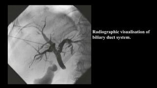
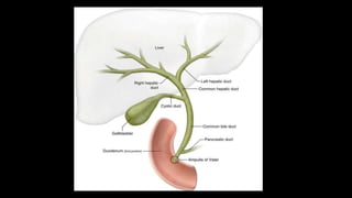
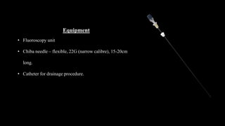
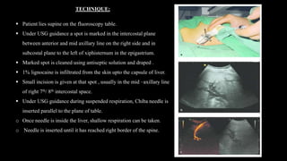
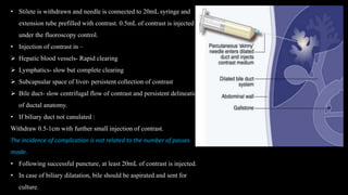
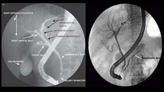
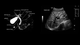
PERCUTANEOUS TRANSHEPATIC
CHOLANGIOGRAPHY (PTC)
Radiographic visualisation of
biliary duct system.
Indications
Diagnostic:
• Prior to therapeutic intervention- biliary
drainage procedure/ stenting
• Distinguish intrahepatic cholestasis and
extrahepatic obstruction ( calculi, stricture,
malignancy).
• Biliary diseases.
Therapeutic:
• Place a percutaneous biliary stent
• Drain infected bile
Contraindications
• Bleeding diathesis :
Platelet count < 1,00,000 /mm3 and
Prothrombin time <60% of control value.
• Biliary sepsis [need of appropriate
antibiotic cover, small volume contrast,
establish drainage]
• Contrast hypersensitivity
• Severe cardiovascular and respiratory
compromise
• Severe jaundice, ascites anemia and poor
general condition of the patient.
Patient Preparation
Preliminary Investigation:
• Ultrasound / CT – assessing the biliary anatomy and cause and level of
obstruction.
• HIV and HBsAg testing
• Check Haemoglobin, platelet count and prothrombin time.
Patient Preparation:
• Informed consent
• Maintain hydration
• NPO for 4hours
• Prophylactic antibiotics
Equipment
• Fluoroscopy unit
• Chiba needle – flexible, 22G (narrow calibre), 15-20cm
long.
• Catheter for drainage procedure.
TECHNIQUE:
 Patient lies supine on the fluoroscopy table.
 Under USG guidance a spot is marked in the intercostal plane
between anterior and mid axillary line on the right side and in
subcostal plane to the left of xiphisternum in the epigastrium.
 Marked spot is cleaned using antiseptic solution and draped .
 1% lignocaine is infiltrated from the skin upto the capsule of liver.
 Small incision is given at that spot , usually in the mid –axillary line
of right 7th/ 8th intercostal space.
 Under USG guidance during suspended respiration, Chiba needle is
inserted parallel to the plane of table.
o Once needle is inside the liver, shallow respiration can be taken.
o Needle is inserted until it has reached right border of the spine.
• Stilete is withdrawn and needle is connected to 20mL syringe and
extension tube prefilled with contrast. 0.5mL of contrast is injected
under the fluoroscopy control.
• Injection of contrast in –
 Hepatic blood vessels- Rapid clearing
 Lymphatics- slow but complete clearing
 Subcapsular space of liver- persistent collection of contrast
 Bile duct- slow centrifugal flow of contrast and persistent delineation
of ductal anatomy.
• If biliary duct not canulated :
Withdraw 0.5-1cm with further small injection of contrast.
The incidence of complication is not related to the number of passes
made.
• Following successful puncture, at least 20mL of contrast is injected.
• In case of biliary dilatation, bile should be aspirated and sent for
culture.
Films:
• AP
• Lateral
• Obliques – RAO, LAO
Delayed images taken for –
• Visualisation of Gall bladder
• Site of CBD obstruction
Erect film
• Demonstration for site of obstruction which is actually lower than what is visualised in supine position.
After care:
• Bed rest
• Monitoring Pulse, Blood pressure and temperature for at least 24hours.
Complications:
1. Cholangitis
2. Bile leak in to peritoneum- Biliary peritonitis
3. Haemorrhage
4. Sepsis
Endoscopic Retrograde
Cholangiopancreatography
(ERCP)
Indications:
• Diagnostic in patients unsuitable/intolerant of MRCP.
• Management of bile duct stones, biliary stricture
• Investigation of diffuse biliary disease
• Evaluation of ampullary lesion
Contraindications:
• Pyloric stenosis, Gastric outlet obstruction
• Thoracic aorta aneurysm
• Severe cardiac/ pulmonary disease
• H/o pseudocyst/ acute pancreatitis episode within 4 weeks
Equipment:
• Side viewing endoscope
• Polythene catheters
• Fluoroscopy unit with spot film facility
Patient Preparation
• NPO 6hours prior to procedure
• Antibiotic cover for biliary obstruction, pseudocyst.
Pancreatic duct
Preliminary Film
Prone AP and LAO of upper abdomen – gallstone, pancreatic calcification/ calculi
Technique
• Xylocaine spray to anaesthetize pharynx
• In left lateral position, endoscope is introduced via the mouth reaching upto the duodenum and ampulla of Vater is
located
• Polythene catheter prefilled with contrast is inserted into the ampulla.
• Test dose of contrast medium is injected to check the position of canula
• First canulate the bile duct and then pancreatic duct . Both are opacified with contrast.
• Take sample of bile if e/o biliary obstruction.
Films:
Prone (straight and posterior obliques) and supine (straight, both obliques, Trendeleburg)
After removal of endoscope
Delayed for the gall bladder and emptying of CBD.
Complications:
Damage by the endoscope
Acute Pancreatitis
Sepsis
After Care:
Nil oral until sensation of pharynx has returned
Monitor Pulse, BP, temperature for 6hpurs
Maintain antibiotics in cases of biliary obstruction.
MYELOGRAPHY
Evaluation of subarachnoid
space within the spinal canal.
Indications :
Limited role because of advent of CT/ MR myelography.
Done in cases where CT/ MR cant be performed.
• Sciatica syndrome with suspected nerve root compression by prolapse of intervertebral disc.
• Cervical spondylolysis
• SOL of spinal cord, its nerve and meninges
Contraindications:
• Skin sepsis over the site of spinal puncture site
• Infection of subarachnoid space
• Contrast hypersensitivity
• Past H/o myelography procedure within 7 days.
• In epilepsy cases, antiepileptic should precede the study.
Patient Preparation :
• Informed Consent
• NPO 3 hours
• Maintain Hydration
• Remove the dentures.
• No sedatives , if necessary diazepam 5mg can be given oral /iv
• Xray LS spine – frontal and lateral view
22 G, 9cm needle is recommended
Site:
L3/L4 intervertebral space
Contrast medium : Iohexol , Iopamidol
Procedure:
• Aspesis maintained
• Lumbar Puncture done
• Injection of contrast medium smoothly over 30-60s.
• Jerky or rapid movement of patient avoided
• Radiography should be started within few minutes of contrast injection
Radiographic Projections:
1. Prone with foot end of table tilted 45⁰ down.
2. Lateral view of lower lumbar spine and LS junction
3. Tilt the table horizontal to obtain view of mid and upper lumbar spine- frontal,obliuq and lateral views.
4. Head down tilt of 5-10⁰ to show lower dorsal subarachnoid space- frontal, oblique and lateral views.
After Care:
1. The patient must be well hydrated.
2. Patient should be allowed to remain ambulant.
bsc.pptx
bsc.pptx

bsc.pptx

  • 1.
  • 2.
  • 4.
    Indications Diagnostic: • Prior totherapeutic intervention- biliary drainage procedure/ stenting • Distinguish intrahepatic cholestasis and extrahepatic obstruction ( calculi, stricture, malignancy). • Biliary diseases. Therapeutic: • Place a percutaneous biliary stent • Drain infected bile Contraindications • Bleeding diathesis : Platelet count < 1,00,000 /mm3 and Prothrombin time <60% of control value. • Biliary sepsis [need of appropriate antibiotic cover, small volume contrast, establish drainage] • Contrast hypersensitivity • Severe cardiovascular and respiratory compromise • Severe jaundice, ascites anemia and poor general condition of the patient.
  • 5.
    Patient Preparation Preliminary Investigation: •Ultrasound / CT – assessing the biliary anatomy and cause and level of obstruction. • HIV and HBsAg testing • Check Haemoglobin, platelet count and prothrombin time. Patient Preparation: • Informed consent • Maintain hydration • NPO for 4hours • Prophylactic antibiotics
  • 6.
    Equipment • Fluoroscopy unit •Chiba needle – flexible, 22G (narrow calibre), 15-20cm long. • Catheter for drainage procedure.
  • 7.
    TECHNIQUE:  Patient liessupine on the fluoroscopy table.  Under USG guidance a spot is marked in the intercostal plane between anterior and mid axillary line on the right side and in subcostal plane to the left of xiphisternum in the epigastrium.  Marked spot is cleaned using antiseptic solution and draped .  1% lignocaine is infiltrated from the skin upto the capsule of liver.  Small incision is given at that spot , usually in the mid –axillary line of right 7th/ 8th intercostal space.  Under USG guidance during suspended respiration, Chiba needle is inserted parallel to the plane of table. o Once needle is inside the liver, shallow respiration can be taken. o Needle is inserted until it has reached right border of the spine.
  • 8.
    • Stilete iswithdrawn and needle is connected to 20mL syringe and extension tube prefilled with contrast. 0.5mL of contrast is injected under the fluoroscopy control. • Injection of contrast in –  Hepatic blood vessels- Rapid clearing  Lymphatics- slow but complete clearing  Subcapsular space of liver- persistent collection of contrast  Bile duct- slow centrifugal flow of contrast and persistent delineation of ductal anatomy. • If biliary duct not canulated : Withdraw 0.5-1cm with further small injection of contrast. The incidence of complication is not related to the number of passes made. • Following successful puncture, at least 20mL of contrast is injected. • In case of biliary dilatation, bile should be aspirated and sent for culture.
  • 9.
    Films: • AP • Lateral •Obliques – RAO, LAO Delayed images taken for – • Visualisation of Gall bladder • Site of CBD obstruction Erect film • Demonstration for site of obstruction which is actually lower than what is visualised in supine position.
  • 10.
    After care: • Bedrest • Monitoring Pulse, Blood pressure and temperature for at least 24hours. Complications: 1. Cholangitis 2. Bile leak in to peritoneum- Biliary peritonitis 3. Haemorrhage 4. Sepsis
  • 11.
  • 12.
    Indications: • Diagnostic inpatients unsuitable/intolerant of MRCP. • Management of bile duct stones, biliary stricture • Investigation of diffuse biliary disease • Evaluation of ampullary lesion Contraindications: • Pyloric stenosis, Gastric outlet obstruction • Thoracic aorta aneurysm • Severe cardiac/ pulmonary disease • H/o pseudocyst/ acute pancreatitis episode within 4 weeks Equipment: • Side viewing endoscope • Polythene catheters • Fluoroscopy unit with spot film facility Patient Preparation • NPO 6hours prior to procedure • Antibiotic cover for biliary obstruction, pseudocyst.
  • 14.
  • 15.
    Preliminary Film Prone APand LAO of upper abdomen – gallstone, pancreatic calcification/ calculi Technique • Xylocaine spray to anaesthetize pharynx • In left lateral position, endoscope is introduced via the mouth reaching upto the duodenum and ampulla of Vater is located • Polythene catheter prefilled with contrast is inserted into the ampulla. • Test dose of contrast medium is injected to check the position of canula • First canulate the bile duct and then pancreatic duct . Both are opacified with contrast. • Take sample of bile if e/o biliary obstruction. Films: Prone (straight and posterior obliques) and supine (straight, both obliques, Trendeleburg) After removal of endoscope Delayed for the gall bladder and emptying of CBD.
  • 16.
    Complications: Damage by theendoscope Acute Pancreatitis Sepsis After Care: Nil oral until sensation of pharynx has returned Monitor Pulse, BP, temperature for 6hpurs Maintain antibiotics in cases of biliary obstruction.
  • 18.
  • 19.
    Evaluation of subarachnoid spacewithin the spinal canal.
  • 20.
    Indications : Limited rolebecause of advent of CT/ MR myelography. Done in cases where CT/ MR cant be performed. • Sciatica syndrome with suspected nerve root compression by prolapse of intervertebral disc. • Cervical spondylolysis • SOL of spinal cord, its nerve and meninges Contraindications: • Skin sepsis over the site of spinal puncture site • Infection of subarachnoid space • Contrast hypersensitivity • Past H/o myelography procedure within 7 days. • In epilepsy cases, antiepileptic should precede the study.
  • 21.
    Patient Preparation : •Informed Consent • NPO 3 hours • Maintain Hydration • Remove the dentures. • No sedatives , if necessary diazepam 5mg can be given oral /iv • Xray LS spine – frontal and lateral view 22 G, 9cm needle is recommended Site: L3/L4 intervertebral space
  • 23.
    Contrast medium :Iohexol , Iopamidol Procedure: • Aspesis maintained • Lumbar Puncture done • Injection of contrast medium smoothly over 30-60s. • Jerky or rapid movement of patient avoided • Radiography should be started within few minutes of contrast injection Radiographic Projections: 1. Prone with foot end of table tilted 45⁰ down. 2. Lateral view of lower lumbar spine and LS junction 3. Tilt the table horizontal to obtain view of mid and upper lumbar spine- frontal,obliuq and lateral views. 4. Head down tilt of 5-10⁰ to show lower dorsal subarachnoid space- frontal, oblique and lateral views.
  • 25.
    After Care: 1. Thepatient must be well hydrated. 2. Patient should be allowed to remain ambulant.