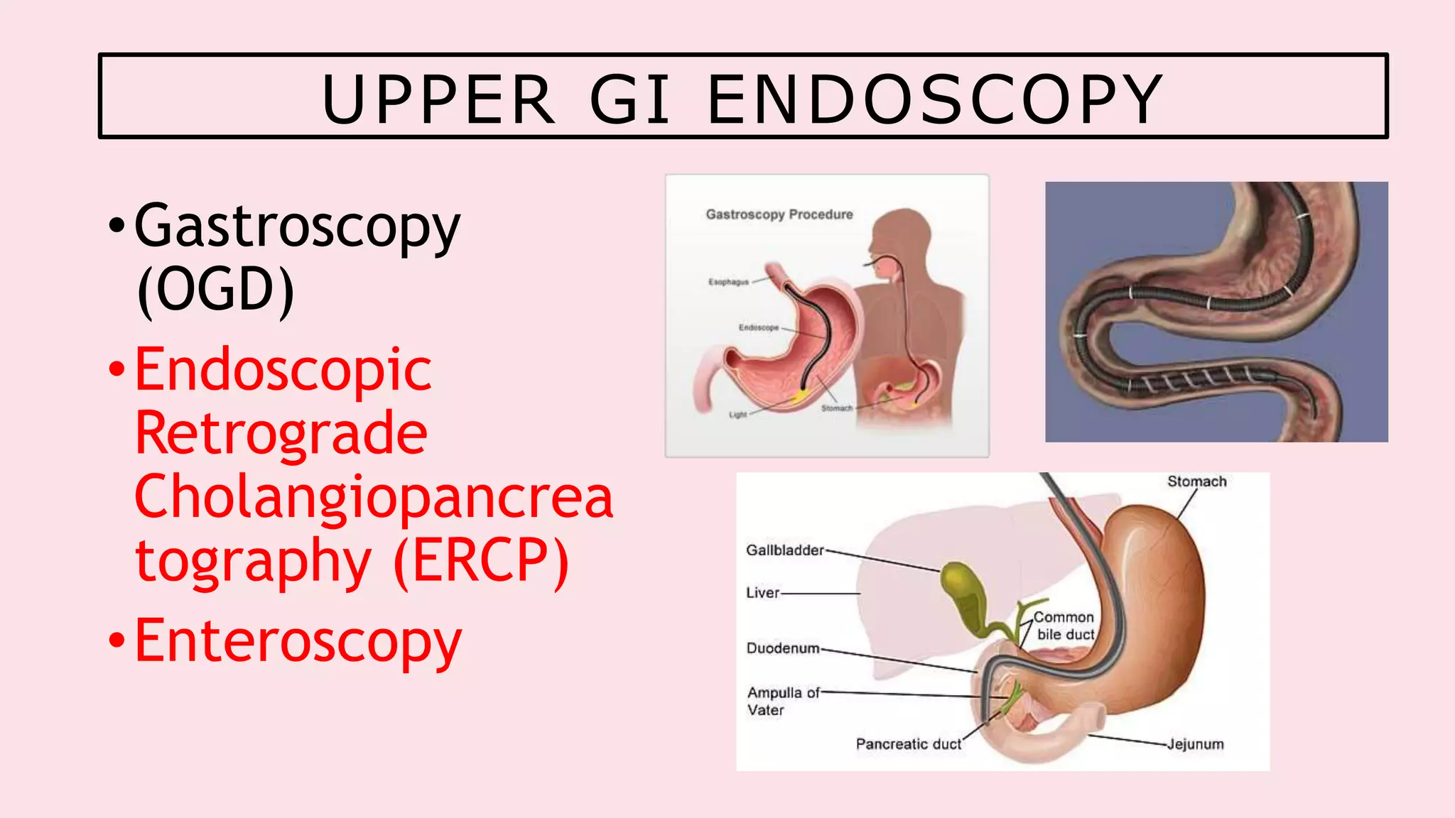
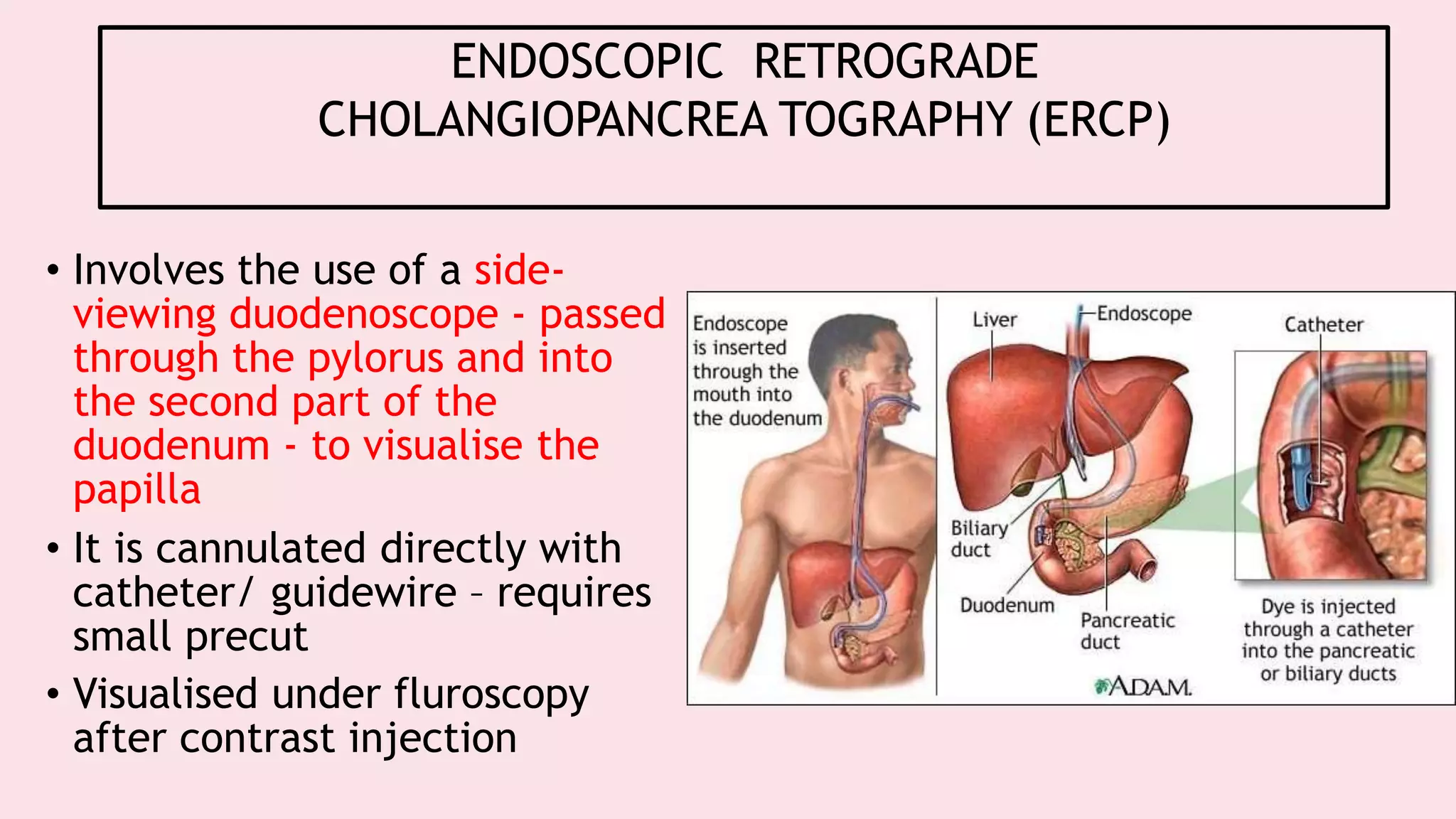
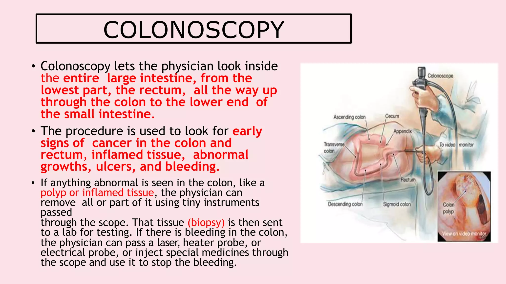
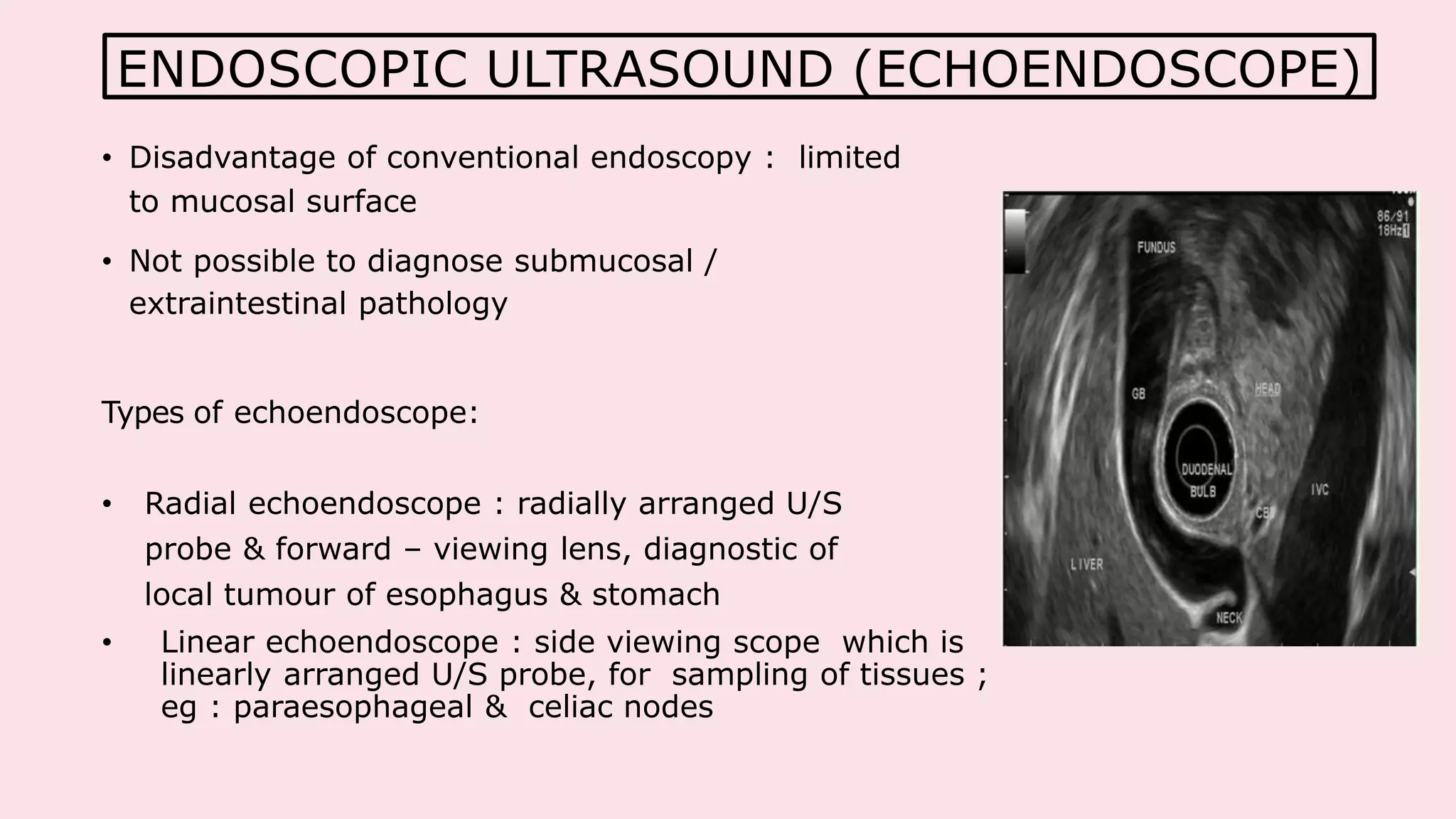
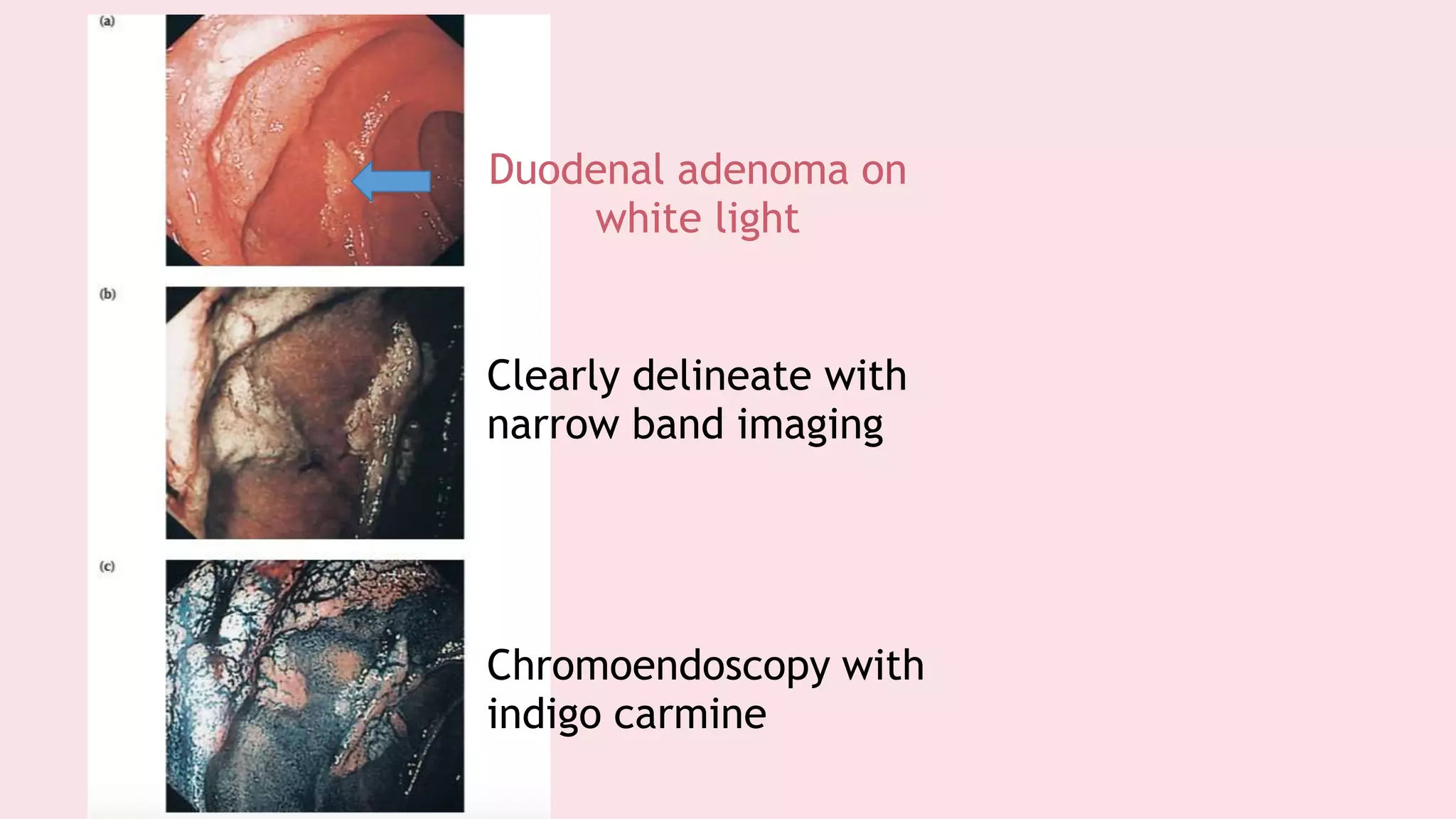
Endoscopy allows examination of body cavities using an endoscope. This document discusses various types of endoscopy including upper GI endoscopy (gastroscopy, ERCP), lower GI endoscopy (colonoscopy, sigmoidoscopy), and therapeutic endoscopy procedures. It describes the anatomy of the digestive tract, techniques, indications, and potential complications of different endoscopic procedures. New imaging technologies such as chromoendoscopy, narrow band imaging, and magnification endoscopy are also mentioned to improve tissue characterization during endoscopy.
Safe Laparoscopic Cholecystectomy Techniques that are discussed here are based on current literature and Evidence Based Medicine guidelines and reviews.
It has not changed the nature of disease
The basic principles of good surgery still apply,including appropriate case selection, excellent exposure,adequate retraction and a high level technical expertise
If a procedure makes no sense with conventional access, it will make no sense with a minimal access approach
The cleaner and gentler the act of operation, the less the patient suffers, the smoother and quicker his convalescence,the more exquisite his healed wound.
We actually do not know what is there stored for us, but we believe that laparoscopy is trending towards advancement and nano and robotic technology is going to replace in future.
3D cameras have come into existence and various newer technologies are being invented.
Brief description of hepatectomy with indications, procedure, pre operative, intra operative and post operative management of the patient. Also describes the various techniques and instrument available for liver resection.
Endoscopy: Types, Preparation, Diagnosis, Procedure and RisksYashodaHospitals
An endoscopy is a diagnostic tool. Find out what it is used for, what happens during an endoscopy, various types of procedure available and endoscopy risks.
ENDOSCOPY IN MEDICAL SPHERE HAS AN UTMOST IMPORTANCE . BE IT DIAGNOSTIC , THERAPEUTIC OR RESEARCH . ITS A BRIEF ATTEMPT TO ACKNOWLEDGE THE BOON OF SCIENCE
Foreign Body Obstruction - Esophagus.pptxVaibhavRamesh
Foreign Body Obstruction of Esophagus - this presentation covers the key aspects of the condition where a foreign body (anything ranging from a blade to a stone) is stuck on the esophagus or the food pipe causing an obstruction of it.
Webinar on Advances in endoscopy - HInduja HospitalHinduja Hospital
The subject of Gastroenterology has advanced in leaps and bounds. The most exciting developments have occurred in the branch of endoscopy. Procedures which could only be performed by long and complex surgeries can now be safely done with an endoscope. Parts of our anatomy which were earlier thought to be in-accessible can now be clearly visualized after swallowing a capsule or with the help of an endoscopic ultrasound.
To know more about some of these fascinating modalities, read on Advances in Endoscopy by our Consultant Gastroenterology, Dr. Tarun Gupta.
Report Back from SGO 2024: What’s the Latest in Cervical Cancer?bkling
Are you curious about what’s new in cervical cancer research or unsure what the findings mean? Join Dr. Emily Ko, a gynecologic oncologist at Penn Medicine, to learn about the latest updates from the Society of Gynecologic Oncology (SGO) 2024 Annual Meeting on Women’s Cancer. Dr. Ko will discuss what the research presented at the conference means for you and answer your questions about the new developments.
Pulmonary Thromboembolism - etilogy, types, medical- Surgical and nursing man...VarunMahajani
Disruption of blood supply to lung alveoli due to blockage of one or more pulmonary blood vessels is called as Pulmonary thromboembolism. In this presentation we will discuss its causes, types and its management in depth.
Title: Sense of Taste
Presenter: Dr. Faiza, Assistant Professor of Physiology
Qualifications:
MBBS (Best Graduate, AIMC Lahore)
FCPS Physiology
ICMT, CHPE, DHPE (STMU)
MPH (GC University, Faisalabad)
MBA (Virtual University of Pakistan)
Learning Objectives:
Describe the structure and function of taste buds.
Describe the relationship between the taste threshold and taste index of common substances.
Explain the chemical basis and signal transduction of taste perception for each type of primary taste sensation.
Recognize different abnormalities of taste perception and their causes.
Key Topics:
Significance of Taste Sensation:
Differentiation between pleasant and harmful food
Influence on behavior
Selection of food based on metabolic needs
Receptors of Taste:
Taste buds on the tongue
Influence of sense of smell, texture of food, and pain stimulation (e.g., by pepper)
Primary and Secondary Taste Sensations:
Primary taste sensations: Sweet, Sour, Salty, Bitter, Umami
Chemical basis and signal transduction mechanisms for each taste
Taste Threshold and Index:
Taste threshold values for Sweet (sucrose), Salty (NaCl), Sour (HCl), and Bitter (Quinine)
Taste index relationship: Inversely proportional to taste threshold
Taste Blindness:
Inability to taste certain substances, particularly thiourea compounds
Example: Phenylthiocarbamide
Structure and Function of Taste Buds:
Composition: Epithelial cells, Sustentacular/Supporting cells, Taste cells, Basal cells
Features: Taste pores, Taste hairs/microvilli, and Taste nerve fibers
Location of Taste Buds:
Found in papillae of the tongue (Fungiform, Circumvallate, Foliate)
Also present on the palate, tonsillar pillars, epiglottis, and proximal esophagus
Mechanism of Taste Stimulation:
Interaction of taste substances with receptors on microvilli
Signal transduction pathways for Umami, Sweet, Bitter, Sour, and Salty tastes
Taste Sensitivity and Adaptation:
Decrease in sensitivity with age
Rapid adaptation of taste sensation
Role of Saliva in Taste:
Dissolution of tastants to reach receptors
Washing away the stimulus
Taste Preferences and Aversions:
Mechanisms behind taste preference and aversion
Influence of receptors and neural pathways
Impact of Sensory Nerve Damage:
Degeneration of taste buds if the sensory nerve fiber is cut
Abnormalities of Taste Detection:
Conditions: Ageusia, Hypogeusia, Dysgeusia (parageusia)
Causes: Nerve damage, neurological disorders, infections, poor oral hygiene, adverse drug effects, deficiencies, aging, tobacco use, altered neurotransmitter levels
Neurotransmitters and Taste Threshold:
Effects of serotonin (5-HT) and norepinephrine (NE) on taste sensitivity
Supertasters:
25% of the population with heightened sensitivity to taste, especially bitterness
Increased number of fungiform papillae
Ethanol (CH3CH2OH), or beverage alcohol, is a two-carbon alcohol
that is rapidly distributed in the body and brain. Ethanol alters many
neurochemical systems and has rewarding and addictive properties. It
is the oldest recreational drug and likely contributes to more morbidity,
mortality, and public health costs than all illicit drugs combined. The
5th edition of the Diagnostic and Statistical Manual of Mental Disorders
(DSM-5) integrates alcohol abuse and alcohol dependence into a single
disorder called alcohol use disorder (AUD), with mild, moderate,
and severe subclassifications (American Psychiatric Association, 2013).
In the DSM-5, all types of substance abuse and dependence have been
combined into a single substance use disorder (SUD) on a continuum
from mild to severe. A diagnosis of AUD requires that at least two of
the 11 DSM-5 behaviors be present within a 12-month period (mild
AUD: 2–3 criteria; moderate AUD: 4–5 criteria; severe AUD: 6–11 criteria).
The four main behavioral effects of AUD are impaired control over
drinking, negative social consequences, risky use, and altered physiological
effects (tolerance, withdrawal). This chapter presents an overview
of the prevalence and harmful consequences of AUD in the U.S.,
the systemic nature of the disease, neurocircuitry and stages of AUD,
comorbidities, fetal alcohol spectrum disorders, genetic risk factors, and
pharmacotherapies for AUD.
The prostate is an exocrine gland of the male mammalian reproductive system
It is a walnut-sized gland that forms part of the male reproductive system and is located in front of the rectum and just below the urinary bladder
Function is to store and secrete a clear, slightly alkaline fluid that constitutes 10-30% of the volume of the seminal fluid that along with the spermatozoa, constitutes semen
A healthy human prostate measures (4cm-vertical, by 3cm-horizontal, 2cm ant-post ).
It surrounds the urethra just below the urinary bladder. It has anterior, median, posterior and two lateral lobes
It’s work is regulated by androgens which are responsible for male sex characteristics
Generalised disease of the prostate due to hormonal derangement which leads to non malignant enlargement of the gland (increase in the number of epithelial cells and stromal tissue)to cause compression of the urethra leading to symptoms (LUTS
Explore natural remedies for syphilis treatment in Singapore. Discover alternative therapies, herbal remedies, and lifestyle changes that may complement conventional treatments. Learn about holistic approaches to managing syphilis symptoms and supporting overall health.
TEST BANK for Operations Management, 14th Edition by William J. Stevenson, Ve...kevinkariuki227
TEST BANK for Operations Management, 14th Edition by William J. Stevenson, Verified Chapters 1 - 19, Complete Newest Version.pdf
Prix Galien International 2024 Forum ProgramLevi Shapiro
June 20, 2024, Prix Galien International and Jerusalem Ethics Forum in ROME. Detailed agenda including panels:
- ADVANCES IN CARDIOLOGY: A NEW PARADIGM IS COMING
- WOMEN’S HEALTH: FERTILITY PRESERVATION
- WHAT’S NEW IN THE TREATMENT OF INFECTIOUS,
ONCOLOGICAL AND INFLAMMATORY SKIN DISEASES?
- ARTIFICIAL INTELLIGENCE AND ETHICS
- GENE THERAPY
- BEYOND BORDERS: GLOBAL INITIATIVES FOR DEMOCRATIZING LIFE SCIENCE TECHNOLOGIES AND PROMOTING ACCESS TO HEALTHCARE
- ETHICAL CHALLENGES IN LIFE SCIENCES
- Prix Galien International Awards Ceremony
These lecture slides, by Dr Sidra Arshad, offer a quick overview of physiological basis of a normal electrocardiogram.
Learning objectives:
1. Define an electrocardiogram (ECG) and electrocardiography
2. Describe how dipoles generated by the heart produce the waveforms of the ECG
3. Describe the components of a normal electrocardiogram of a typical bipolar leads (limb II)
4. Differentiate between intervals and segments
5. Enlist some common indications for obtaining an ECG
Study Resources:
1. Chapter 11, Guyton and Hall Textbook of Medical Physiology, 14th edition
2. Chapter 9, Human Physiology - From Cells to Systems, Lauralee Sherwood, 9th edition
3. Chapter 29, Ganong’s Review of Medical Physiology, 26th edition
4. Electrocardiogram, StatPearls - https://www.ncbi.nlm.nih.gov/books/NBK549803/
5. ECG in Medical Practice by ABM Abdullah, 4th edition
6. ECG Basics, http://www.nataliescasebook.com/tag/e-c-g-basics
2. LEARNING OBJECTIVES
At the end of this seminar, we should be able to
understand :
• The definition of endoscopy and endoscope
• Upper & lower GI endoscopy
• The indication for diagnostic and therapeutic endoscopy/ colonoscopy/
endoscopic retrograde cholangiopancreatography
• The recognition and management of complications
• Briefly on other types of endoscopy,
• Endoscopic ultrasound
• Respiratory
• Cystoscopy
3. • Endoscopy Greek Word ‘Endo’ = Inside ‘scopy ‘= to see
ENDOSCOPY
Examination of the interior of a canal or hollow viscus by means
of a special instrument, such as an endoscope
ENDOSCOPE
Device using fiber optics and powerful lens systems to provide
lighting and visualization of the interior of cavity. The portion of
the endoscope inserted into the body may be rigid or flexible,
depending upon the medical procedure.
INTRODUCTION
4. The digestive tract consists of the followings :
• Mouth
• Throat
• Esophagus
• Stomach
• Duodenum
• Small bowel
• Colon
• Rectum
• Anus
• And other GI organs .
INTRODUCTION
6. • Involves the use of a side-
viewing duodenoscope - passed
through the pylorus and into
the second part of the
duodenum - to visualise the
papilla
• It is cannulated directly with
catheter/ guidewire – requires
small precut
• Visualised under fluroscopy
after contrast injection
ENDOSCOPIC RETROGRADE
CHOLANGIOPANCREA TOGRAPHY (ERCP)
9. Indication :
•Relief biliary obstruction – gallstone & biliary stricture
If there is delay in reliefing – require percutaneous drainage
Adequate biliary sphincterotomy normally performed over well positioned guidewire
Gallstones <1 cm will pass spontaneously in days & weeks
Most endoscopists prefer to ensure duct clearance at initial procedure to reduce
risk of impaction, cholangitis / pancreatitis
Trawling of duct using balloon catheter / extraction using wire basket
If standard techniques fail – mechanical lithotripsy is done – placement of
removable plastic stent
Correct stent placement confirmed by flow of bile after release & presence of air in
biliary tree on follow up radiographs
10.
11. Other indications :
• Pancreatic disease
• Pancreatic stone extraction
• Dilatation of pancreatic duct strictures
• Transgastric drainage of pancreatic pseudocysts
• Assessment of biliary dysmotility (sphincter of Oddi
dysfunction)
THERAPEUTIC ERCP
12. • Duodenal perforation (1.3%)/hemorrhage after scope insertion/
sphincterotomy (1.4%)
• Pancreatitis (4.3%)
• Sepsis (3-30%)
• Mortality (1%)
Post sphincterotomy complications :
• CT scan required in patients with pain, tachycardia / hypotension
postprocedure
• Can be severe – extensive pancreatic necrosis
• Recommended to administered per-rectal indomethacin/diclofenac
immediately before / after procedure – to decrease post ERCP pancreatitis
COMPLICATIONS OF ERCP
13. RISK FACTORS FOR POST – ERCP PANCREATITIS
Definite
Suspected SOD
Young age
Normal bilirubin
Prior ERCP – related pancreatitis
Difficult cannulation
Pancreatic duct
contrast injection
Balloon dilatation of biliary
sphincter
Possible
Female
Low volume of ERCP performed
Absent CBD stone
14. ENDOSCOPIC ASSESSMENT OF SMALL BOWEL
(ENTEROSCOPE )
Indications :
• GI blood loss –with recurrent iron deficiency anemia (occult
hemorrhage)/recurrent overt blood loss per rectum (cryptic
hemorrhage) in pt with normal OGD (with duodenal biopsies) &
colonoscopy
• Malabsorption
• Exclusion of cryptic IBD- Crohn disease
• Targeting lesions in imaging
• Inherited polyposis syndrome
15. TECHNIQUE
1) STANDARD ENTEROSCOPE – may reach in proximal small
bowel
• Requires 45 minutes
• May be uncomfortable – requires sedation
• Long thin endoscope inserted transnasally into stomach &
pushed through pylorus with gastroscope passed through mouth
• Carried distally by peristalsis propels balloon inflated at tip
• Limitations – long examination time (6-8 hours), discomfort to
patient, danger of perforation
16.
17. • If area of interest was outside reach of standard
enteroscope , direct access via enterotomy
• Advance technique of small bowel assessment :
• Capsule endoscope allows diagnostic mucosal view of entire
small bowel
• Single/double-balloon enteroscopy – endoscopic access to
entire small bowel for biopsy & therapeutics
20. • Provides good visualisation from mouth to colon with
high diagnostic yield
• ‘Gold standard’ for GI bleeding
• Contraindications :
• Small bowel strictures – may cause acute obstruction
requiring retrieval at laparotomy / via laparoscopy
• Gastroparesis
• Pseudo-obstruction
21. 3) SINGLE/DOUBLE-BALLOON ENTEROSCOPY
• Direct visualisation & therapeutic intervention for entire small
bowel via oral / rectal route
• Developed in Japan 2001 – thin enteroscope & overtube both
fitted with a balloon
22.
23. Technique Advantages Disadvantages
Traditional enteroscopy • Simple technique with wide
availability
• Full range of therapeutics
available
• Performed under sedation
• Some discomfort
• Can only access proximal small
bowel
Capsule endoscopy • Able to visualise the entire
small
• Preferable
• No sedation
• Painless
• No biopsies
• Not controllable & no accurate
localisation
• Variable transit
• Incomplete studies due to
battery life
• Not suitable for patients with
strictures
• Large capsule to swallow
Double/single-balloon
enteroscopy
• Able to visualise the entire
small bowel
• Full range of therapeutic
• Requires sedation/general
anaesthesia
• Patient discomfort
• May take 3-4 hours, require
admission
• Complications - perforation
25. COLONOSCOPY
• Colonoscopy lets the physician look inside
the entire large intestine, from the
lowest part, the rectum, all the way up
through the colon to the lower end of
the small intestine.
• The procedure is used to look for early
signs of cancer in the colon and
rectum, inflamed tissue, abnormal
growths, ulcers, and bleeding.
• If anything abnormal is seen in the colon, like a
polyp or inflamed tissue, the physician can
remove all or part of it using tiny instruments
passed
through the scope. That tissue (biopsy) is then sent
to a lab for testing. If there is bleeding in the colon,
the physician can pass a laser, heater probe, or
electrical probe, or inject special medicines through
the scope and use it to stop the bleeding.
26. INDICATIONS OF COLONOSCOPY
• Rectal bleeding with looser/more frequent +/- abdominal pain
related to bowel action
• Iron deficiency anemia (after biochemical confirmation +/-
negative coeliac serology) : oesophagogastroduodenoscopy &
colonoscopy
• Right iliac fossa mass if U/S suggest colonic origin
• Change in bowel habit associated with fever/elevated
inflammatory response
• Chronic diarrhoea (6 weeks) after sigmoidoscopy/rectal
biopsy & negative coeliac serology
• Follow-up of colorectal cancer & polyps
• Screening of patients with family history of colorectal cancer
• Assessment/removal of a lesion seen in radiological examination
• Assessment of ulcerative colitis/Crohn extent & activity
• Surveillance of IBD
• Surveillance of acromegaly/ureterosigmoidostomy
27.
28. COMPLICATIONS OF COLONOSCOPY
Absolute :
Suspected perforation of intestine
Signs of peritonitis in toxic patient
Relative :
Severe acute colitis
Gross acute gastrointestinal bleeding
Poor bowel preparation
Recent surgical anastomosis
Partial or complete intestinal obstruction
Abdominal or iliac aneurysm
29. SIGMOIDOSCOPY
• To look at the inside of the large intestine from the rectum
through the last part of the colon : sigmoid or
descending colon
Indications :
• To find the cause of diarrhea, abdominal pain, or constipation
• To look for early signs of cancer in the descending colon and
rectum. With flexible sigmoidoscopy, the physician can see
bleeding, inflammation, abnormal growths, and ulcers in
the descending colon and rectum.
• Flexible sigmoidoscopy is not sufficient to detect polyps or
cancer in the ascending or transverse colon two-thirds of the
colon
30.
31.
32. ENDOSCOPIC ULTRASOUND (ECHOENDOSCOPE)
• Disadvantage of conventional endoscopy : limited
to mucosal surface
• Not possible to diagnose submucosal /
extraintestinal pathology
Types of echoendoscope:
• Radial echoendoscope : radially arranged U/S
probe & forward – viewing lens, diagnostic of
local tumour of esophagus & stomach
• Linear echoendoscope : side viewing scope which is
linearly arranged U/S probe, for sampling of tissues ;
eg : paraesophageal & celiac nodes
33. INDICATIONS FOR ENDOSCOPIC ULTRASOUND
Diagnostic
Staging of esophageal/gastric malignancy
Staging of hepatobiliary malignancy
Diagnosis of choledochal microlithiasis
Therapeutic
Biopsy of paraesophageal LN
Biopsy of submucosal upper GI lesions
Biopsy of pancreaticobiliary mass
Biopsy of portal lymphadenopathy
Biopsy of left adrenal & left liver massess
Transgastric drainage of pancreatic pseudocyst
Celiac plexus block
34. IMPROVED ENDOSCOPIC IMAGING
Chromoendoscopy, narrow band imaging & high
resolution magnification endoscopy
• Chromoendoscopy – topical application of stains / pigments to
improve tissue localisation, characterisation / diagnosis
• Agents that is used : methylene blue (vital), indigo carmine
(contrast) & India ink (tattooing), acetic acid & Lugol’s iodine
• Narrow band imaging – uses 2 discrete band of lights to
increase contrast image of tissue surface
• High resolution magnification endoscopy – to achieve near
cellular definition of mucosa for surveillance of neoplasia
35. Duodenal adenoma on
white light
Clearly delineate with
narrow band imaging
Chromoendoscopy with
indigo carmine
37. BRONCHOSCOPY
• A bronchoscope is a tube with a tiny
camera on the end which is inserted
through the nose (or mouth) into
the lungs. During a bronchoscopy
procedure, a scope will be inserted
through the nostril until it passes
through the throat into the trachea
and bronchi. A bronchoscope is used
to provide a view of the airways of the
lung. The scope also allows the doctor
to collect lung secretions and lung
tissue for biopsy for tissue
specimens.
39. CYSTOSCOPY
• Cystoscopy is a procedure that
uses a flexible fiber optic scope
inserted through the urethra into
the urinary bladder.
• The bladder is filled with water
and the interior of the bladder is
inspected.
• The image seen through the
cystoscope may also be viewed
on a color monitor and recorded
on videotape for later evaluation.
40. SUMMARY
• The definition of endoscopy and endoscope
• Upper & lower GI endoscopy
• The indication for diagnostic and therapeutic endoscopy/ colonoscopy/
endoscopic retrograde cholangiopancreatography
• The recognition and management of complications
• Briefly on other types of endoscopy
• Endoscopic ultrasound
• Respiratory
• Cystoscopy
41. References
• Bailey & Love’s H. B., & M. L. (2018). Short Practice of
Surgery(27th ed., Vol. 1). Boca Ratonca : CRC Press
• M., S. B. (2016). SRBs Manual of Surgery : Jaypee Brothers
Medical P.