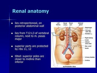
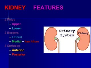
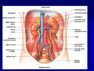
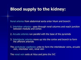
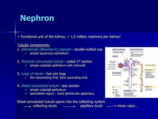
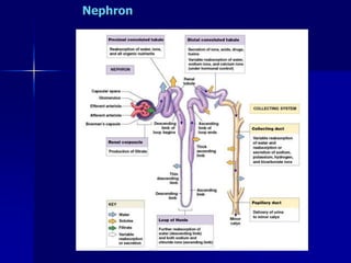
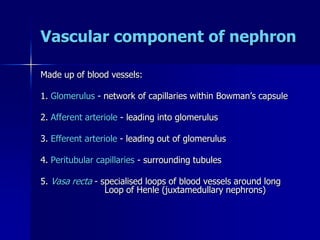
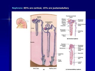
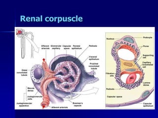
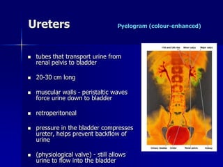
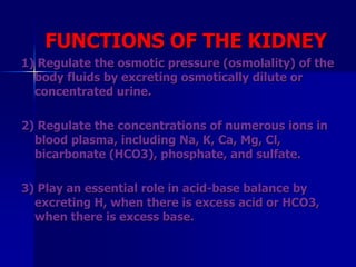
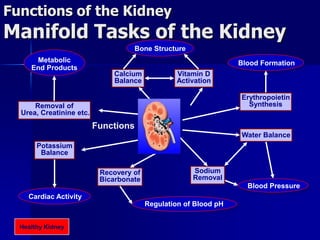
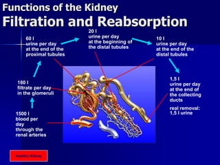
1. Renal System Anatomy
& PHYSIOLOGY
DR. AMAR UL-ALA BUTT
CONSULTANT UROLOGIST
06-R-M-12967 (MOH 88/458)
BURAIDAH CENTRAL HOSPITAL(7/1)
2. Renal system summary
Functions:
1. Remove wastes from the body (urine)
2. Regulates fluid balance, maintains homeostasis
Structures:
2 kidneys - filter blood, produce urine
2 ureters - transport urine (kidneys to bladder)
bladder - reservoir for urine
urethra - transport of urine
3. Renal anatomy
lies retroperitoneal, on
posterior abdominal wall
lies from T12-L3 of vertebral
column, next to m. psoas
major
superior parts are protected
by ribs 11, 12
tilted: superior poles are
closer to midline than
inferior
4. KIDNEY FEATURES
2 Poles
– Upper
– Lower
2 Borders
– Lateral
– Medial – has hilum
2 Surfaces
– Anterior
– Posterior
8. Renal anatomy
R kidney lower than L
(due to R lobe of liver)
~ 12cm long x 5cm
wide x 2.5cm thick
bean-shaped , reddish-
brown organs
renal hilum
kidney levels change
during respiration and
postural changes
9.
10. External surface – 3 layers of tissue
1. renal fascia (outer)
– CT attaches kidney to
posterior abdominal wall –
flexible, allows kidney to
move with respiration
– covered by layer of fat
2. perirenal fat (middle layer)
– protective cushion
3. renal capsule (innermost)
– layer of collagen fibres -
barrier against
trauma, infection etc.
– kidney hangs suspended by
collagen fibres and packed
in soft cushion of adipose
tissue
11. Internal anatomy
outer cortex, inner medulla
6-18 conical renal pyramids
apex - renal papilla projects
into the renal sinus
renal columns
extend from cortex inward
to renal sinus between
adjacent renal pyramids
granular texture similar to
that of the cortex
12. Internal anatomy
ureter enters renal sinus, it
expands to form a chamber
called - renal pelvis
pelvis branches to form 2-3
major calyces
branch further to form 6-8
minor calyces
Each minor calyx surrounds
the papilla of a renal pyramid
ducts within papilla connect to
wall of the calyx and discharge
urine produced in the cortex
and medulla
Urine passes through the
calyces into the ureter
15. Blood supply to the kidney:
Renal arteries from abdominal aorta enter hilum and branch:
1. Interlobar arteries - pass through renal columns and reach junction
between medulla and cortex
2. Arcuate arteries run parallel with the base of the pyramids
3. Interlobular arteries move up into the cortex and branch to form
the afferent arteriole
The peritubular capillaries unite to form the interlobular veins, arcuate
vein, interlobar vein, renal vein
The renal vein exits at hilus and joins the IVC
19. Vascular component of nephron
Made up of blood vessels:
1. Glomerulus - network of capillaries within Bowman’s capsule
2. Afferent arteriole - leading into glomerulus
3. Efferent arteriole - leading out of glomerulus
4. Peritubular capillaries - surrounding tubules
5. Vasa recta - specialised loops of blood vessels around long
Loop of Henle (juxtamedullary nephrons)
22. Ureters Pyelogram (colour-enhanced)
tubes that transport urine from
renal pelvis to bladder
20-30 cm long
muscular walls - peristaltic waves
force urine down to bladder
retroperitoneal
pressure in the bladder compresses
ureter, helps prevent backflow of
urine
(physiological valve) - still allows
urine to flow into the bladder
23. RIGHT URETER, RELATIONS
–Anteriorly
Duodenum
Terminal part of ileum
Right colic & iliocolic vessels
Right gonadal vessels
Root of mesentry
–Posteriorly
Right psoas major muscle
Bifurcation of right common iliac A
24. LEFT URETER, RELATIONS
–Anteriorly
Sigmoid colon
Sigmoid mesocolon
Left colic vessels
Left gonadal vessels
–Posteriorly
Left psoas major muscle
Bifurcation of left common iliac A
25. URETER CONSTRICTIONS
1. Where it joins
the renal pelvis
2. Where it crosses
the pelvic brim
3. Where it pierces
the bladder wall
26. Bladder
hollow muscular organ
retroperitoneal, posterior to
pubic symphysis
Capacity ~ 300-400 ml
(max = 1000 ml)
empty: looks like a deflated
balloon, rugae
full spherical rises above
abdominal cavity
Males: anterior to rectum,
above prostate
Females: inferior to uterus,
anterior to vagina
27. URINARY BLADDER FEATURES
Apex: points anteriorly---median umbilical
lig.
Base: (posterior surface)
Superior surface
2 Inferiolateral surfaces
Neck: rests on prostate
28. Support of bladder
superior surfaces - peritoneum
middle umbilical ligament -
superior border to umbilicus
lateral umbilical ligaments -
sides of bladder to umbilicus
At base, tough ligamentous
bands anchor bladder to pelvic
and pubic bones
Trigone : triangular area bounded
by openings of ureters and exit
to urethra
cystitis - inflammation of the
bladder wall
29. Cross section of ureter & bladder
lined by transitional epithelium to
allow for stretch of wall
lamina propria -
mucous membrane
– elastin fibres
smooth muscle (m. detrusor)
– inner and outer longitudinal
– middle circular
contraction compresses
bladder and expels contents
into urethra
internal sphincter -
involuntary control
30. Urethra
Female ~ 4cm long
– opens to exterior between
clitoris and vaginal opening
Male ~ 20 cm long
– passes through prostate
gland
– pierces urogenital
diaphragm
– enters penis and extends
throughout length
– opens at urethral orifice
31. MALE URETHRA PARTS
Prostatic part:
– The widest part
– About 3 cm
Membranous part:
– In the urogenital diaphragm
– About 1.25 cm
Penile part:
– In the penis
– About 15 – 16 cm long
32. FUNCTIONS OF THE KIDNEY
1) Regulate the osmotic pressure (osmolality) of the
body fluids by excreting osmotically dilute or
concentrated urine.
2) Regulate the concentrations of numerous ions in
blood plasma, including Na, K, Ca, Mg, Cl,
bicarbonate (HCO3), phosphate, and sulfate.
3) Play an essential role in acid-base balance by
excreting H, when there is excess acid or HCO3,
when there is excess base.
33. FUNCTIONS OF THE KIDNEY
4) Regulate the volume of the ECF by controlling Na
and water excretion.
5) Help to regulate arterial blood pressure by adjusting
Na excretion and producing various substances
(e.g.,renin) that can affect blood pressure.
6) Eliminate the waste products of metabolism,
including urea (the main nitrogen-containing end-
product of protein metabolism in humans), uric acid
(an end-product of purine metabolism), and
creatinine (an end-product of muscle metabolism).
34. FUNCTIONS OF THE KIDNEY
7) Remove many drugs (e.g.penicillin) and foreign
or toxic compounds.
8) Are the major production sites of certain
hormones, including erythropoietin and 1,25-
dihydroxy vitamin D3
9) Degrade several polypeptide hormones, including
insulin, glucagon, and parathyroid hormone
10) Synthesize ammonia, which plays a role in acid-
base balance
35. FUNCTIONS OF THE
KIDNEY
11) Synthesize substances that affect renal blood
flow and Na excretion, including arachidonic
acid derivatives (prostaglandins, thromboxane
A2) and kallikrein (a proteolytic enzyme that
results in the production of kinins).
When the kidneys fail, a host of problems
ensue. Dialysis and kidney transplantation
are commonly used treatments for advanced
(end-stage) renal failure
36. Bone Structure
Vitamin D
Activation
Calcium
Balance
Blood Formation
Erythropoietin
Synthesis
Cardiac Activity
Potassium
Balance
Regulation of Blood pH
Recovery of
Bicarbonate
Blood Pressure
Water Balance
Sodium
Removal
Metabolic
End Products
Removal of
Urea, Creatinine etc.
Functions of the Kidney
Manifold Tasks of the Kidney
Functions
Healthy Kidney
37. Functions of the Kidney
Filtration and Reabsorption
1500 l
blood per
day
through the
renal arteries
180 l
filtrate per day
in the glomeruli
60 l
urine per day
at the end of the
proximal tubules
20 l
urine per day
at the beginning of
the distal tubules
10 l
urine per day
at the end of the
distal tubules
1,5 l
urine per day
at the end of
the collecting
ducts
real removal:
1,5 l urine
Healthy Kidney
38. Micturition reflex:
Stretch receptors stimulated when filled to ~
220 ml
afferent fibres to spinal cord
motor neurons to sm in bladder wall
contracts m. detrusor and increases
pressure
need to relax both internal and external
sphincter - external under voluntary
control
if external sphincter does not
relax, internal sphincter remains closed &
sm in bladder wall relaxes again
Once volume exceeds 500 ml, micturition
reflex may generate enough P to open
internal sphincter
leads to reflexive relaxation of external
sphincter