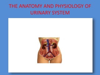
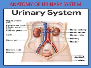
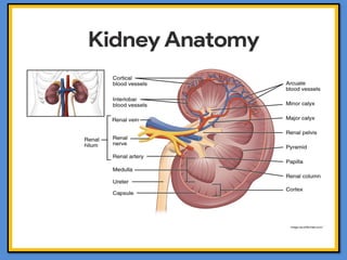
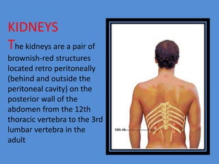
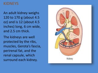
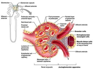
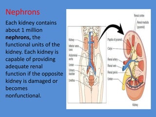
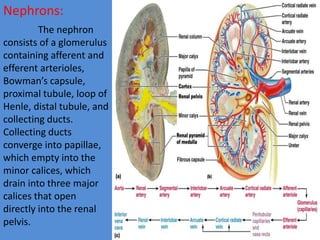
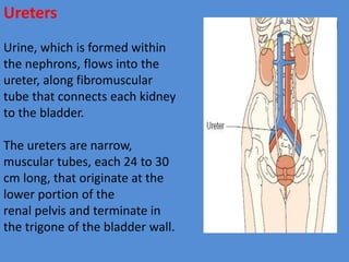
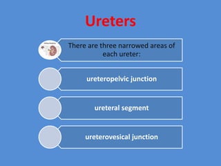
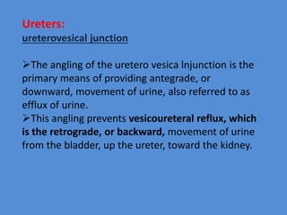
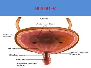
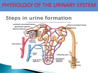
THE
GENITO-URINARY
SYSTEM
G. RAJA
1ST YEAR MSC (NURSING)
MSAJ COLLEGE OF NURSING
THE ANATOMY AND PHYSIOLOGY OF
URINARY SYSTEM
ANATOMY OF URINARY SYSTEM
ANATOMY
The urinary system comprises the
kidneys, ureters , bladder, and
urethra
KIDNEYS
The kidneys are a pair of
brownish-red structures
located retro peritoneally
(behind and outside the
peritoneal cavity) on the
posterior wall of the
abdomen from the 12th
thoracic vertebra to the 3rd
lumbar vertebra in the
adult
KIDNEYS
An adult kidney weighs
120 to 170 g (about 4.5
oz) and is 12 (about 4.5
inches) long, 6 cm wide,
and 2.5 cm thick.
The kidneys are well
protected by the ribs,
muscles, Gerota’s fascia,
perirenal fat, and the
renal capsule, which
surround each kidney.
The kidney consists of two distinct
regions:
Renal Parenchyma
Renal Pelvis
Renal Parenchyma
The renal parenchyma is divided into the cortex and the medulla.
CORTEX:
The cortex contains the
glomeruli , proximal and
distal tubules, and cortical
collecting ducts and their
adjacent peritubular
capillaries.
MEDULLA:
The medulla resembles
conical pyramids. The
pyramids are situated with
the base facing the concave
surface of the kidney and
the apex facing the hilum,
or pelvis
Renal Pelvis
The hilum, or pelvis, is the concave portion of the
kidney through which the renal artery enters and the
renal vein exits. The renal artery (arising from the
abdominal aorta) divides into smaller and smaller
vessels, eventually forming the afferent arteriole.
The afferent arteriole branches to form the
glomerulus, which is the capillary bed responsible for
glomerular filtration. Blood leaves the glomerulus
through the efferent arteriole and flows back to the
inferior vena cava through a network of capillaries and
veins.
KIDNEY:
The glomerular membrane
normally allows filtration of fluid
and small molecules yet limits
passage of larger molecules, such as
blood cells and albumin. Kidney
function begins to decrease at a
rate of approximately 1% each year
beginning at approximately age 30.
The glomerulus is
composed of three
filtering
layers: the capillary
endothelium, the
basement
membrane, and the
epithelium.
Kidneys
• Urine formation
• Excretion of waste products
• Regulation of electrolytes
• Regulation of acid–base balance
• Control of water balance
• Control of blood pressure
• Renal clearance
• Regulation of red blood cell production
• Synthesis of vitamin D to active form
• Secretion of prostaglandins
Nephrons
Each kidney contains
about 1 million
nephrons, the
functional units of the
kidney. Each kidney is
capable of providing
adequate renal
function if the opposite
kidney is damaged or
becomes
nonfunctional.
Nephrons:
The nephron
consists of a glomerulus
containing afferent and
efferent arterioles,
Bowman’s capsule,
proximal tubule, loop of
Henle, distal tubule, and
collecting ducts.
Collecting ducts
converge into papillae,
which empty into the
minor calices, which
drain into three major
calices that open
directly into the renal
pelvis.
Nephrons are struturally divided into two types:
cortical
juxtamedullary
Ureters
Urine, which is formed within
the nephrons, flows into the
ureter, along fibromuscular
tube that connects each kidney
to the bladder.
The ureters are narrow,
muscular tubes, each 24 to 30
cm long, that originate at the
lower portion of the
renal pelvis and terminate in
the trigone of the bladder wall.
Ureters
There are three narrowed areas of
each ureter:
ureteropelvic junction
ureteral segment
ureterovesical junction
Ureters:
ureterovesical junction
The angling of the uretero vesica lnjunction is the
primary means of providing antegrade, or
downward, movement of urine, also referred to as
efflux of urine.
This angling prevents vesicoureteral reflux, which
is the retrograde, or backward, movement of urine
from the bladder, up the ureter, toward the kidney.
During voiding (micturition), increased intravesical
pressure keeps the uretero vesical junction closed
and keeps urine within the ureters. As soon as
micturition is completed, intravesical pressure
returns to its
normal low baseline value, allowing efflux of urine to
resume.
Therefore, the only time that the bladder is
completely empty is in the last seconds of micturition
before efflux of urine resumes.
Did YOU
know?
“The left ureter is slightly
shorter than the right”
Ureters
The lining of the ureters is made up of transitional
cell epithelium called urothelium. As in the
bladder, the urothelium prevents reabsorption of
urine.
The movement of urine from the renal pelves
through the ureters into the bladder is facilitated
by peristaltic waves (occurring about one to five
times per minute) from contraction of the smooth
muscle in the ureter wall (Walsh, Retik, Vaughan &
Wein, (1998).
Ureters
Ureters functions as tubes that actively convey
urine from the kidneys to the bladder.
Size. The ureters are two slender tubes
each 25 to 30 cm (10 to 12 inches) long and 6
mm (1/4 inch) in diameter
BLADDER
Bladder
Adult bladder capacity is about 300 to 600 mL of urine. In infancy, the
bladder is found within the abdomen. In adolescence and through
adulthood, the bladder assumes its position in the true pelvis.
The urinary bladder is a muscular, hollow sac located just behind the
pubic bone.
The bladder is characterized by its central, hollow area called the
vesicle, which has two inlets (the ureters) and one outlet (the
urethrovesical junction), which is surrounded by the bladder neck.
The bladder neck contains bundles of involuntary smooth muscle that
form a portion of the urethral sphincter known as the internal sphincter.
The portion of the sphincteric mechanism that is under voluntary
control is the external urinary sphincter at the anterior urethra, the
segment most distal from the bladder (Walsh et al., 1998).
The urinary bladder functions as a muscular sac that expands as urine is
produced by the kidneys to allow storage of urine until voiding is
convenient
adventitia
detrusor
lamina propria
urothelium
Bladder
The wall of the bladder comprises four layers:
. URETHRA
Urethra
The urethra arises from the base
of the bladder: In the male, it
passes through the penis; in the
female, it opens just anterior to
the vagina.
In the male, the prostate gland,
which lies just below the bladder
neck, surrounds the urethra
posteriorly and laterally.
The urethra is a muscular tube
that drains urine from the body; it
is 3–4 cm long in females, but
closer to 20 cm in males.
Anatomy of the Upper and Lower Urinary
Tracts
FUNCTION:
The urinary system—the structures of which
precisely maintainthe internal chemical
environment of the body—perform various
excretory, regulatory, and secretary functions. A
thorough understanding of the urinary system is
necessary for assessing individuals with acute or
chronic urinary dysfunction and implementing
appropriate nursing care
PHYSIOLOGY OF THE URINARY SYSTEM
Every day, the kidneys filter gallons of the
fluid from the bloodstream. The normal
physiology that takes place in the urinary
system are as follows.
URINE FORMATION
Urine formation is a result of the three processes:
Glomerular filtration. Water and solutes smaller
than proteins are forced through the capillary walls
and pores of the glomerular capsule into the renal
tubule.
Tubular reabsorption. Water, glucose, amino acids,
and needed ions are transported out of the filtrate
into the tubule cells and then enter the capillary
blood..
Tubular secretion. Hydrogen, potassium, creatinine,
and drugs are removed from the peritubular blood
and secreted by the tubule cells into the filtrate.
Characteristics of Urine
In 24 hours, the marvelously complex kidneys filter some 150 to 180 liters of blood
plasma through their glomeruli into the tubules.
Daily volume. In 24 hours, only about 1.0 to 1.8 liters of urine are produced.
Components. Urine contains nitrogenous wastes and unneeded substances.
Color. Freshly voided urine is generally clear and pale to deep yellow.
Odor. When formed, urine is sterile and slightly aromatic, but if allowed to stand, it
takes on an ammonia odor caused by the action of bacteria on the urine solutes.
pH. Urine pH is usually slightly acidic (around 6), but changes in body metabolism and
certain foods may cause it to be much more acidic or basic.
Specific gravity. Whereas the specific gravity of pure water is 1.0, the specific gravity of
urine usually ranges from 1.001 to 1.035.
Solutes. Solutes normally found in urine include sodium and potassium ions, urea, uric
acid, creatinine, ammonia, bicarbonate ions, and various other ions.
Micturition or voiding
Micturition or voiding is the act of emptying the bladder.
Accumulation. Ordinarily, the bladder continues to collect urine until about 200 ml
have accumulated.
Activation. At about this point, stretching of the bladder wall activates stretch
receptors.
Transmission. Impulses transmitted to the sacral region of the spinal cord and then
back to the bladder via the pelvic splanchnic nerves cause the bladder to go into reflex
contractions.
Passage. As the contractions become stronger, stored urine is forced past the internal
urethral sphincter into the upper part of the urethra.
External sphincter. Because the lower external sphincter is skeletal muscle and
voluntarily controlled, we can choose to keep it closed or it can be relaxed so that
urine is flushed from the body
GENITOURINARY
ASSESSMENT
Urinary System - Male and Female
• Frequency of urination
• Amount of urine (large or small)
• Urgency (client’s sense that he or she must void now, cannot wait)
• Dysuria and its timing during voiding (at beginning or end, throughout)
• Nocturia (new onset or increase in usual pattern)
• Urinary Retention
• Incontinence (including urge and stress)
• Change in color and odor of urine
• Presence of stones or sediment in the urine
• Hematuria
• Pain in costovertebral angle, flank or abdomen
• Suprapubic pain
• Perineal, genital, groin or low-back pain
• Painful intercourse
•Sexual history and practices, including risk
behaviours and contraception
(unprotected oral, anal or vaginal
intercourse, multiple partners)
• History of STIs including hepatitis B and
HIV
• Enlarged, painful nodes (axilla, groin)
• Recent antibiotic or steroid use
Male-specific GU related symptoms
• Difficulty in starting or stopping urinary stream
• Voluntary bearing down (straining) to urinate • Nature of stream (speed,
strength, volume)
• Post-void dribbling or post-void fullness
• Discharge from penis, itching
• Lesions on the external genitalia
• Genital, groin, suprapubic or low-back pain
• Testicular pain or swelling
• Torsion of the testes
• Testicular self exam (frequency regularity)
• History of hydrocele, epididymitis, prostatism,varicocele, hernia, undescended
testis, spermatocele, recent vasectomy, erectile dysfunction
Other Associated Symptoms
• Fever, chills, rigors, malaise
• Nausea, vomiting
• Diarrhea, constipation
• Decrease in appetite
• Change in sleep pattern
• Weight loss
Female-specific GU related symptoms
• Urinary symptoms, (pain, burning, malaise, abdominal pain, back pain, fever)
• Urethral discharge
• Menstrual History (menarche, interval, regularity, duration and amount of flow,
dysmenorrhea)
• Date of most recent menstrual period (normal)
• Premenstrual symptoms (swelling, headache, mood swings, pain)
• Abnormal uterine bleeding
• Symptoms of menopause
• Age at menopause
• Postmenopausal bleeding
• Obstetric History: Gravida, Term, Para, Abortion, Live, (GTPAL) difficulties with
pregnancies, deliveries
• Infertility
• Post coital bleeding
• Vaginal discharge (onset, colour, odour, consistency, quantity)
• Sense of pelvic relaxation (pelvic organs feel as though they are falling down or out)
• Lesions or persistent ulcerations to the external genitalia
Medical History (Specific to Genitourinary System)
• Cystitis, pyelonephritis
• Renal disease
• Congenital structural abnormalities in the genitourinary tract
• Renal stones
• Recent onset of or increase in sexual activity
• Recent GU tract instrumentation (e.g., catheter, urethral dilatation, cystoscopy. colposcopy)
• Recent gynaecological procedures
• Menopause (with no hormone replacement therapy)
• Diabetes mellitus
• Immuno-compromise
• Sexually transmitted infections
• Pelvic inflammatory disease
• Human papilloma virus
• Sexual abuse
• Allergies
• Exposure to chemical irritants
• Medications currently used, prescription and over the counter (e.g., immuno-suppressants,
oral contraceptives, antihypertensives,
anti-psychotics)
• Herbal preparations
• Risk behaviours (e.g., unprotected sex, alcohol or drug abuse, use of illicit injection drugs)
Personal and Social History (Specific to Genitourinary System)
• Personal hygiene, toileting habits
• Sexual practices (risk behaviours, sexual orientation)
• Sexual or physical abuse
• Symptomatic sexual partner
• Use of contraceptive creams, foam, condoms, diaphragms etc.
• Use of bubble bath, douches
• Tight-fitting underwear or other clothing
• Multiple sexual partners
• Disruption in sex life (from GU symptoms)
• Smoking (associated risk of bladder cancer)
• Fear, embarrassment, anxiety
• Missing work, school or social functions because of GU symptoms (e.g.,
incontinence)
PHYSICAL ASSESSMENT
General
• Apparent state of health
• Appearance of comfort or distress
• Color (e.g., flushed, pale)
• Nutritional status (emaciated or obese)
• Match between appearance and stated age
Vital Signs
• Temperature
• Heart rate
• Respiratory rate
• Blood pressure
Genitourinary System – Male
Inspection
• Abdominal or flank surgical scars
• Edema (facial, peripheral)
• Penis, scrotum and pubic area: inflammation, discharge, lesions, swelling,
asymmetry, changes in hair distribution, nits, warts
• Rectum: lesions, discharge, swelling, haemorrhoids
• Inguinal and femoral areas (for hernia)
Palpation
• Suprapubic tenderness
• Bladder distension
• Abdominal tenderness or masses
• Costovertebral angle tenderness
• Enlargement of kidney (normal kidneys are usually not palpable unless
client is thin)
• Inguinal and femoral nodes for swellings and hernia
• Superficial inguinal ring (for hernia)
• Penis: tenderness, induration, nodules, lesions
• Testes and scrotal contents: size, position, atrophy of testes, tenderness,
swelling, warmth, masses, hydrocele
• Rectum: anal sphincter tone, rectal wall tumors, prostate gland
• Prostate: size, shape, contour, consistency, tenderness or nodules
Percussion
• Costovertebral angle tenderness
• Bladder distension
Note: Remember to also examine the following areas as
part of your assessment:
• Head, eyes, ears, nose, throat: assess for pharyngitis and
conjunctivitis (chlamydial infection, gonorrhea)
• Skin: assess for skin lesions, rashes, polyarthralgias of
systemic
Genitourinary System - female
Inspection
• External genitalia: labia majora and labia minora: lesions, ulcerations, masses,
induration, and areas of different color, hair distribution
• Perineum: lesions, ulcerations, masses, induration, scars
• Clitoris: size, lesions, ulcerations
• Urethra: discharge, lesions, ulcerations
• Vagina: speculum exam- inflammation, atrophy, discharge, lesions, ulcerations,
excoriation
• Vagina: speculum exam-masses, induration or nodularity, relaxation of
perineum (ask client to bear down and observe for any bulging of vaginal walls)
• Cervix: speculum exam- position, color, shape, size, consistency discharge,
erosions, ulcerations
• Os: multipara or nullipara
Palpation
• Lymph nodes: enlargement, tenderness, mobility and consistency
(supraclavicular, infraclavicular, axilla, epitrochlear, inguinal)
• Skene’s and Bartholin’s glands: masses, discharge, tenderness
• Cervix: cervical tenderness, bleeding after contact, consistency of
cervical tissue (normal cervix is pink and feels firm, like the tip of the
nose; in pregnancy, the cervix is bluish and feels softer, like the lips of
the mouth)
• Uterus: position, size, contour, consistency of uterine tissue, mobility
on movement
• Adnexa: ovaries for tenderness, masses, consistency, contour,
mobility, pain on movement (Chandelier’s sign)
• Anus: lesions, ulcerations, tenderness, fissures, hemorrhoids
Percussion
• Costovertebral angle tenderness
• Bladder distension
CLINICAL REASONING AND CLINICAL JUDGMENT
The first step is to distinguish between GU symptoms requiring urgent
attention and those that can be managed by the certified practice nurse.
The following signs and symptoms require immediate referral to a
physician or nurse practitioner:
• Bleeding from the urethra, male or female
• Urinary retention
• Urethral discharge
• Severe genitourinary pain (consider PID or ectopic pregnancy)
• Scrotal swelling
• Erectile dysfunction (priaprism)
• Systemic symptoms (sepsis)
• Incontinence (new onset)
• Recent urologic/renal surgery
• Immuno-compromised
DIAGNOSTIC TESTS
The certified practice nurse may consider the following
diagnostic tests to support clinical decision-making:
• Urinalysis
• C&S
• Dipstick testing: blood, protein, white blood cells (WBC),
nitrites, pH
• Routine and Microscopic (spun urine): white and red blood
cells, bacteria or casts, epithelial cells
• Culture and sensitivity of urethral discharge or prostatic
secretions
• Pregnancy test
• Sexually transmitted infections
• HIV if required
• Pap
Gentiourinary system ANATOMY AND PHYSIOLOGY & ASSESSMENT

Gentiourinary system ANATOMY AND PHYSIOLOGY & ASSESSMENT

  • 1.
    THE GENITO-URINARY SYSTEM G. RAJA 1ST YEARMSC (NURSING) MSAJ COLLEGE OF NURSING
  • 2.
    THE ANATOMY ANDPHYSIOLOGY OF URINARY SYSTEM
  • 3.
  • 4.
    ANATOMY The urinary systemcomprises the kidneys, ureters , bladder, and urethra
  • 6.
    KIDNEYS The kidneys area pair of brownish-red structures located retro peritoneally (behind and outside the peritoneal cavity) on the posterior wall of the abdomen from the 12th thoracic vertebra to the 3rd lumbar vertebra in the adult
  • 7.
    KIDNEYS An adult kidneyweighs 120 to 170 g (about 4.5 oz) and is 12 (about 4.5 inches) long, 6 cm wide, and 2.5 cm thick. The kidneys are well protected by the ribs, muscles, Gerota’s fascia, perirenal fat, and the renal capsule, which surround each kidney.
  • 8.
    The kidney consistsof two distinct regions: Renal Parenchyma Renal Pelvis
  • 9.
    Renal Parenchyma The renalparenchyma is divided into the cortex and the medulla. CORTEX: The cortex contains the glomeruli , proximal and distal tubules, and cortical collecting ducts and their adjacent peritubular capillaries. MEDULLA: The medulla resembles conical pyramids. The pyramids are situated with the base facing the concave surface of the kidney and the apex facing the hilum, or pelvis
  • 10.
    Renal Pelvis The hilum,or pelvis, is the concave portion of the kidney through which the renal artery enters and the renal vein exits. The renal artery (arising from the abdominal aorta) divides into smaller and smaller vessels, eventually forming the afferent arteriole. The afferent arteriole branches to form the glomerulus, which is the capillary bed responsible for glomerular filtration. Blood leaves the glomerulus through the efferent arteriole and flows back to the inferior vena cava through a network of capillaries and veins.
  • 11.
    KIDNEY: The glomerular membrane normallyallows filtration of fluid and small molecules yet limits passage of larger molecules, such as blood cells and albumin. Kidney function begins to decrease at a rate of approximately 1% each year beginning at approximately age 30.
  • 12.
    The glomerulus is composedof three filtering layers: the capillary endothelium, the basement membrane, and the epithelium.
  • 14.
    Kidneys • Urine formation •Excretion of waste products • Regulation of electrolytes • Regulation of acid–base balance • Control of water balance • Control of blood pressure • Renal clearance • Regulation of red blood cell production • Synthesis of vitamin D to active form • Secretion of prostaglandins
  • 15.
    Nephrons Each kidney contains about1 million nephrons, the functional units of the kidney. Each kidney is capable of providing adequate renal function if the opposite kidney is damaged or becomes nonfunctional.
  • 16.
    Nephrons: The nephron consists ofa glomerulus containing afferent and efferent arterioles, Bowman’s capsule, proximal tubule, loop of Henle, distal tubule, and collecting ducts. Collecting ducts converge into papillae, which empty into the minor calices, which drain into three major calices that open directly into the renal pelvis.
  • 17.
    Nephrons are struturallydivided into two types: cortical juxtamedullary
  • 18.
    Ureters Urine, which isformed within the nephrons, flows into the ureter, along fibromuscular tube that connects each kidney to the bladder. The ureters are narrow, muscular tubes, each 24 to 30 cm long, that originate at the lower portion of the renal pelvis and terminate in the trigone of the bladder wall.
  • 19.
    Ureters There are threenarrowed areas of each ureter: ureteropelvic junction ureteral segment ureterovesical junction
  • 20.
    Ureters: ureterovesical junction The anglingof the uretero vesica lnjunction is the primary means of providing antegrade, or downward, movement of urine, also referred to as efflux of urine. This angling prevents vesicoureteral reflux, which is the retrograde, or backward, movement of urine from the bladder, up the ureter, toward the kidney.
  • 21.
    During voiding (micturition),increased intravesical pressure keeps the uretero vesical junction closed and keeps urine within the ureters. As soon as micturition is completed, intravesical pressure returns to its normal low baseline value, allowing efflux of urine to resume. Therefore, the only time that the bladder is completely empty is in the last seconds of micturition before efflux of urine resumes.
  • 22.
    Did YOU know? “The leftureter is slightly shorter than the right”
  • 23.
    Ureters The lining ofthe ureters is made up of transitional cell epithelium called urothelium. As in the bladder, the urothelium prevents reabsorption of urine. The movement of urine from the renal pelves through the ureters into the bladder is facilitated by peristaltic waves (occurring about one to five times per minute) from contraction of the smooth muscle in the ureter wall (Walsh, Retik, Vaughan & Wein, (1998).
  • 24.
    Ureters Ureters functions astubes that actively convey urine from the kidneys to the bladder. Size. The ureters are two slender tubes each 25 to 30 cm (10 to 12 inches) long and 6 mm (1/4 inch) in diameter
  • 25.
  • 26.
    Bladder Adult bladder capacityis about 300 to 600 mL of urine. In infancy, the bladder is found within the abdomen. In adolescence and through adulthood, the bladder assumes its position in the true pelvis. The urinary bladder is a muscular, hollow sac located just behind the pubic bone. The bladder is characterized by its central, hollow area called the vesicle, which has two inlets (the ureters) and one outlet (the urethrovesical junction), which is surrounded by the bladder neck. The bladder neck contains bundles of involuntary smooth muscle that form a portion of the urethral sphincter known as the internal sphincter. The portion of the sphincteric mechanism that is under voluntary control is the external urinary sphincter at the anterior urethra, the segment most distal from the bladder (Walsh et al., 1998). The urinary bladder functions as a muscular sac that expands as urine is produced by the kidneys to allow storage of urine until voiding is convenient
  • 27.
  • 28.
  • 29.
    Urethra The urethra arisesfrom the base of the bladder: In the male, it passes through the penis; in the female, it opens just anterior to the vagina. In the male, the prostate gland, which lies just below the bladder neck, surrounds the urethra posteriorly and laterally. The urethra is a muscular tube that drains urine from the body; it is 3–4 cm long in females, but closer to 20 cm in males.
  • 30.
    Anatomy of theUpper and Lower Urinary Tracts FUNCTION: The urinary system—the structures of which precisely maintainthe internal chemical environment of the body—perform various excretory, regulatory, and secretary functions. A thorough understanding of the urinary system is necessary for assessing individuals with acute or chronic urinary dysfunction and implementing appropriate nursing care
  • 31.
    PHYSIOLOGY OF THEURINARY SYSTEM
  • 32.
    PHYSIOLOGY OF THEURINARY SYSTEM Every day, the kidneys filter gallons of the fluid from the bloodstream. The normal physiology that takes place in the urinary system are as follows.
  • 33.
    URINE FORMATION Urine formationis a result of the three processes: Glomerular filtration. Water and solutes smaller than proteins are forced through the capillary walls and pores of the glomerular capsule into the renal tubule. Tubular reabsorption. Water, glucose, amino acids, and needed ions are transported out of the filtrate into the tubule cells and then enter the capillary blood.. Tubular secretion. Hydrogen, potassium, creatinine, and drugs are removed from the peritubular blood and secreted by the tubule cells into the filtrate.
  • 34.
    Characteristics of Urine In24 hours, the marvelously complex kidneys filter some 150 to 180 liters of blood plasma through their glomeruli into the tubules. Daily volume. In 24 hours, only about 1.0 to 1.8 liters of urine are produced. Components. Urine contains nitrogenous wastes and unneeded substances. Color. Freshly voided urine is generally clear and pale to deep yellow. Odor. When formed, urine is sterile and slightly aromatic, but if allowed to stand, it takes on an ammonia odor caused by the action of bacteria on the urine solutes. pH. Urine pH is usually slightly acidic (around 6), but changes in body metabolism and certain foods may cause it to be much more acidic or basic. Specific gravity. Whereas the specific gravity of pure water is 1.0, the specific gravity of urine usually ranges from 1.001 to 1.035. Solutes. Solutes normally found in urine include sodium and potassium ions, urea, uric acid, creatinine, ammonia, bicarbonate ions, and various other ions.
  • 35.
    Micturition or voiding Micturitionor voiding is the act of emptying the bladder. Accumulation. Ordinarily, the bladder continues to collect urine until about 200 ml have accumulated. Activation. At about this point, stretching of the bladder wall activates stretch receptors. Transmission. Impulses transmitted to the sacral region of the spinal cord and then back to the bladder via the pelvic splanchnic nerves cause the bladder to go into reflex contractions. Passage. As the contractions become stronger, stored urine is forced past the internal urethral sphincter into the upper part of the urethra. External sphincter. Because the lower external sphincter is skeletal muscle and voluntarily controlled, we can choose to keep it closed or it can be relaxed so that urine is flushed from the body
  • 36.
  • 37.
    Urinary System -Male and Female • Frequency of urination • Amount of urine (large or small) • Urgency (client’s sense that he or she must void now, cannot wait) • Dysuria and its timing during voiding (at beginning or end, throughout) • Nocturia (new onset or increase in usual pattern) • Urinary Retention • Incontinence (including urge and stress) • Change in color and odor of urine • Presence of stones or sediment in the urine • Hematuria • Pain in costovertebral angle, flank or abdomen • Suprapubic pain • Perineal, genital, groin or low-back pain • Painful intercourse
  • 38.
    •Sexual history andpractices, including risk behaviours and contraception (unprotected oral, anal or vaginal intercourse, multiple partners) • History of STIs including hepatitis B and HIV • Enlarged, painful nodes (axilla, groin) • Recent antibiotic or steroid use
  • 39.
    Male-specific GU relatedsymptoms • Difficulty in starting or stopping urinary stream • Voluntary bearing down (straining) to urinate • Nature of stream (speed, strength, volume) • Post-void dribbling or post-void fullness • Discharge from penis, itching • Lesions on the external genitalia • Genital, groin, suprapubic or low-back pain • Testicular pain or swelling • Torsion of the testes • Testicular self exam (frequency regularity) • History of hydrocele, epididymitis, prostatism,varicocele, hernia, undescended testis, spermatocele, recent vasectomy, erectile dysfunction
  • 40.
    Other Associated Symptoms •Fever, chills, rigors, malaise • Nausea, vomiting • Diarrhea, constipation • Decrease in appetite • Change in sleep pattern • Weight loss
  • 41.
    Female-specific GU relatedsymptoms • Urinary symptoms, (pain, burning, malaise, abdominal pain, back pain, fever) • Urethral discharge • Menstrual History (menarche, interval, regularity, duration and amount of flow, dysmenorrhea) • Date of most recent menstrual period (normal) • Premenstrual symptoms (swelling, headache, mood swings, pain) • Abnormal uterine bleeding • Symptoms of menopause • Age at menopause • Postmenopausal bleeding • Obstetric History: Gravida, Term, Para, Abortion, Live, (GTPAL) difficulties with pregnancies, deliveries • Infertility • Post coital bleeding • Vaginal discharge (onset, colour, odour, consistency, quantity) • Sense of pelvic relaxation (pelvic organs feel as though they are falling down or out) • Lesions or persistent ulcerations to the external genitalia
  • 42.
    Medical History (Specificto Genitourinary System) • Cystitis, pyelonephritis • Renal disease • Congenital structural abnormalities in the genitourinary tract • Renal stones • Recent onset of or increase in sexual activity • Recent GU tract instrumentation (e.g., catheter, urethral dilatation, cystoscopy. colposcopy) • Recent gynaecological procedures • Menopause (with no hormone replacement therapy) • Diabetes mellitus • Immuno-compromise • Sexually transmitted infections • Pelvic inflammatory disease • Human papilloma virus • Sexual abuse • Allergies • Exposure to chemical irritants • Medications currently used, prescription and over the counter (e.g., immuno-suppressants, oral contraceptives, antihypertensives, anti-psychotics) • Herbal preparations • Risk behaviours (e.g., unprotected sex, alcohol or drug abuse, use of illicit injection drugs)
  • 43.
    Personal and SocialHistory (Specific to Genitourinary System) • Personal hygiene, toileting habits • Sexual practices (risk behaviours, sexual orientation) • Sexual or physical abuse • Symptomatic sexual partner • Use of contraceptive creams, foam, condoms, diaphragms etc. • Use of bubble bath, douches • Tight-fitting underwear or other clothing • Multiple sexual partners • Disruption in sex life (from GU symptoms) • Smoking (associated risk of bladder cancer) • Fear, embarrassment, anxiety • Missing work, school or social functions because of GU symptoms (e.g., incontinence)
  • 44.
    PHYSICAL ASSESSMENT General • Apparentstate of health • Appearance of comfort or distress • Color (e.g., flushed, pale) • Nutritional status (emaciated or obese) • Match between appearance and stated age Vital Signs • Temperature • Heart rate • Respiratory rate • Blood pressure
  • 45.
    Genitourinary System –Male Inspection • Abdominal or flank surgical scars • Edema (facial, peripheral) • Penis, scrotum and pubic area: inflammation, discharge, lesions, swelling, asymmetry, changes in hair distribution, nits, warts • Rectum: lesions, discharge, swelling, haemorrhoids • Inguinal and femoral areas (for hernia) Palpation • Suprapubic tenderness • Bladder distension • Abdominal tenderness or masses • Costovertebral angle tenderness • Enlargement of kidney (normal kidneys are usually not palpable unless client is thin) • Inguinal and femoral nodes for swellings and hernia • Superficial inguinal ring (for hernia) • Penis: tenderness, induration, nodules, lesions • Testes and scrotal contents: size, position, atrophy of testes, tenderness, swelling, warmth, masses, hydrocele • Rectum: anal sphincter tone, rectal wall tumors, prostate gland • Prostate: size, shape, contour, consistency, tenderness or nodules
  • 46.
    Percussion • Costovertebral angletenderness • Bladder distension Note: Remember to also examine the following areas as part of your assessment: • Head, eyes, ears, nose, throat: assess for pharyngitis and conjunctivitis (chlamydial infection, gonorrhea) • Skin: assess for skin lesions, rashes, polyarthralgias of systemic
  • 47.
    Genitourinary System -female Inspection • External genitalia: labia majora and labia minora: lesions, ulcerations, masses, induration, and areas of different color, hair distribution • Perineum: lesions, ulcerations, masses, induration, scars • Clitoris: size, lesions, ulcerations • Urethra: discharge, lesions, ulcerations • Vagina: speculum exam- inflammation, atrophy, discharge, lesions, ulcerations, excoriation • Vagina: speculum exam-masses, induration or nodularity, relaxation of perineum (ask client to bear down and observe for any bulging of vaginal walls) • Cervix: speculum exam- position, color, shape, size, consistency discharge, erosions, ulcerations • Os: multipara or nullipara
  • 48.
    Palpation • Lymph nodes:enlargement, tenderness, mobility and consistency (supraclavicular, infraclavicular, axilla, epitrochlear, inguinal) • Skene’s and Bartholin’s glands: masses, discharge, tenderness • Cervix: cervical tenderness, bleeding after contact, consistency of cervical tissue (normal cervix is pink and feels firm, like the tip of the nose; in pregnancy, the cervix is bluish and feels softer, like the lips of the mouth) • Uterus: position, size, contour, consistency of uterine tissue, mobility on movement • Adnexa: ovaries for tenderness, masses, consistency, contour, mobility, pain on movement (Chandelier’s sign) • Anus: lesions, ulcerations, tenderness, fissures, hemorrhoids
  • 49.
    Percussion • Costovertebral angletenderness • Bladder distension
  • 50.
    CLINICAL REASONING ANDCLINICAL JUDGMENT The first step is to distinguish between GU symptoms requiring urgent attention and those that can be managed by the certified practice nurse. The following signs and symptoms require immediate referral to a physician or nurse practitioner: • Bleeding from the urethra, male or female • Urinary retention • Urethral discharge • Severe genitourinary pain (consider PID or ectopic pregnancy) • Scrotal swelling • Erectile dysfunction (priaprism) • Systemic symptoms (sepsis) • Incontinence (new onset) • Recent urologic/renal surgery • Immuno-compromised
  • 51.
    DIAGNOSTIC TESTS The certifiedpractice nurse may consider the following diagnostic tests to support clinical decision-making: • Urinalysis • C&S • Dipstick testing: blood, protein, white blood cells (WBC), nitrites, pH • Routine and Microscopic (spun urine): white and red blood cells, bacteria or casts, epithelial cells • Culture and sensitivity of urethral discharge or prostatic secretions • Pregnancy test • Sexually transmitted infections • HIV if required • Pap