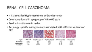
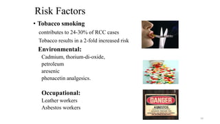
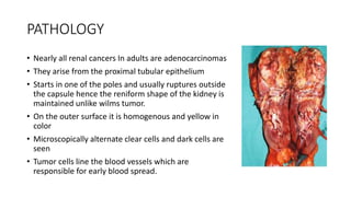
60 year old male came with
complains of painless hematuria
and a palpable abdominal lump
ANATOMY
- Retroperitoneal organ
- Right kidney L1-L3
- Left kidney T12-L2
Left kidney is little nearer to median plane than right
The kidneys are mobile organs that move vertically within
the retroperitoneum during normal respiration
external features
Shape:bean shaped
2 poles: upper & lower
2 Border: medial & lateral
2 Surface: anterior & posterior
Size: approx. 11–14 cm in length, 6 cm wide and 3cm thick
Weight: around 150 gm. in males & 135 gm. in females
RELATIONS
Anterior posterior
Coverings and Hilum of the kidney
Blood Supply
• Arterial supply by the renal arteries which are direct branches from abdominal aorta
• Entire arterial system is composed of end artries hence prone for infarction
• Venous drainage through the renal veins which drains into the inferior vena cava.
• veins anastomose freely
• Lymph Drainage
• The right kidney drains predominantly into the paracaval and interaortocaval lymph nodes
• left kidney drains exclusively to the para-aortic lymph nodes
• Nerve Supply:
• Through renal sympathetic plexus (T10 – L1) fibres, mainly vasomotor.
• Afferent nerves T10 to T12 thoracic nerves.
• Functions
• Excretion of wastes,
• Acid-base homeostasis,
• Osmolality regulation, Blood pressure regulation and Hormone secretion
INTERNAL STRUCTURES
DIFFERENTIAL DIAGNOSIS OF HEMATURIA
KEY POINT
-painless hematuria (papilloma and
carcinoma)
-Age –elederly (RCC)
-abdominal lump (polycyctic kidney ,
wilms tumor,
Renal cell carcinoma)
NEOPLAMS OF THE KIDNEY
• Benign
• Adenoma
• Papilloma arising from pelvis
• Hemangioma
• Malignant
• Renal Cell Carcinoma (Adenocarcinoma of Kidney)-MOST COMMON
• Transistional cell carcinoma
• Squamous cell carcinoma –EXTREMELY RARE
RENAL CELL CARCINOMA
• It is also called Hypernephroma or Grawitz tumor
• Commonly found in age group of 40 to 60 years
• Predominantly seen in males
• Aetiology :specific oncogenes are associated with different variants of
RCC
Clear cell
75%
Type
Incidence
Associated
mutations VHL
Papillary type 1
5%
c-Met
Papillary type 2
10%
FH
Chromophobe
5%
BHD
Oncocytoma
5%
BHD
Risk Factors
• Tobacco smoking
contributes to 24-30% of RCC cases
Tobacco results in a 2-fold increased risk
Environmental:
Cadmium, thorium-di-oxide,
petroleum
aresenic
phenacetin analgesics.
Occupational:
Leather workers
Asbestos workers
10
PATHOLOGY
• Nearly all renal cancers In adults are adenocarcinomas
• They arise from the proximal tubular epithelium
• Starts in one of the poles and usually ruptures outside
the capsule hence the reniform shape of the kidney is
maintained unlike wilms tumor.
• On the outer surface it is homogenous and yellow in
color
• Microscopically alternate clear cells and dark cells are
seen
• Tumor cells line the blood vessels which are
responsible for early blood spread.
THANK YOU
• Gauri Kulkarni

Renal cell carcinoma

  • 1.
    60 year oldmale came with complains of painless hematuria and a palpable abdominal lump
  • 2.
    ANATOMY - Retroperitoneal organ -Right kidney L1-L3 - Left kidney T12-L2 Left kidney is little nearer to median plane than right The kidneys are mobile organs that move vertically within the retroperitoneum during normal respiration external features Shape:bean shaped 2 poles: upper & lower 2 Border: medial & lateral 2 Surface: anterior & posterior Size: approx. 11–14 cm in length, 6 cm wide and 3cm thick Weight: around 150 gm. in males & 135 gm. in females
  • 3.
  • 4.
    Coverings and Hilumof the kidney
  • 5.
    Blood Supply • Arterialsupply by the renal arteries which are direct branches from abdominal aorta • Entire arterial system is composed of end artries hence prone for infarction • Venous drainage through the renal veins which drains into the inferior vena cava. • veins anastomose freely • Lymph Drainage • The right kidney drains predominantly into the paracaval and interaortocaval lymph nodes • left kidney drains exclusively to the para-aortic lymph nodes • Nerve Supply: • Through renal sympathetic plexus (T10 – L1) fibres, mainly vasomotor. • Afferent nerves T10 to T12 thoracic nerves. • Functions • Excretion of wastes, • Acid-base homeostasis, • Osmolality regulation, Blood pressure regulation and Hormone secretion
  • 6.
  • 7.
    DIFFERENTIAL DIAGNOSIS OFHEMATURIA KEY POINT -painless hematuria (papilloma and carcinoma) -Age –elederly (RCC) -abdominal lump (polycyctic kidney , wilms tumor, Renal cell carcinoma)
  • 8.
    NEOPLAMS OF THEKIDNEY • Benign • Adenoma • Papilloma arising from pelvis • Hemangioma • Malignant • Renal Cell Carcinoma (Adenocarcinoma of Kidney)-MOST COMMON • Transistional cell carcinoma • Squamous cell carcinoma –EXTREMELY RARE
  • 9.
    RENAL CELL CARCINOMA •It is also called Hypernephroma or Grawitz tumor • Commonly found in age group of 40 to 60 years • Predominantly seen in males • Aetiology :specific oncogenes are associated with different variants of RCC Clear cell 75% Type Incidence Associated mutations VHL Papillary type 1 5% c-Met Papillary type 2 10% FH Chromophobe 5% BHD Oncocytoma 5% BHD
  • 10.
    Risk Factors • Tobaccosmoking contributes to 24-30% of RCC cases Tobacco results in a 2-fold increased risk Environmental: Cadmium, thorium-di-oxide, petroleum aresenic phenacetin analgesics. Occupational: Leather workers Asbestos workers 10
  • 11.
    PATHOLOGY • Nearly allrenal cancers In adults are adenocarcinomas • They arise from the proximal tubular epithelium • Starts in one of the poles and usually ruptures outside the capsule hence the reniform shape of the kidney is maintained unlike wilms tumor. • On the outer surface it is homogenous and yellow in color • Microscopically alternate clear cells and dark cells are seen • Tumor cells line the blood vessels which are responsible for early blood spread.
  • 12.