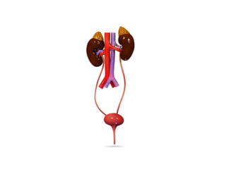
• WHAT ISURINARY SYSTEM?
• The urinary system, also known as the renal
system or urinary tract, consists of
• The kidneys (two)
• Ureters (two)
• Bladder (one)
• The urethra(one)
• The purpose of the urinary system is to eliminate waste
from the body, regulate blood volume and blood
pressure, control levels of electrolytes and metabolites,
and regulate blood pH.
• The urinary tract is the body's drainage system for the
eventual removal of urine.
5.
• There areseveral functions of the Urinary
System:
• Removal of waste product from the body
(mainly urea and uric acid)
• Regulation of electrolyte balance (e.g.
sodium, potassium and calcium)
• Regulation of acid-base homeostasis.
• Controlling blood volume and maintaining
blood pressure.
6.
• THE KIDNEYS
•The kidneys are two bean
shaped organs found on the left and right sides of
the body in vertebrates.
• They are located at the back of the abdominal
cavity in the retroperitoneal space.
• The kidneys are located high in the abdominalcavity,
one on each side of the spine, and lie in
a retroperitoneal position at a slightly oblique angle.
• The asymmetry within the abdominal cavity, caused
by the position of the liver, typically results in the
right kidney being slightly lower and smaller than
the left, and being placed slightly more to the
middle than the left kidney.
7.
• RELATIONSHIP
• Rightkidney
• Anteriorly:the duodenum,hepatic flexure of the
colon & right lobe of the liver.
• Posteriorly: Diaphragm,muscles of posterior
abdominal wall
• Superiorly: the right adrenal gland
• Left kidney
• Anteriorly:the spleen & splenic vessels,jejunum
splenic flexure of the colon,pancreas & stomach.
• Posteriorly: Diaphragm,muscles of posterior
abdominal wall
• Superiorly: the left adrenal gland
9.
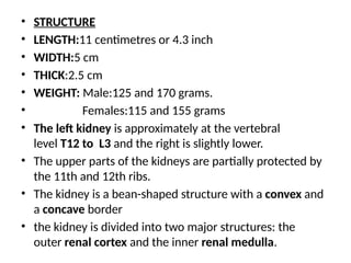
• STRUCTURE
• LENGTH:11centimetres or 4.3 inch
• WIDTH:5 cm
• THICK:2.5 cm
• WEIGHT: Male:125 and 170 grams.
• Females:115 and 155 grams
• The left kidney is approximately at the vertebral
level T12 to L3 and the right is slightly lower.
• The upper parts of the kidneys are partially protected by
the 11th and 12th ribs.
• The kidney is a bean-shaped structure with a convex and
a concave border
• the kidney is divided into two major structures: the
outer renal cortex and the inner renal medulla.
12.
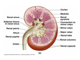
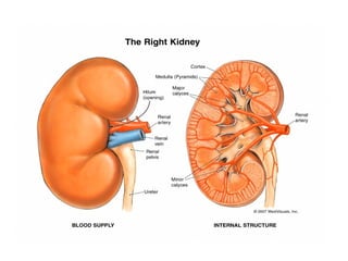
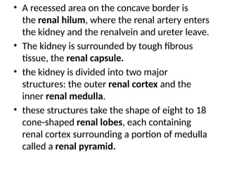
• A recessedarea on the concave border is
the renal hilum, where the renal artery enters
the kidney and the renalvein and ureter leave.
• The kidney is surrounded by tough fibrous
tissue, the renal capsule.
• the kidney is divided into two major
structures: the outer renal cortex and the
inner renal medulla.
• these structures take the shape of eight to 18
cone-shaped renal lobes, each containing
renal cortex surrounding a portion of medulla
called a renal pyramid.
13.
• Between therenal pyramids are projections of
cortex called renal columns.
• The nephron is the structural and functional
unit of the kidney,span the cortex and
medulla.
• Each adult kidney contains around one million
nephrons
• The initial filtering portion of a nephron is
the renal corpuscle which is located in the
cortex.
• This is followed by a renal tubule that passes
from the cortex deep into the medullary
pyramids.
14.
• Part ofthe renal cortex, a medullary ray is a
collection of renal tubules that drain into a
single collecting duct.
• each pyramid empties urine into a minor calyx;
minor calyces empty into major calyces, and major
calyces empty into the renal pelvis.
• This becomes the ureter.
• At the hilum, the ureter and renal vein exit the
kidney and the renal artery enters.
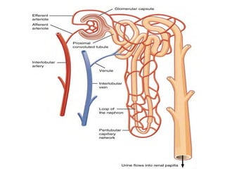
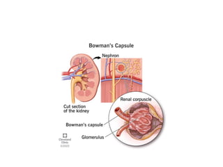
• THE NEPHRON
• The nephron is the microscopic structural and
functional unit of the kidney.
• It is composed of a renal corpuscle and a renal
tubule.
15.
• The renalcorpuscle consists of a tuft
of capillaries called a glomerulus and an
encompassing Bowman's capsule.
• A healthy adult has 0.8 to 1.5 million nephrons in
each kidney.
• they cleanse the blood and balance the
constituents of the
• circulation. The afferent arterioles form a tuft of
high-pressure capillaries about 200 μm in
diameter, the glomerulus.
• After passing through the renal
• corpuscle, the capillaries form a second arteriole,
the efferent arteriole
18.
• In adissected kidney, it is easy to identify the
cortex; it appears lighter in color compared to
the rest of the kidney.
• All of the renal corpuscles as well as both the
proximal convoluted tubules (PCTs) and distal
convoluted tubules are
• found here. Some nephrons have a short loop
of Henle that does not dip beyond the cortex.
• URINE FORMATION
• Nephrons take a simple filtrate of the blood
and modify it into urine. Many changes take
place in the different parts of the
• nephron before urine is created for disposal.
19.
• The principletask of the nephron population is to
balance the plasma to homeostatic set points and
excrete potential toxins in the urine.
• They do this by accomplishing three principle
functions—filtration, reabsorption, and secretion.
• They also have additional secondary functions that
exert control in three areas: blood pressure (via
production of renin),
• red blood cell production (via the hormone EPO),
• and calcium absorption (via conversion of calcidiol
into calcitriol,the active form of vitamin D).
20.
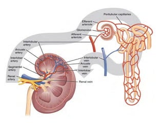
• Renal Corpuscle
•The renal corpuscle consists of a tuft of
capillaries called the glomerulus that is largely
surrounded by Bowman’s (glomerular) capsule.
• The glomerulus is a high-pressure capillary bed
between afferent and efferent arterioles.
• As blood passes through the glomerulus, 10 to
20 percent of the plasma filters between these
sieve-like fingers to be captured by Bowman’s
capsule and funneled to the PCT (proximal
convoluted tubules).
22.
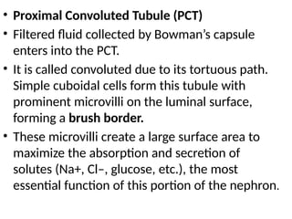
• Proximal ConvolutedTubule (PCT)
• Filtered fluid collected by Bowman’s capsule
enters into the PCT.
• It is called convoluted due to its tortuous path.
Simple cuboidal cells form this tubule with
prominent microvilli on the luminal surface,
forming a brush border.
• These microvilli create a large surface area to
maximize the absorption and secretion of
solutes (Na+, Cl–, glucose, etc.), the most
essential function of this portion of the nephron.
23.
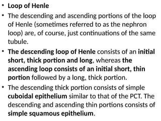
• Loop ofHenle
• The descending and ascending portions of the loop
of Henle (sometimes referred to as the nephron
loop) are, of course, just continuations of the same
tubule.
• The descending loop of Henle consists of an initial
short, thick portion and long, whereas the
ascending loop consists of an initial short, thin
portion followed by a long, thick portion.
• The descending thick portion consists of simple
cuboidal epithelium similar to that of the PCT. The
descending and ascending thin portions consists of
simple squamous epithelium.
24.
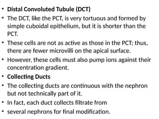
• Distal ConvolutedTubule (DCT)
• The DCT, like the PCT, is very tortuous and formed by
simple cuboidal epithelium, but it is shorter than the
PCT.
• These cells are not as active as those in the PCT; thus,
there are fewer microvilli on the apical surface.
• However, these cells must also pump ions against their
concentration gradient.
• Collecting Ducts
• The collecting ducts are continuous with the nephron
but not technically part of it.
• In fact, each duct collects filtrate from
• several nephrons for final modification.
25.
• They arelined with simple squamous epithelium with
receptors for ADH antidiuretic hormone or Vasopressin.
• Glomerular Filtration Rate (GFR)
• The volume of filtrate formed by both kidneys per
minute is termed the glomerular filtration rate (GFR).
• The heart pumps about 5 L blood per min under resting
conditions.
• Approximately 20 percent or one liter enters the kidneys
to be filtered.
• On average, this liter results in the production of about
125 mL/min filtrate produced in men (range of 90 to 140
mL/min) and 105 mL/min filtrate produced in women
(range of 80 to 125 mL/min).
26.
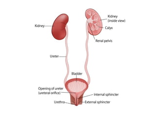
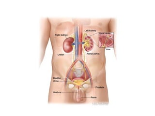
• URETERS
• Thekidneys and ureters are completely retroperitoneal,
and the bladder has a peritoneal covering only over the
dome.
• As urine is formed, it drains into the calyces of the
kidney, which merge to form the funnel-shaped renal
pelvis in the hilum of each kidney.
• The hilum narrows to become the ureter of each kidney.
• As they approach the bladder, they turn medially and
pierce the bladder wall obliquely.
• This is important because it creates an one-way valve
that allows urine into the bladder but prevents reflux of
urine from the bladder back into the ureter.
27.
• Children bornlacking this oblique course of the
ureter through the bladder wall are susceptible
to “vesicoureteral reflux,” which dramatically
increases their risk of serious UTI.
• Pregnancy also increases the likelihood of reflux
and UTI.
• The ureters are approximately 30 cm long.
• The muscular layer of the ureter consists of
longitudinal and circular smooth muscles that
create the peristaltic contractions to move the
urine into the bladder without the aid of gravity.
28.
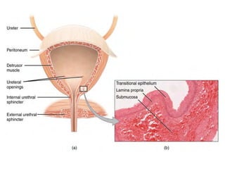
• URINARY BLADDER
•The urinary bladder collects urine from both ureters.
• The bladder lies anterior to the uterus in
females,posterior to the pubic bone and anterior to
the rectum.
• During late pregnancy, its capacity is reduced due to
compression by the enlarging uterus, resulting in
increased frequency of urination.
• The bladder is partially retroperitoneal (outside the
peritoneal cavity)
• Volumes in adults can range from nearly zero to
500–600 mL.
30.
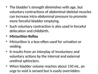
• The bladder’sstrength diminishes with age, but
voluntary contractions of abdominal skeletal muscles
can increase intra-abdominal pressure to promote
more forceful bladder emptying.
• Such voluntary contraction is also used in forceful
defecation and childbirth.
• Micturition Reflex
• Micturition is a less-often used for urination or
voiding.
• It results from an interplay of involuntary and
voluntary actions by the internal and external
urethral sphincters.
• When bladder volume reaches about 150 mL, an
urge to void is sensed but is easily overridden.
31.
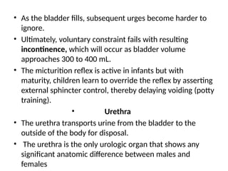
• As thebladder fills, subsequent urges become harder to
ignore.
• Ultimately, voluntary constraint fails with resulting
incontinence, which will occur as bladder volume
approaches 300 to 400 mL.
• The micturition reflex is active in infants but with
maturity, children learn to override the reflex by asserting
external sphincter control, thereby delaying voiding (potty
training).
• Urethra
• The urethra transports urine from the bladder to the
outside of the body for disposal.
• The urethra is the only urologic organ that shows any
significant anatomic difference between males and
females
33.
• Voiding isregulated by an involuntary autonomic nervous
system-controlled internal urinary sphincter, consisting of
smooth muscle and voluntary skeletal muscle that forms the
external urinary sphincter below it.
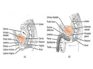
• Female Urethra
• The external urethral orifice is embedded in the anterior
vaginal wall inferior to the clitoris, superior to the vaginal
opening and medial to the labia minora.
• Its short length, about 4 cm, is less of a barrier to fecal
bacteria than the longer male urethra and the best
explanation for the greater incidence of UTI in women.
• Male Urethra
• The male urethra passes through the prostate gland
immediately inferior to the bladder before passing below the
pubic symphysis.
34.
• The lengthof the male urethra varies between men but
averages 20 cm in length.
• Filtration, Reabsorption, Secretion: The Three Steps of
Urine Formation
• he kidneys filter unwanted substances from the blood and
produce urine to excrete them.
• There are three main steps of urine formation: glomerular
filtration, reabsorption, and secretion.
• These processes ensure that only waste and excess water
are removed from the body.
• 1. The Glomerulus Filters Water and Other Substances
from the Bloodstream
• Each kidney contains over 1 million tiny structures called
nephrons. Each nephron has a glomerulus, the site of
blood filtration.
35.
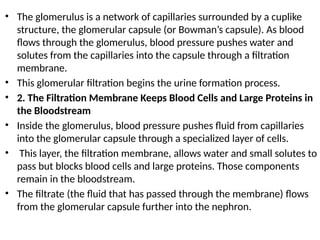
• The glomerulusis a network of capillaries surrounded by a cuplike
structure, the glomerular capsule (or Bowman’s capsule). As blood
flows through the glomerulus, blood pressure pushes water and
solutes from the capillaries into the capsule through a filtration
membrane.
• This glomerular filtration begins the urine formation process.
• 2. The Filtration Membrane Keeps Blood Cells and Large Proteins in
the Bloodstream
• Inside the glomerulus, blood pressure pushes fluid from capillaries
into the glomerular capsule through a specialized layer of cells.
• This layer, the filtration membrane, allows water and small solutes to
pass but blocks blood cells and large proteins. Those components
remain in the bloodstream.
• The filtrate (the fluid that has passed through the membrane) flows
from the glomerular capsule further into the nephron.
37.
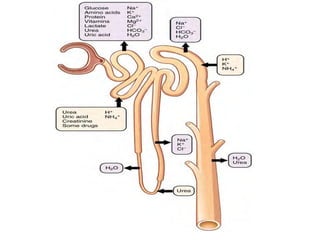
• 3. ReabsorptionMoves Nutrients and Water Back into
the Bloodstream
• The glomerulus filters water and small solutes out of the
bloodstream.
• The resulting filtrate contains waste, but also other
substances the body needs: essential ions, glucose,
amino acids, and smaller proteins. When the filtrate
exits the glomerulus, it flows into a duct in the nephron
called the renal tubule.
• As it moves, the needed substances and some water are
reabsorbed through the tube wall into adjacent
capillaries.
• This reabsorption of vital nutrients from the filtrate is
the second step in urine creation.
38.
• 4. WasteIons and Hydrogen Ions Secreted from the
Blood Complete the Formation of Urine
• The filtrate absorbed in the glomerulus flows through
the renal tubule, where nutrients and water are
reabsorbed into capillaries.
• At the same time, waste ions and hydrogen ions pass
from the capillaries into the renal tubule.
• This process is called secretion.
• The secreted ions combine with the remaining filtrate
and become urine.
• The urine flows out of the nephron tubule into a
collecting duct. It passes out of the kidney through the
renal pelvis, into the ureter, and down to the bladder.
39.
• Physical Characteristicsof Urine
• The urinary system’s ability to filter the blood resides
in about 2 to 3 million tufts of specialized capillaries
—the glomeruli—distributed more or less equally
between the two kidneys.
• The glomeruli create about 200 liters of this filtrate
every day, yet you excrete less than two liters of
waste you call urine.
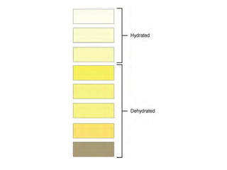
• Some of the characteristics such as color and odor
are rough descriptors of your state of hydration.
• For example, if you exercise or work outside, and
sweat a great deal, your urine will turn darker and
produce a slight odor, even if you drink plenty of
water. Athletes are often advised to consume water
until their urine is clear.
40.
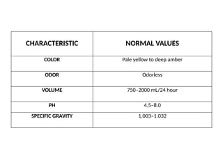
CHARACTERISTIC NORMAL VALUES
COLORPale yellow to deep amber
ODOR Odorless
VOLUME 750–2000 mL/24 hour
PH 4.5–8.0
SPECIFIC GRAVITY 1.003–1.032
42.
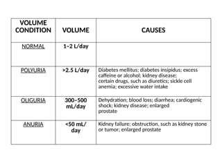
VOLUME
CONDITION VOLUME CAUSES
NORMAL1–2 L/day
POLYURIA >2.5 L/day Diabetes mellitus; diabetes insipidus; excess
caffeine or alcohol; kidney disease;
certain drugs, such as diuretics; sickle cell
anemia; excessive water intake
OLIGURIA 300–500
mL/day
Dehydration; blood loss; diarrhea; cardiogenic
shock; kidney disease; enlarged
prostate
ANURIA <50 mL/
day
Kidney failure; obstruction, such as kidney stone
or tumor; enlarged prostate
43.
• Specific gravityis a measure of the quantity of
solutes per unit volume of a solution.
• Urine will always have a specific gravity
greater than pure water (water = 1.0) due to
the presence of solutes.
• Endocrine Regulation of Kidney Function
• Several hormones have specific, important
roles in regulating kidney function.
• They act to stimulate or inhibit blood flow.
• Renin–Angiotensin–Aldosterone
• Antidiuretic Hormone (ADH)
44.
• Regulation ofFluid Volume and Composition
• The major hormones influencing total body water are
ADH, aldosterone.
• Blood volume is important in maintaining sufficient
blood pressure.
• Diuretics and Fluid Volume
• A diuretic is a compound that increases urine volume.
• Regulation of Extracellular Na+
• Sodium has a very strong osmotic effect and attracts
water.
• It plays a larger role in the osmolarity of the plasma
than any other circulating component of the blood.
45.
• If thereis too much Na+ present, either due to
poor control or excess dietary consumption, a
series of metabolic problems ensue.
• There is an increase in total volume of water,
which leads to hypertension (high blood pressure).
• Over a long period, this increases the risk of
serious complications such as heart
attacks,strokes, and aneurysms.
• It can also contribute to system-wide edema
(swelling).
• Mechanisms for regulating Na+ concentration
include the renin–angiotensin–aldosterone system
and ADH.