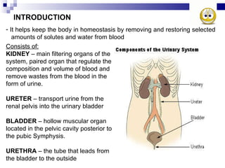
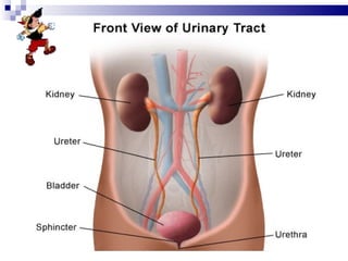
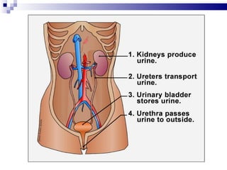
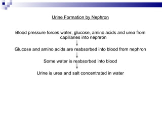
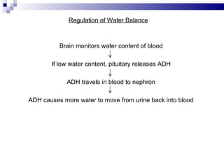
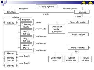
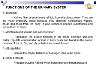
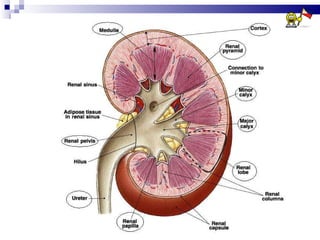
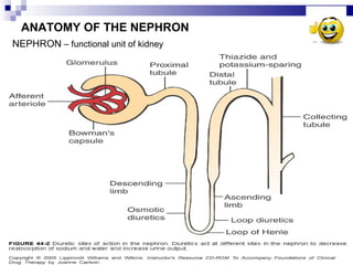
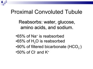
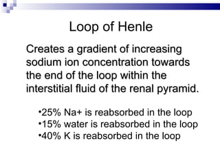
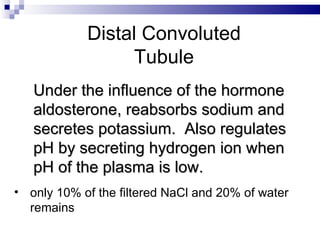
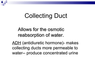
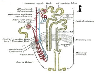
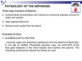
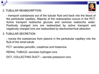
Urinary System Edison D. Ramos, RMT, MPH Assistant Professor II College of Medical Technology Manila Central University
INTRODUCTION It helps keep the body in homeostasis by removing and restoring selected  amounts of solutes and water from blood Consists of: KIDNEY  – main filtering organs of the system, paired organ that regulate the composition and volume of blood and remove wastes from the blood in the form of urine. URETER  – transport urine from the renal pelvis into the urinary bladder BLADDER  – hollow muscular organ located in the pelvic cavity posterior to the pubic Symphysis. URETHRA  – the tube that leads from the bladder to the outside
 
 
Urine Formation by Nephron Blood pressure forces water, glucose, amino acids and urea from capillaries into nephron Glucose and amino acids are reabsorbed into blood from nephron Some water is reabsorbed into blood Urine is urea and salt concentrated in water
Regulation of Water Balance Brain monitors water content of blood If low water content, pituitary releases ADH ADH travels in blood to nephron ADH causes more water to move from urine back into blood
Urinary System Has specific Performs specific Structure Function enables Kidney includes Nephron Collecting Ducts Minor Calyces Major Calyces Renal Pelvis Ureters Urinary Bladder Urethra Lead to Urine flows to Urine flows to Urine flows to Urine flows to Urine flows to Urine flows to Urine elimination includes Waste and foreign substance Urine storage Urine formation Tubular secretion Tubular reabsorption Glomerular filtration by
FUNCTIONS OF THE URINARY SYSTEM 1.  Excretion - Kidney filter large amounts of fluid from the bloodstream. They are the major excretory organ because they eliminate nitrogenous wastes, drugs and toxin from the body. It also reabsorb needed substances and return them to blood. 2.  Maintain blood volume and concentration - Regulating the proper balance in the blood between salt and water, regulate concentration of ions in body fluids and blood so the proper balance of Na, K, Ca, and phosphate ions is maintained. 3.  pH regulation - Control the proper balance of hydrogen ions in the blood 4.  Blood pressure - Produce enzyme RENIN which helps maintain blood pressure
5. Erythrocyte concentration - Produce the protein hormone ERYTHROPOIETIN which stimulates red blood cell production 6. Vitamin D production - It converts vitamin D to calciferol which is its active form. Kidney participate along with liver and skin in vitamin D synthesis.
EXTERNAL ANATOMY OF THE KIDNEY - Kidneys are paired organs that are reddish in color and resemble kidney beans in shape They are about the size of a closed fist The right kidney is slightly lower than the left because of the large area occupied by the liver. HILUM  – notch in the concave center of the kidney through which a ureters leaves the kidney and blood vessels, nerve, lymph vessels enter and exit the kidney Three layers surrounding each kidney Renal capsule – innermost layer, act as a barrier against infection and trauma. Adipose capsule – second layer, mass of fatty tissue that protect the kidney from blows Renal fascia – outermost layer, anchors the kidney to the abdominal wall.
 
CORTEX – outer area of kidney MEDULLA – inner area of kidney RENAL PYRAMID – striated, triangular structure within the medulla whose bases face the cortex and tips point to the center of kidney (renal papillae) RENAL COLUMN – cortical material that extends between the pyramids MINOR CALYX – funnel-shaped structures that surround the tip of renal pyramid MAJOR CALYX – union of minor calyces RENAL PELVIS – union of major calyces, large collecting funnel which narrows to form ureter
 
ANATOMY OF THE NEPHRON NEPHRON  – functional unit of kidney
BOWMAN’S CAPSULE – double-walled globe where nephron begins. GLOMERULUS – capillary network surrounded by bowman’s capsule PROXIMAL CONVOLUTED TUBULE – first part of the renal tubule DESCENDING LIMB OF HENLE – name given to PCT as it dips into the medulla LOOP OF HENLE – U-shaped structure of the limb of Henle ASCENDING LIMB OF HENLE – name given to loop of Henle as it ascends toward the cortex DISTAL CONVOLUTED TUBULE – name given to the ascending limb of Henle as it enters the cortex and becomes convoluted COLLECTING DUCT – connects with the distal tubules of the other nephron
 
Proximal Convoluted Tubule Reabsorbs: water, glucose, amino acids, and sodium. 65% of Na +  is reabsorbed 65% of H 2 O is reabsorbed 90% of filtered bicarbonate (HCO 3 - ) 50% of Cl -  and K +
Loop of Henle Creates a gradient of increasing sodium ion concentration towards the end of the loop within the interstitial fluid of the renal pyramid. 25% Na+ is reabsorbed in the loop 15% water is reabsorbed in the loop 40% K is reabsorbed in the loop
Distal Convoluted Tubule Under the influence of the hormone aldosterone, reabsorbs sodium and secretes potassium.  Also regulates pH by secreting hydrogen ion when pH of the plasma is low. only 10% of the filtered NaCl and 20% of water remains
Collecting Duct Allows for the osmotic reabsorption of water. ADH  (antidiuretic hormone)- makes collecting ducts more permeable to water-- produce concentrated urine
BLOOD AND NERVE SUPPLY TO THE NEURON Nephrons are mainly responsible for removing wastes from the blood and regulating its electrolytes and fluid content, they are richly supplied with blood vessels. RIGHT and LEFT ARTERIES – transport 1200ml of blood to the kidneys every minute The arteries that branch and pass between the renal pyramids in the renal column as the INTERLOBAR ARTERIES AFFERENT ARTERIOLE – takes blood from the renal artery to bowman’s capsule, where the arteriole divides into tangled capillary network known as GLOMERULUS EFFERENT ARTERIOLE – formed from union of glomerulus, carries blood away from the bowman’s capsule. Each efferent arteriole further divides to form a network of capillaries called PERITUBULAR CAPILLARIES PERITUBULAR CAPILLARIES reunite to form INTERLOBULAR VEIN
 
PHYSIOLOGY OF THE NEPHRONS Three major functions of Nephron Control blood concentration and volume by removing selected amount of water and solutes Help regulate blood pH Remove toxic waste from the blood Formation of Urine 1. GLOMERULAR FILTRATION - removes water and dissolved substances from the plasma of blood (Na, K, Ca, Mg, Cl, Sulfate, Phosphate, glucose, urea, uric acid) 90% of the fluid gets reabsorb in the renal tubules and reenters the plasma. The remaining small portion leaves the kidney as urine
2. TUBULAR REABSORPTION - transport substances out of the tubular fluid and back into the blood of the peritubular capillary. Majority of the reabsorption occurs in the PCT. Active transport reabsorbs glucose and osmosis reabsorbs water. Positively charged ions are reabsorbed by active transport and negatively charged ions are reabsorbed by electrochemical attraction 3. TUBULAR SECRETION - moves the substances from plasma in the peritubular capillary into the fluid of the renal tubule PCT: secretes penicillin, creatinine and histamine RENAL TUBULE: secretes hydrogen ions DCT, COLLECTING DUCT – secrete potassium ions
 
ANATOMY AND FUNCTIONS OF URETER Body has 2 ureters each one descending from a kidney Each ureter is an extension of the pelvis of the kidney and extends down to the urinary bladder FUNCTION: transport urine from the renal pelvis into the urinary bladder - The urine is carried through the ureters by peristaltic contraction of smooth muscle walls but  gravity and hydrostatic pressure also contribute URINARY BLADDER Hollow muscular organ located in the pelvic cavity. It is a movable organ held in position by folds of peritoneum When empty, it resembles a deflated balloon. When full of urine, it is spherical shape. As urine volume increases, it becomes pear-shaped and ascends into abdominal cavity
Urine is expelled from the bladder by an act known as MICTURITION commonly referred to as urination or voiding. The average capacity of bladder is 700 to 800ml Ureter Bladder
PROBLEMS OF THE URINARY TRACT Incontinence (urine leakage) Causes of Incontinence Stress incontinence:  leaking small amounts of urine when coughing, lifting, or exercising Urge incontinence: the bladder suddenly and unexpectedly contracts and expels urine Overflow incontinence:  bladder cannot completely empty so urine dribbles
Treatments for Incontinence  Kegel exercises to strengthen the urinary sphincter Medicines that increase the sphincter’s ability to contract Surgery to strengthen the pelvic muscles or to lift the bladder Retrain the bladder to increase its storage capacity (allowing 3-4 hours between urinating) Drugs to prevent urge incontinence Surgery to remove part of prostate gland if responsible for overflow incontinence
Kidney stones form in the kidney pelvis.  There are 4 types of stones. Calcium stones  (most common) Uric acid stones Bacteria caused stones Cystein stones
Treatments for Kidney Stones  Small stones may pass with no pain Larger stones may pass but cause extreme of pain, requiring a lot pain medication Stones that are too large to pass may require surgical treatment including: using a ureteroscope to go up and snare the stone using a nephroscope to crush the stone and retrieve it using shock wave lithotripsy where a person is submerged in water containing shock waves to pulverize the stones
Urinary Tract Infection (UTI) Second most common infection  following respiratory infections  UTI occur when bacteria ( E. coli ) from the digestive tract get into the opening of the urinary tract and multiply Bacteria first infect the urethra, then move to the bladder and finally to the kidneys  UTI tend to occur more in women than men
Women may have more UTIs than men because:  they have a shorter urethra, allowing quicker access to the bladder the urethral opening is nearer the anus intercourse may result in UTIs in women
Symptoms of UTIs Urge to urinate but only small amount of urine produced Pain and burning sensation in bladder Fever  Blood in urine
Diagnosis and Treatment Doctors check urine for white and red blood cells and bacteria Bacteria grown in culture to determine which antibiotic will work the best UTIs are treated with antibiotics and are often cured within 1 or 2 days
The End

Anaphysio ch 12 urinary

  • 1.
    Urinary System EdisonD. Ramos, RMT, MPH Assistant Professor II College of Medical Technology Manila Central University
  • 2.
    INTRODUCTION It helpskeep the body in homeostasis by removing and restoring selected amounts of solutes and water from blood Consists of: KIDNEY – main filtering organs of the system, paired organ that regulate the composition and volume of blood and remove wastes from the blood in the form of urine. URETER – transport urine from the renal pelvis into the urinary bladder BLADDER – hollow muscular organ located in the pelvic cavity posterior to the pubic Symphysis. URETHRA – the tube that leads from the bladder to the outside
  • 3.
  • 4.
  • 5.
    Urine Formation byNephron Blood pressure forces water, glucose, amino acids and urea from capillaries into nephron Glucose and amino acids are reabsorbed into blood from nephron Some water is reabsorbed into blood Urine is urea and salt concentrated in water
  • 6.
    Regulation of WaterBalance Brain monitors water content of blood If low water content, pituitary releases ADH ADH travels in blood to nephron ADH causes more water to move from urine back into blood
  • 7.
    Urinary System Hasspecific Performs specific Structure Function enables Kidney includes Nephron Collecting Ducts Minor Calyces Major Calyces Renal Pelvis Ureters Urinary Bladder Urethra Lead to Urine flows to Urine flows to Urine flows to Urine flows to Urine flows to Urine flows to Urine elimination includes Waste and foreign substance Urine storage Urine formation Tubular secretion Tubular reabsorption Glomerular filtration by
  • 8.
    FUNCTIONS OF THEURINARY SYSTEM 1. Excretion - Kidney filter large amounts of fluid from the bloodstream. They are the major excretory organ because they eliminate nitrogenous wastes, drugs and toxin from the body. It also reabsorb needed substances and return them to blood. 2. Maintain blood volume and concentration - Regulating the proper balance in the blood between salt and water, regulate concentration of ions in body fluids and blood so the proper balance of Na, K, Ca, and phosphate ions is maintained. 3. pH regulation - Control the proper balance of hydrogen ions in the blood 4. Blood pressure - Produce enzyme RENIN which helps maintain blood pressure
  • 9.
    5. Erythrocyte concentration- Produce the protein hormone ERYTHROPOIETIN which stimulates red blood cell production 6. Vitamin D production - It converts vitamin D to calciferol which is its active form. Kidney participate along with liver and skin in vitamin D synthesis.
  • 10.
    EXTERNAL ANATOMY OFTHE KIDNEY - Kidneys are paired organs that are reddish in color and resemble kidney beans in shape They are about the size of a closed fist The right kidney is slightly lower than the left because of the large area occupied by the liver. HILUM – notch in the concave center of the kidney through which a ureters leaves the kidney and blood vessels, nerve, lymph vessels enter and exit the kidney Three layers surrounding each kidney Renal capsule – innermost layer, act as a barrier against infection and trauma. Adipose capsule – second layer, mass of fatty tissue that protect the kidney from blows Renal fascia – outermost layer, anchors the kidney to the abdominal wall.
  • 11.
  • 12.
    CORTEX – outerarea of kidney MEDULLA – inner area of kidney RENAL PYRAMID – striated, triangular structure within the medulla whose bases face the cortex and tips point to the center of kidney (renal papillae) RENAL COLUMN – cortical material that extends between the pyramids MINOR CALYX – funnel-shaped structures that surround the tip of renal pyramid MAJOR CALYX – union of minor calyces RENAL PELVIS – union of major calyces, large collecting funnel which narrows to form ureter
  • 13.
  • 14.
    ANATOMY OF THENEPHRON NEPHRON – functional unit of kidney
  • 15.
    BOWMAN’S CAPSULE –double-walled globe where nephron begins. GLOMERULUS – capillary network surrounded by bowman’s capsule PROXIMAL CONVOLUTED TUBULE – first part of the renal tubule DESCENDING LIMB OF HENLE – name given to PCT as it dips into the medulla LOOP OF HENLE – U-shaped structure of the limb of Henle ASCENDING LIMB OF HENLE – name given to loop of Henle as it ascends toward the cortex DISTAL CONVOLUTED TUBULE – name given to the ascending limb of Henle as it enters the cortex and becomes convoluted COLLECTING DUCT – connects with the distal tubules of the other nephron
  • 16.
  • 17.
    Proximal Convoluted TubuleReabsorbs: water, glucose, amino acids, and sodium. 65% of Na + is reabsorbed 65% of H 2 O is reabsorbed 90% of filtered bicarbonate (HCO 3 - ) 50% of Cl - and K +
  • 18.
    Loop of HenleCreates a gradient of increasing sodium ion concentration towards the end of the loop within the interstitial fluid of the renal pyramid. 25% Na+ is reabsorbed in the loop 15% water is reabsorbed in the loop 40% K is reabsorbed in the loop
  • 19.
    Distal Convoluted TubuleUnder the influence of the hormone aldosterone, reabsorbs sodium and secretes potassium. Also regulates pH by secreting hydrogen ion when pH of the plasma is low. only 10% of the filtered NaCl and 20% of water remains
  • 20.
    Collecting Duct Allowsfor the osmotic reabsorption of water. ADH (antidiuretic hormone)- makes collecting ducts more permeable to water-- produce concentrated urine
  • 21.
    BLOOD AND NERVESUPPLY TO THE NEURON Nephrons are mainly responsible for removing wastes from the blood and regulating its electrolytes and fluid content, they are richly supplied with blood vessels. RIGHT and LEFT ARTERIES – transport 1200ml of blood to the kidneys every minute The arteries that branch and pass between the renal pyramids in the renal column as the INTERLOBAR ARTERIES AFFERENT ARTERIOLE – takes blood from the renal artery to bowman’s capsule, where the arteriole divides into tangled capillary network known as GLOMERULUS EFFERENT ARTERIOLE – formed from union of glomerulus, carries blood away from the bowman’s capsule. Each efferent arteriole further divides to form a network of capillaries called PERITUBULAR CAPILLARIES PERITUBULAR CAPILLARIES reunite to form INTERLOBULAR VEIN
  • 22.
  • 23.
    PHYSIOLOGY OF THENEPHRONS Three major functions of Nephron Control blood concentration and volume by removing selected amount of water and solutes Help regulate blood pH Remove toxic waste from the blood Formation of Urine 1. GLOMERULAR FILTRATION - removes water and dissolved substances from the plasma of blood (Na, K, Ca, Mg, Cl, Sulfate, Phosphate, glucose, urea, uric acid) 90% of the fluid gets reabsorb in the renal tubules and reenters the plasma. The remaining small portion leaves the kidney as urine
  • 24.
    2. TUBULAR REABSORPTION- transport substances out of the tubular fluid and back into the blood of the peritubular capillary. Majority of the reabsorption occurs in the PCT. Active transport reabsorbs glucose and osmosis reabsorbs water. Positively charged ions are reabsorbed by active transport and negatively charged ions are reabsorbed by electrochemical attraction 3. TUBULAR SECRETION - moves the substances from plasma in the peritubular capillary into the fluid of the renal tubule PCT: secretes penicillin, creatinine and histamine RENAL TUBULE: secretes hydrogen ions DCT, COLLECTING DUCT – secrete potassium ions
  • 25.
  • 26.
    ANATOMY AND FUNCTIONSOF URETER Body has 2 ureters each one descending from a kidney Each ureter is an extension of the pelvis of the kidney and extends down to the urinary bladder FUNCTION: transport urine from the renal pelvis into the urinary bladder - The urine is carried through the ureters by peristaltic contraction of smooth muscle walls but gravity and hydrostatic pressure also contribute URINARY BLADDER Hollow muscular organ located in the pelvic cavity. It is a movable organ held in position by folds of peritoneum When empty, it resembles a deflated balloon. When full of urine, it is spherical shape. As urine volume increases, it becomes pear-shaped and ascends into abdominal cavity
  • 27.
    Urine is expelledfrom the bladder by an act known as MICTURITION commonly referred to as urination or voiding. The average capacity of bladder is 700 to 800ml Ureter Bladder
  • 28.
    PROBLEMS OF THEURINARY TRACT Incontinence (urine leakage) Causes of Incontinence Stress incontinence: leaking small amounts of urine when coughing, lifting, or exercising Urge incontinence: the bladder suddenly and unexpectedly contracts and expels urine Overflow incontinence: bladder cannot completely empty so urine dribbles
  • 29.
    Treatments for Incontinence Kegel exercises to strengthen the urinary sphincter Medicines that increase the sphincter’s ability to contract Surgery to strengthen the pelvic muscles or to lift the bladder Retrain the bladder to increase its storage capacity (allowing 3-4 hours between urinating) Drugs to prevent urge incontinence Surgery to remove part of prostate gland if responsible for overflow incontinence
  • 30.
    Kidney stones formin the kidney pelvis. There are 4 types of stones. Calcium stones (most common) Uric acid stones Bacteria caused stones Cystein stones
  • 31.
    Treatments for KidneyStones Small stones may pass with no pain Larger stones may pass but cause extreme of pain, requiring a lot pain medication Stones that are too large to pass may require surgical treatment including: using a ureteroscope to go up and snare the stone using a nephroscope to crush the stone and retrieve it using shock wave lithotripsy where a person is submerged in water containing shock waves to pulverize the stones
  • 32.
    Urinary Tract Infection(UTI) Second most common infection following respiratory infections UTI occur when bacteria ( E. coli ) from the digestive tract get into the opening of the urinary tract and multiply Bacteria first infect the urethra, then move to the bladder and finally to the kidneys UTI tend to occur more in women than men
  • 33.
    Women may havemore UTIs than men because: they have a shorter urethra, allowing quicker access to the bladder the urethral opening is nearer the anus intercourse may result in UTIs in women
  • 34.
    Symptoms of UTIsUrge to urinate but only small amount of urine produced Pain and burning sensation in bladder Fever Blood in urine
  • 35.
    Diagnosis and TreatmentDoctors check urine for white and red blood cells and bacteria Bacteria grown in culture to determine which antibiotic will work the best UTIs are treated with antibiotics and are often cured within 1 or 2 days
  • 36.