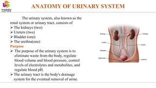
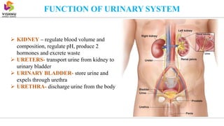
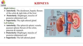
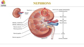
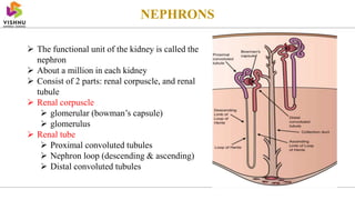
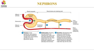
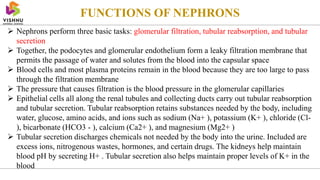
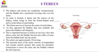
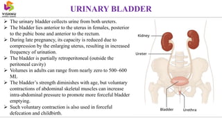
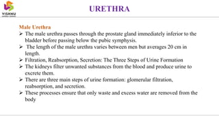
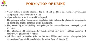
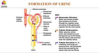
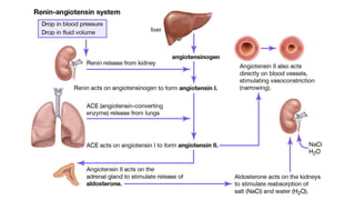
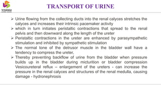
URINARY SYSTEM
Shri Vishnu College of Pharmacy (Autonomous)
Affiliated to Andhra Univ., Visakhapatnam; Approved by AICTE and PCI, New Delhi, and recognised by APSCHE
Prepared by
Grandhi Sandeep Ganesh
Dept. of Pharmacology
ANATOMY OF URINARY SYSTEM
The urinary system, also known as the
renal system or urinary tract, consists of
The kidneys (two)
Ureters (two)
Bladder (one)
The urethra(one)
Purpose
 The purpose of the urinary system is to
eliminate waste from the body, regulate
blood volume and blood pressure, control
levels of electrolytes and metabolites, and
regulate blood pH.
The urinary tract is the body's drainage
system for the eventual removal of urine.
FUNCTION OF URINARY SYSTEM
 KIDNEY – regulate blood volume and
composition, regulate pH, produce 2
hormones and excrete waste
 URETERS- transport urine from kidney to
urinary bladder
 URINARY BLADDER- store urine and
expels through urethra
 URETHRA- discharge urine from the body
KIDNEYS
The kidneys are two bean shaped organs found on the left and right sides of the
body in vertebrates
They are located at the back of the abdominal cavity in the retroperitoneal space
The kidneys are located high in the abdominal cavity, one on each side of the
spine, and lie in a retroperitoneal position at a slightly oblique angle
The asymmetry within the abdominal cavity, caused by the position of the liver,
typically results in the right kidney being slightly lower and smaller than the left,
and being placed slightly more to the middle than the left kidney.
Internally the kidneys consist of renal cortex, renal medulla, renal pyramids,
renal columns, major and minor calyces and renal pelvis
Blood enters the kidney through the renal artery and leaves through the renal
vein
Right kidney
 Anteriorly: The duodenum, hepatic flexure
of the colon & right lobe of the liver
 Posteriorly: Diaphragm, muscles of
posterior abdominal wall
 Superiorly: The right adrenal gland
Left kidney
 Anteriorly: The spleen & splenic vessels,
jejunum splenic flexure of the colon,
pancreas & stomach.
 Posteriorly: Diaphragm, muscles of
posterior abdominal wall
 Superiorly: The left adrenal gland
KIDNEYS
KIDNEYS
LENGTH: 11 centimeter or 4.3 inch
WIDTH: 5 cm
THICK: 2.5 cm
WEIGHT: Male:125 and 170 grams.
Females: 115 and 155 grams
The left kidney is approximately at the vertebral level T12 to L3 and the right
kidney is slightly lower
The upper parts of the kidneys are partially protected by the 11th and 12th ribs
The kidney is a bean-shaped structure with a convex and a concave border
The kidney is divided into two major structures: the outer renal cortex and the
inner renal medulla.
NEPHRONS
NEPHRONS
 The functional unit of the kidney is called the
nephron
 About a million in each kidney
 Consist of 2 parts: renal corpuscle, and renal
tubule
 Renal corpuscle
 glomerular (bowman’s capsule)
 glomerulus
 Renal tube
 Proximal convoluted tubules
 Nephron loop (descending & ascending)
 Distal convoluted tubules
NEPHRONS
Renal Corpuscle
 The renal corpuscle consists of a tuft of capillaries called the glomerulus that is largely
surrounded by Bowman’s (glomerular) capsule
 The glomerulus is a high-pressure capillary bed between afferent and efferent arterioles.
 As blood passes through the glomerulus, 10 to 20 percent of the plasma filters between
these sieve- like fingers to be captured by Bowman’s capsule and funneled to the PCT
(proximal convoluted tubules).
Proximal Convoluted Tubule (PCT)
 Filtered fluid collected by Bowman’s capsule enters into the PCT
 It is called convoluted due to its tortuous path. Simple cuboidal cells form this tubule
with prominent microvilli on the luminal surface, forming a brush border
 These microvilli create a large surface area to maximize the absorption and secretion of
solutes (Na+, Cl–, glucose, etc.), the most essential function of this portion of the
nephron.
NEPHRONS
NEPHRONS
Loop of Henle
 The descending and ascending portions of the loop of Henle (sometimes
referred to as the nephron loop) are, of course, just continuations of the same
tubule.
 The descending loop of Henle consists of an initial short, thick portion and
long, whereas the ascending loop consists of an initial short, thin portion
followed by a long, thick portion.
 The descending thick portion consists of simple cuboidal epithelium similar to
that of the PCT. The descending and ascending thin portions consists of simple
squamous epithelium.
Distal Convoluted Tubule (DCT)
 The DCT, like the PCT, is very tortuous and formed by simple cuboidal
epithelium, but it is shorter than the PCT.
 These cells are not as active as those in the PCT; thus, there are fewer microvilli
on the apical surface.
 However, these cells must also pump ions against their concentration gradient.
Collecting Ducts
 The collecting ducts are continuous with the nephron but not technically part of
it.
 In fact, each duct collects filtrate from
 several nephrons for final modification
 They are lined with simple squamous epithelium with receptors for ADH
antidiuretic hormone or Vasopressin.
NEPHRONS
Glomerular Filtration Rate (GFR)
The volume of filtrate formed by both kidneys per minute is termed the
glomerular filtration rate (GFR)
 The heart pumps about 5 L blood per min under resting conditions.
 Approximately 20 percent or one liter enters the kidneys to be filtered.
 On average, this liter results in the production of about 125 mL/min filtrate
produced in men (range of 90 to 140 mL/min) and 105 mL/min filtrate produced
in women (range of 80 to 125 mL/min).
NEPHRONS
FUNCTIONS OF NEPHRONS
 Nephrons perform three basic tasks: glomerular filtration, tubular reabsorption, and tubular
secretion
 Together, the podocytes and glomerular endothelium form a leaky filtration membrane that
permits the passage of water and solutes from the blood into the capsular space
 Blood cells and most plasma proteins remain in the blood because they are too large to pass
through the filtration membrane
 The pressure that causes filtration is the blood pressure in the glomerular capillaries
 Epithelial cells all along the renal tubules and collecting ducts carry out tubular reabsorption
and tubular secretion. Tubular reabsorption retains substances needed by the body, including
water, glucose, amino acids, and ions such as sodium (Na+ ), potassium (K+ ), chloride (Cl-
), bicarbonate (HCO3 - ), calcium (Ca2+ ), and magnesium (Mg2+ )
 Tubular secretion discharges chemicals not needed by the body into the urine. Included are
excess ions, nitrogenous wastes, hormones, and certain drugs. The kidneys help maintain
blood pH by secreting H+ . Tubular secretion also helps maintain proper levels of K+ in the
blood
 The kidneys and ureters are completely retroperitoneal,
and the bladder has a peritoneal covering only over the
dome.
 As urine is formed, it drains into the calyces of the
kidney, which merge to form the funnel-shaped renal
pelvis in the hilum of each kidney.
 The hilum narrows to become the ureter of each kidney.
 As they approach the bladder, they turn medially and
pierce the bladder wall obliquely.
 This is important because it creates an one-way valve that
allows urine into the bladder but prevents reflux of urine
from the bladder back into the ureter.
 The ureters are approximately 30 cm long.
 The muscular layer of the ureter consists of longitudinal
and circular smooth muscles that create the peristaltic
contractions to move the urine into the bladder without
the aid of gravity.
UTEREUS
 The urinary bladder collects urine from both ureters.
 The bladder lies anterior to the uterus in females, posterior
to the pubic bone and anterior to the rectum.
 During late pregnancy, its capacity is reduced due to
compression by the enlarging uterus, resulting in increased
frequency of urination.
 The bladder is partially retroperitoneal (outside the
peritoneal cavity)
 Volumes in adults can range from nearly zero to 500–600
ML
 The bladder’s strength diminishes with age, but voluntary
contractions of abdominal skeletal muscles can increase
intra-abdominal pressure to promote more forceful bladder
emptying.
 Such voluntary contraction is also used in forceful
defecation and childbirth.
URINARY BLADDER
URETHRA
 The urethra transports urine from the bladder to the outside of the body for
disposal
 The urethra is the only urologic organ that shows any significant anatomic
difference between males and females
 Voiding is regulated by an involuntary autonomic nervous system-controlled
internal urinary sphincter, consisting of smooth muscle and voluntary skeletal
muscle that forms the external urinary sphincter below it.
Female Urethra
 The external urethral orifice is embedded in the anterior vaginal wall inferior to
the clitoris, superior to the vaginal opening and medial to the labia minora.
 Its short length, about 4 cm, is less of a barrier to fecal bacteria than the longer
male urethra and the best explanation for the greater incidence of UTI in women.
Male Urethra
 The male urethra passes through the prostate gland immediately inferior to the
bladder before passing below the pubic symphysis.
 The length of the male urethra varies between men but averages 20 cm in
length.
 Filtration, Reabsorption, Secretion: The Three Steps of Urine Formation
 The kidneys filter unwanted substances from the blood and produce urine to
excrete them.
 There are three main steps of urine formation: glomerular filtration,
reabsorption, and secretion.
 These processes ensure that only waste and excess water are removed from the
body
URETHRA
FORMATION OF URINE
 Nephrons take a simple filtrate of the blood and modify it into urine. Many changes
take place in the different parts of the
 Nephron before urine is created for disposal.
 The principle task of the nephron population is to balance the plasma to homeostatic
set points and excrete potential toxins in the urine.
 They do this by accomplishing three principle functions—filtration, reabsorption, and
secretion.
 They also have additional secondary functions that exert control in three areas: blood
pressure (via production of renin),
 red blood cell production (via the hormone EPO), and calcium absorption (via
conversion of calcidiol into calcitriol, the active form of vitamin D)
FORMATION OF URINE
JUXTAGLOMERULAR APPARATUS
JUXTAGLOMERULAR (JGA)
It is a combination of specialized tubular and vascular cells located near
the glomerulus of each nephron
JGA is formed by three different structures
Macula densa cells
Extra glomerular mesangial cells
Juxtaglomerular cells
JUXTAGLOMERULAR APPARATUS
Macula densa cells
 These are specialized renal tubular epithelial cells
 Situated between afferent and efferent arterioles of the same nephron
 Macula densa plays important role in tubuloglomerual feedback mechanism
 These cells are not innervated
Extraglomerular mesangial cells
 These cells are also called agranular cells, lacis cells
 These cells are situated in the triangular region bound by afferent arteriole,
efferent arteriole & macula densa cells
 These cells play important role in the regulation of glomerular filteration by
their contractile property
 These cells are phagocytic in nature
JUXTAGLOMERULAR APPARATUS
Juxtaglomerular cells
 Myoepithelial cells (modified vascular smooth muscle cells) called JG cells
 Also called granular cells because presence of secretory granules in their
cytoplasm
 JG cells are well developed golgi apparatus and endoplasmic reticulum and
ribosomes
 JG cells act as baroreceptor and innervated by sympathetic nerve fibres
Function of JGA
 Primary function of JGA is secretion of hormones
 It also regulates blood flow and GFR
 Secretes hormones
 Renin
 Prostaglandins
Micturition is the process by which the urinary bladder empties when it
becomes filled
This involves two main steps:
 First, the bladder fills progressively until the tension in its walls rises above
a threshold level;
 This elicits the second step, which is a nervous reflex called the micturition
reflex that empties the bladder or, if this fails, at least causes a conscious
desire to urinate.
 Although the micturition reflex is an autonomic spinal cord reflex, it can also be
inhibited or facilitated by centers in the cerebral cortex or brain stem
MICTURITION
 The urinary bladder is a smooth muscle
chamber composed of two main parts:
 The body, which is the major part of the
bladder in which urine collects, and
 the neck, which is a funnel-shaped
extension of the body, passing inferiorly
and anteriorly into the urogenital triangle
and connecting with the urethra.
 The smooth muscle of the bladder is
called the detrusor muscle - contraction
of the detrusor muscle is a major step in
emptying the bladder
MICTURITION
 Urine flowing from the collecting ducts into the renal calyces stretches the
calyces and increases their intrinsic pacemaker activity
 which in turn initiates peristaltic contractions that spread to the renal
pelvis and then downward along the length of the ureter
 Peristaltic contractions in the ureter are enhanced by parasympathetic
stimulation and inhibited by sympathetic stimulation
 The normal tone of the detrusor muscle in the bladder wall have a
tendency to compress the ureter,
 Thereby preventing backflow of urine from the bladder when pressure
builds up in the bladder during micturition or bladder compression
Vesicoureteral reflux – enlargement of the ureters - can increase the
pressure in the renal calyces and structures of the renal medulla, causing
damage - hydronephrosis
TRANSPORT OF URINE
TESTS
TESTS
TESTS
TESTS

URINARY SYSTEM

  • 1.
    URINARY SYSTEM Shri VishnuCollege of Pharmacy (Autonomous) Affiliated to Andhra Univ., Visakhapatnam; Approved by AICTE and PCI, New Delhi, and recognised by APSCHE Prepared by Grandhi Sandeep Ganesh Dept. of Pharmacology
  • 2.
    ANATOMY OF URINARYSYSTEM The urinary system, also known as the renal system or urinary tract, consists of The kidneys (two) Ureters (two) Bladder (one) The urethra(one) Purpose  The purpose of the urinary system is to eliminate waste from the body, regulate blood volume and blood pressure, control levels of electrolytes and metabolites, and regulate blood pH. The urinary tract is the body's drainage system for the eventual removal of urine.
  • 3.
    FUNCTION OF URINARYSYSTEM  KIDNEY – regulate blood volume and composition, regulate pH, produce 2 hormones and excrete waste  URETERS- transport urine from kidney to urinary bladder  URINARY BLADDER- store urine and expels through urethra  URETHRA- discharge urine from the body
  • 4.
    KIDNEYS The kidneys aretwo bean shaped organs found on the left and right sides of the body in vertebrates They are located at the back of the abdominal cavity in the retroperitoneal space The kidneys are located high in the abdominal cavity, one on each side of the spine, and lie in a retroperitoneal position at a slightly oblique angle The asymmetry within the abdominal cavity, caused by the position of the liver, typically results in the right kidney being slightly lower and smaller than the left, and being placed slightly more to the middle than the left kidney. Internally the kidneys consist of renal cortex, renal medulla, renal pyramids, renal columns, major and minor calyces and renal pelvis Blood enters the kidney through the renal artery and leaves through the renal vein
  • 5.
    Right kidney  Anteriorly:The duodenum, hepatic flexure of the colon & right lobe of the liver  Posteriorly: Diaphragm, muscles of posterior abdominal wall  Superiorly: The right adrenal gland Left kidney  Anteriorly: The spleen & splenic vessels, jejunum splenic flexure of the colon, pancreas & stomach.  Posteriorly: Diaphragm, muscles of posterior abdominal wall  Superiorly: The left adrenal gland KIDNEYS
  • 6.
    KIDNEYS LENGTH: 11 centimeteror 4.3 inch WIDTH: 5 cm THICK: 2.5 cm WEIGHT: Male:125 and 170 grams. Females: 115 and 155 grams The left kidney is approximately at the vertebral level T12 to L3 and the right kidney is slightly lower The upper parts of the kidneys are partially protected by the 11th and 12th ribs The kidney is a bean-shaped structure with a convex and a concave border The kidney is divided into two major structures: the outer renal cortex and the inner renal medulla.
  • 7.
  • 8.
    NEPHRONS  The functionalunit of the kidney is called the nephron  About a million in each kidney  Consist of 2 parts: renal corpuscle, and renal tubule  Renal corpuscle  glomerular (bowman’s capsule)  glomerulus  Renal tube  Proximal convoluted tubules  Nephron loop (descending & ascending)  Distal convoluted tubules
  • 9.
  • 10.
    Renal Corpuscle  Therenal corpuscle consists of a tuft of capillaries called the glomerulus that is largely surrounded by Bowman’s (glomerular) capsule  The glomerulus is a high-pressure capillary bed between afferent and efferent arterioles.  As blood passes through the glomerulus, 10 to 20 percent of the plasma filters between these sieve- like fingers to be captured by Bowman’s capsule and funneled to the PCT (proximal convoluted tubules). Proximal Convoluted Tubule (PCT)  Filtered fluid collected by Bowman’s capsule enters into the PCT  It is called convoluted due to its tortuous path. Simple cuboidal cells form this tubule with prominent microvilli on the luminal surface, forming a brush border  These microvilli create a large surface area to maximize the absorption and secretion of solutes (Na+, Cl–, glucose, etc.), the most essential function of this portion of the nephron. NEPHRONS
  • 11.
    NEPHRONS Loop of Henle The descending and ascending portions of the loop of Henle (sometimes referred to as the nephron loop) are, of course, just continuations of the same tubule.  The descending loop of Henle consists of an initial short, thick portion and long, whereas the ascending loop consists of an initial short, thin portion followed by a long, thick portion.  The descending thick portion consists of simple cuboidal epithelium similar to that of the PCT. The descending and ascending thin portions consists of simple squamous epithelium.
  • 12.
    Distal Convoluted Tubule(DCT)  The DCT, like the PCT, is very tortuous and formed by simple cuboidal epithelium, but it is shorter than the PCT.  These cells are not as active as those in the PCT; thus, there are fewer microvilli on the apical surface.  However, these cells must also pump ions against their concentration gradient. Collecting Ducts  The collecting ducts are continuous with the nephron but not technically part of it.  In fact, each duct collects filtrate from  several nephrons for final modification  They are lined with simple squamous epithelium with receptors for ADH antidiuretic hormone or Vasopressin. NEPHRONS
  • 13.
    Glomerular Filtration Rate(GFR) The volume of filtrate formed by both kidneys per minute is termed the glomerular filtration rate (GFR)  The heart pumps about 5 L blood per min under resting conditions.  Approximately 20 percent or one liter enters the kidneys to be filtered.  On average, this liter results in the production of about 125 mL/min filtrate produced in men (range of 90 to 140 mL/min) and 105 mL/min filtrate produced in women (range of 80 to 125 mL/min). NEPHRONS
  • 14.
    FUNCTIONS OF NEPHRONS Nephrons perform three basic tasks: glomerular filtration, tubular reabsorption, and tubular secretion  Together, the podocytes and glomerular endothelium form a leaky filtration membrane that permits the passage of water and solutes from the blood into the capsular space  Blood cells and most plasma proteins remain in the blood because they are too large to pass through the filtration membrane  The pressure that causes filtration is the blood pressure in the glomerular capillaries  Epithelial cells all along the renal tubules and collecting ducts carry out tubular reabsorption and tubular secretion. Tubular reabsorption retains substances needed by the body, including water, glucose, amino acids, and ions such as sodium (Na+ ), potassium (K+ ), chloride (Cl- ), bicarbonate (HCO3 - ), calcium (Ca2+ ), and magnesium (Mg2+ )  Tubular secretion discharges chemicals not needed by the body into the urine. Included are excess ions, nitrogenous wastes, hormones, and certain drugs. The kidneys help maintain blood pH by secreting H+ . Tubular secretion also helps maintain proper levels of K+ in the blood
  • 15.
     The kidneysand ureters are completely retroperitoneal, and the bladder has a peritoneal covering only over the dome.  As urine is formed, it drains into the calyces of the kidney, which merge to form the funnel-shaped renal pelvis in the hilum of each kidney.  The hilum narrows to become the ureter of each kidney.  As they approach the bladder, they turn medially and pierce the bladder wall obliquely.  This is important because it creates an one-way valve that allows urine into the bladder but prevents reflux of urine from the bladder back into the ureter.  The ureters are approximately 30 cm long.  The muscular layer of the ureter consists of longitudinal and circular smooth muscles that create the peristaltic contractions to move the urine into the bladder without the aid of gravity. UTEREUS
  • 16.
     The urinarybladder collects urine from both ureters.  The bladder lies anterior to the uterus in females, posterior to the pubic bone and anterior to the rectum.  During late pregnancy, its capacity is reduced due to compression by the enlarging uterus, resulting in increased frequency of urination.  The bladder is partially retroperitoneal (outside the peritoneal cavity)  Volumes in adults can range from nearly zero to 500–600 ML  The bladder’s strength diminishes with age, but voluntary contractions of abdominal skeletal muscles can increase intra-abdominal pressure to promote more forceful bladder emptying.  Such voluntary contraction is also used in forceful defecation and childbirth. URINARY BLADDER
  • 17.
    URETHRA  The urethratransports urine from the bladder to the outside of the body for disposal  The urethra is the only urologic organ that shows any significant anatomic difference between males and females  Voiding is regulated by an involuntary autonomic nervous system-controlled internal urinary sphincter, consisting of smooth muscle and voluntary skeletal muscle that forms the external urinary sphincter below it. Female Urethra  The external urethral orifice is embedded in the anterior vaginal wall inferior to the clitoris, superior to the vaginal opening and medial to the labia minora.  Its short length, about 4 cm, is less of a barrier to fecal bacteria than the longer male urethra and the best explanation for the greater incidence of UTI in women.
  • 18.
    Male Urethra  Themale urethra passes through the prostate gland immediately inferior to the bladder before passing below the pubic symphysis.  The length of the male urethra varies between men but averages 20 cm in length.  Filtration, Reabsorption, Secretion: The Three Steps of Urine Formation  The kidneys filter unwanted substances from the blood and produce urine to excrete them.  There are three main steps of urine formation: glomerular filtration, reabsorption, and secretion.  These processes ensure that only waste and excess water are removed from the body URETHRA
  • 19.
    FORMATION OF URINE Nephrons take a simple filtrate of the blood and modify it into urine. Many changes take place in the different parts of the  Nephron before urine is created for disposal.  The principle task of the nephron population is to balance the plasma to homeostatic set points and excrete potential toxins in the urine.  They do this by accomplishing three principle functions—filtration, reabsorption, and secretion.  They also have additional secondary functions that exert control in three areas: blood pressure (via production of renin),  red blood cell production (via the hormone EPO), and calcium absorption (via conversion of calcidiol into calcitriol, the active form of vitamin D)
  • 20.
  • 22.
    JUXTAGLOMERULAR APPARATUS JUXTAGLOMERULAR (JGA) Itis a combination of specialized tubular and vascular cells located near the glomerulus of each nephron JGA is formed by three different structures Macula densa cells Extra glomerular mesangial cells Juxtaglomerular cells
  • 23.
    JUXTAGLOMERULAR APPARATUS Macula densacells  These are specialized renal tubular epithelial cells  Situated between afferent and efferent arterioles of the same nephron  Macula densa plays important role in tubuloglomerual feedback mechanism  These cells are not innervated Extraglomerular mesangial cells  These cells are also called agranular cells, lacis cells  These cells are situated in the triangular region bound by afferent arteriole, efferent arteriole & macula densa cells  These cells play important role in the regulation of glomerular filteration by their contractile property  These cells are phagocytic in nature
  • 24.
    JUXTAGLOMERULAR APPARATUS Juxtaglomerular cells Myoepithelial cells (modified vascular smooth muscle cells) called JG cells  Also called granular cells because presence of secretory granules in their cytoplasm  JG cells are well developed golgi apparatus and endoplasmic reticulum and ribosomes  JG cells act as baroreceptor and innervated by sympathetic nerve fibres Function of JGA  Primary function of JGA is secretion of hormones  It also regulates blood flow and GFR  Secretes hormones  Renin  Prostaglandins
  • 25.
    Micturition is theprocess by which the urinary bladder empties when it becomes filled This involves two main steps:  First, the bladder fills progressively until the tension in its walls rises above a threshold level;  This elicits the second step, which is a nervous reflex called the micturition reflex that empties the bladder or, if this fails, at least causes a conscious desire to urinate.  Although the micturition reflex is an autonomic spinal cord reflex, it can also be inhibited or facilitated by centers in the cerebral cortex or brain stem MICTURITION
  • 26.
     The urinarybladder is a smooth muscle chamber composed of two main parts:  The body, which is the major part of the bladder in which urine collects, and  the neck, which is a funnel-shaped extension of the body, passing inferiorly and anteriorly into the urogenital triangle and connecting with the urethra.  The smooth muscle of the bladder is called the detrusor muscle - contraction of the detrusor muscle is a major step in emptying the bladder MICTURITION
  • 27.
     Urine flowingfrom the collecting ducts into the renal calyces stretches the calyces and increases their intrinsic pacemaker activity  which in turn initiates peristaltic contractions that spread to the renal pelvis and then downward along the length of the ureter  Peristaltic contractions in the ureter are enhanced by parasympathetic stimulation and inhibited by sympathetic stimulation  The normal tone of the detrusor muscle in the bladder wall have a tendency to compress the ureter,  Thereby preventing backflow of urine from the bladder when pressure builds up in the bladder during micturition or bladder compression Vesicoureteral reflux – enlargement of the ureters - can increase the pressure in the renal calyces and structures of the renal medulla, causing damage - hydronephrosis TRANSPORT OF URINE
  • 28.
  • 29.
  • 30.
  • 31.