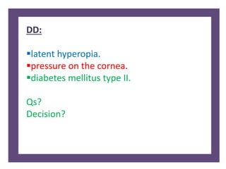
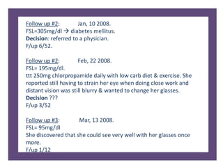
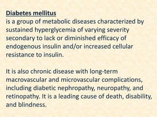
A 41-year-old female presented with difficulty reading small prints and using her cellphone for 6 months. She was diagnosed with hyperopic presbyopia but returned with worsening vision. Her blood sugar was found to be elevated, diagnosing her with diabetes mellitus. Diabetes can cause refractive changes, cataracts, and retinopathy due to physiological changes in the eye. A full ocular examination is important for managing diabetic patients.
Presenters :
Jenil Shelsiya
Sisira PS
Gopika Jyothirmayan
Special Thanks To Sushma Shrestha
and Mentor Deepak Rai (Optometrist).
If any query,Viewers are requested to refer to book for detailed explanation or can ask me question directly in the comment box. Answers will be given from Presenter's side.
Accommodation/ Accommodation of Eye, Measurement of Accommodation of Eye (hea...Bikash Sapkota
CLICK HERE TO DOWNLOAD FULL PPT ❤❤ https://healthkura.com/measurement-of-accommodation-of-eye/ ❤❤
Dear viewers Check Out my other piece of works at ❤❤❤ https://healthkura.com ❤❤❤
Measurement of Accommodation of eye:
Amplitude, Facility,
Relative Accommodation, Fatigue, Lag,
Dynamic Retinoscopy
Presentation Layout:
-Introduction to accommodation of eye
-Mechanism
-Components
-Measurement of accommodation of eye
- Amplitude
- Facility
- Relative accommodation
- Lag
-Dynamic Retinoscopy
Accommodation
-dioptric adjustment of the crystalline lens of the eye
- to obtain clear vision for a given target of regard
-process by which the refractive power of eye is altered
- to ensure a clear retinal image
For further reading
-Clinical Procedures in Optometry by J.D. Bartlett, J.B. Eskridge, J.F. Amos
-Primary Care Optometry by Theodere Grosvenor
-Borish’s Clinical Refraction by W.J. Benjamin
-Clinical Procedures for Ocular examination by Carlson et al
-American Academy of Ophthalmology
-Optometric Clinical Practice Guideline by American Optometric Association
-Internet
Follow me to get in touch with optometric and ophthalmic updates
Central Retinal Artery Occlusion (CRAO) for undergraduate MBBS Students.
Covers the basics of Aetiology, pathophysiology, clinical features, types, associated conditions and management of CRAO.
Also encompasses salient points for PGMEE
Scleral contact lenses , types, uses in various ocular conditions.
An in-depth and unbiased details of these lenses as a therapeutic and also as a drug - delivery system in modern ophthalmology.
A must read for all Ophthalmologists and Optometrists.
Peripheral Neuropathy Diabetic Connection? - A Critical AnalysisGraMedica
Peripheral nerve damage is blamed solely as a result of diabetes. This presentation takes a hard look at the evidence, asks and answers the hard questions to show that diabetes should be the last consideration after all other causes are eliminated.
Learn more at www.GraMedica.com.
Neuropathy and Foot Exam - Diabetes Symposia presented at Hôpital Sacré Coeur in Milot, Haiti.
CRUDEM’s Education Committee (a subcommittee of the Board of Directors) sponsors one-week medical symposia on specific medical topics, i.e. diabetes, infectious disease. The classes are held at Hôpital Sacré Coeur and doctors and nurses come from all over Haiti to attend.
Dry Eye and Ocular surface diseases in diabetes mellitusDhwanit Khetwani
RELATION OF DIABETES WITH DRY EYE AND OTHER OCULAR SURFACE DISEASES, MADE FOR THE PURPOSE PROTOCOL PRESENTATION. MADE BY DR DHWANIT KHETWANI OPHTHALMOLOGY RESIDENT
Acute Congestive Glaucoma / Optic Neuritis / Painful Loss Of Vision by Dr. Mu...Zeeshan Hameed
About Author:
Dr. Muhammad Zeeshan Hameed MBBS,FCPS(Resident Eye Surgeon)
GMC/DHQ Teachng Hosptal, Gujranwala Pakistan.
About Presentation:
This presentation covers the complete topic of acute congestive glaucoma, optic neuritis and ddx of sudden painful loss of vision
Glaucoma is an optic neuropathy characterized by progressive loss of retinal ganglion cells and their axons and a characteristic visual field defect.
It is the 2nd leading cause of blindness worldwide, often diagnosed late. For the foreseeable future, the best hope for glaucoma therefore is early diagnosis and monitoring.
medical management of chronic open angle glaucoma, primary angle closure glaucoma after iridotomy, normotensive glaucoma and acute angle closure attack.
Short summary for what is supposed to be in the Departmental Manual according to the Saudi Central Board for Accreditation of Healthcare Institutions (CBAHI).
Factory Supply Best Quality Pmk Oil CAS 28578–16–7 PMK Powder in Stockrebeccabio
Factory Supply Best Quality Pmk Oil CAS 28578–16–7 PMK Powder in Stock
Telegram: bmksupplier
signal: +85264872720
threema: TUD4A6YC
You can contact me on Telegram or Threema
Communicate promptly and reply
Free of customs clearance, Double Clearance 100% pass delivery to USA, Canada, Spain, Germany, Netherland, Poland, Italy, Sweden, UK, Czech Republic, Australia, Mexico, Russia, Ukraine, Kazakhstan.Door to door service
Hot Selling Organic intermediates
Explore natural remedies for syphilis treatment in Singapore. Discover alternative therapies, herbal remedies, and lifestyle changes that may complement conventional treatments. Learn about holistic approaches to managing syphilis symptoms and supporting overall health.
micro teaching on communication m.sc nursing.pdfAnurag Sharma
Microteaching is a unique model of practice teaching. It is a viable instrument for the. desired change in the teaching behavior or the behavior potential which, in specified types of real. classroom situations, tends to facilitate the achievement of specified types of objectives.
Ozempic: Preoperative Management of Patients on GLP-1 Receptor Agonists Saeid Safari
Preoperative Management of Patients on GLP-1 Receptor Agonists like Ozempic and Semiglutide
ASA GUIDELINE
NYSORA Guideline
2 Case Reports of Gastric Ultrasound
Report Back from SGO 2024: What’s the Latest in Cervical Cancer?bkling
Are you curious about what’s new in cervical cancer research or unsure what the findings mean? Join Dr. Emily Ko, a gynecologic oncologist at Penn Medicine, to learn about the latest updates from the Society of Gynecologic Oncology (SGO) 2024 Annual Meeting on Women’s Cancer. Dr. Ko will discuss what the research presented at the conference means for you and answer your questions about the new developments.
Flu Vaccine Alert in Bangalore Karnatakaaddon Scans
As flu season approaches, health officials in Bangalore, Karnataka, are urging residents to get their flu vaccinations. The seasonal flu, while common, can lead to severe health complications, particularly for vulnerable populations such as young children, the elderly, and those with underlying health conditions.
Dr. Vidisha Kumari, a leading epidemiologist in Bangalore, emphasizes the importance of getting vaccinated. "The flu vaccine is our best defense against the influenza virus. It not only protects individuals but also helps prevent the spread of the virus in our communities," he says.
This year, the flu season is expected to coincide with a potential increase in other respiratory illnesses. The Karnataka Health Department has launched an awareness campaign highlighting the significance of flu vaccinations. They have set up multiple vaccination centers across Bangalore, making it convenient for residents to receive their shots.
To encourage widespread vaccination, the government is also collaborating with local schools, workplaces, and community centers to facilitate vaccination drives. Special attention is being given to ensuring that the vaccine is accessible to all, including marginalized communities who may have limited access to healthcare.
Residents are reminded that the flu vaccine is safe and effective. Common side effects are mild and may include soreness at the injection site, mild fever, or muscle aches. These side effects are generally short-lived and far less severe than the flu itself.
Healthcare providers are also stressing the importance of continuing COVID-19 precautions. Wearing masks, practicing good hand hygiene, and maintaining social distancing are still crucial, especially in crowded places.
Protect yourself and your loved ones by getting vaccinated. Together, we can help keep Bangalore healthy and safe this flu season. For more information on vaccination centers and schedules, residents can visit the Karnataka Health Department’s official website or follow their social media pages.
Stay informed, stay safe, and get your flu shot today!
Couples presenting to the infertility clinic- Do they really have infertility...Sujoy Dasgupta
Dr Sujoy Dasgupta presented the study on "Couples presenting to the infertility clinic- Do they really have infertility? – The unexplored stories of non-consummation" in the 13th Congress of the Asia Pacific Initiative on Reproduction (ASPIRE 2024) at Manila on 24 May, 2024.
The prostate is an exocrine gland of the male mammalian reproductive system
It is a walnut-sized gland that forms part of the male reproductive system and is located in front of the rectum and just below the urinary bladder
Function is to store and secrete a clear, slightly alkaline fluid that constitutes 10-30% of the volume of the seminal fluid that along with the spermatozoa, constitutes semen
A healthy human prostate measures (4cm-vertical, by 3cm-horizontal, 2cm ant-post ).
It surrounds the urethra just below the urinary bladder. It has anterior, median, posterior and two lateral lobes
It’s work is regulated by androgens which are responsible for male sex characteristics
Generalised disease of the prostate due to hormonal derangement which leads to non malignant enlargement of the gland (increase in the number of epithelial cells and stromal tissue)to cause compression of the urethra leading to symptoms (LUTS
2. 41 years old female post graduate
student visited the clinic on 21 oct, 2007
complaining of difficulty in reading small
prints & using her cell phone since 6
months.
3. Clinical findings:
VA sc 6/6 6/6
Cornea: clear, quite
Pupil: round, equal, reactive to light & accommodation.
EOM Full CT ortho T 12mmHg,OU
Confrontational field test: full, OU.
Direct ophthalmoscopy: clear lens and vitreous, OU.
Optic disc: healthy and normal with distinct margins, OU.
Physiological cupping of about 20% OU.
Retina & macula: normal, OU.
4. Refractive findings:
Static retinoscopy:
OD +0.75DS OS +0.75DS
Subjective:
OD +0.50DS 6/5 OS +0.50Ds 6/5 Add+1.00D N5
Keratometry:
OD: 43.00@180/43.00@ 090 OS: 43.00@180/43.00@ 090
Diagnosis:
Hyperopic presbyopia.
Rx:
OD +0.50DS OS +0.50DS Add+1.00D
F/up: 24/12
5. She came back to the clinic on the 8 of January 2008
complaining of poor vision at distance and at near even with
her glasses on. She said this started 2 weeks ago.
Clinical findings:
VAsc 6/24 6/24 VAcc 6/24 6/24
T 10 mmHg 12mmHg
Refractive findings:
Static retinoscopy:
OD + 4.25/-0.50x90 OS + 4.25/-0.25x90
Subjective:
OD + 4.00DS 6/6 OS + 4.00DS 6/6
Add+ 1.00DOU N6
7. Follow up #2: Jan, 10 2008.
FSL=305mg/dl diabetes mellitus.
Decision: referred to a physician.
F/up 6/52.
Follow up #2: Feb, 22 2008.
FSL= 195mg/dl.
ttt 250mg chlorpropamide daily with low carb diet & exercise. She
reported still having to strain her eye when doing close work and
distant vision was still blurry & wanted to change her glasses.
Decision ???
F/up 3/52
Follow up #3: Mar, 13 2008.
FSL= 95mg/dl
She discovered that she could see very well with her glasses once
more.
F/up 1/12
10. Diabetes mellitus
is a group of metabolic diseases characterized by
sustained hyperglycemia of varying severity
secondary to lack or diminished efficacy of
endogenous insulin and/or increased cellular
resistance to insulin.
It is also chronic disease with long-term
macrovascular and microvascular complications,
including diabetic nephropathy, neuropathy, and
retinopathy. It is a leading cause of death, disability,
and blindness.
11. Types:
Type 1 DM results from destruction of beta cells in the
pancreas mostly an immune-mediated disease with
autoimmune markers. Age of onset ˂ 30 yrs.
Type 2 DM underlying causes can vary from
predominant insulin resistance with relative insulin
deficiency to a predominant insulin secretory defect with
insulin resistance. Mostly hereditary.
Most patients with type 2 DM do not initially require
insulin therapy.
13. Conc. of glucose in aqueous //conc. in plasma.
hyperglycemia ↑glucose level in serum and ↑glucose in
aqueous humor.
Facts about glucose:
1. It diffuses through the lens (transport system is not needed).
2. Initial rate of glucose metabolism is determined by hexokinase
(regulatory enzyme).
3. Excessive glucose in lens (˃200 mg/ 100 ml) saturates hexokinase
glucose piles up and is converted into sorbitol and fructose.
Sorbitol: a polyol intermediate that is not able to escape from the
lens & generates high intracellular osmotic pressure.
14. ↑sorbitol draw water (hydration):
1. ↓refractive index of the lens (more plus)
2. Rupture lens fiber vacuolation in lens cortex.
3. Curvature changes in lens capsule.
Decreased refractive index will increase the whole
refractive power of the eye and cause a myopic shift and
vice versa in hypoglycemia.
Another effect of sorbitol in the lens:
(lens protein disruption ) disorganize lens collagen fiber
that was precisely arranged to maintain transparency.
Diabetic cataract.
.
15. ↑ PH of diabetic acidosis dilute the plasma, ↓its
osmotic pressure.
the osmotic pressure of the aqueous will be less
than that of the lens which will encourage the
action of fluid movement to the higher
concentration (the lens); lens hydration.
16. Presbyopia starts earlier in diabetic patients as a
result of several factors like:
1. Glycogen deposition within the ciliary body that
reduces the ciliary body's ability to function and
2. Changes in lens hydration
3. Changes in the lens capsule.
17. Retina:
Poor glucose metabolism:
1. ↑ release of vascular endothelial growth factor (VEGF)
in retina.
2. Damage to the walls of the capillaries (pericytes loss).
3. Formation of tiny aneurysms in the walls of the
capillaries within the retina.
4. Microaneurysms begin to bleed (hemorrhage) and
result in protein & fat deposits into the surrounding
retina " non-proliferative diabetic retinopathy“.
5. When this affects the macula it is called "diabetic
maculopathy“.
18. As VEGF elevates in the stagnated retina
Neovascularization “proliferative diabetic retinopathy”
These new fragile vessels might:
1. Break causing retinal hemorrhage.
2. Grow into the angle of the AC and cause neovascular
glaucoma.
3. Pull the retina causing tractional retinal detachment.
20. Visual (refractive error fluctuations, increased contrast
sensitivity, reduced night vision, increased glare sensitivity,
and early onset presbyopia).
Diabetic retinopathy.
Glaucoma.
Cataract.
Macular oedema.
Tractional retinal detachment.
Other reversible or temporary effects such as: recurrent
chalazion (abnormal fat metabolism secondary to
abnormal glucose metabolism) & recurrent stye (low
immunity).
Reduced corneal sensitivity resulting in dry eye, corneal
abrasion.
Oculomotor nerve palsy.
21. Examination for Ocular Manifestations of Diabetes
Mellitus
Adults and children aged 10 years or older with type 1 DM
should have an initial comprehensive, dilated-pupil eye
examination within 5 years after the onset of DM.
The patient with type 2 DM should have an initial
comprehensive, dilated-pupil eye examination shortly after
the diagnosis of DM.
22. Full examination
1. Visual needs & environments assessment (e.g. driving).
2. Ocular motility assessment, convergence, pupil reflexes.
3. History: personal/ family history of an ocular/ general health,
medication & dosage, and previous optical prescription.
4. Aided and/or unaided vision of each eye (record the specific
prescription used).
5. Internal and external examination of the eye:
6. Slit-lamp biomicroscopy anterior eye, adnexa, AC & lens.
7. Subjective refraction (if plasma glucose level is ˂ 160 mg/dl).
8. Assessment of accommodation (add for intermediate or near
tasks).
9. Binocular balancing and binocular visual acuity.
10. Contrast sensitivity tests.
11. IOP measurement on patients at risk of glaucoma.
12. Visual field assessment on all patients, especially at risk of
glaucoma.
23. Full Exam:
Fundus:
Minimally direct ophthalmoscopy
(undilated).
Inadequate view of the fundus pupil
dilation and/or indirect ophthalmoscopy.
Fundus examination with slit-lamp and
condensing (e.g. Volk) lens and/or
appropriate digital imaging.