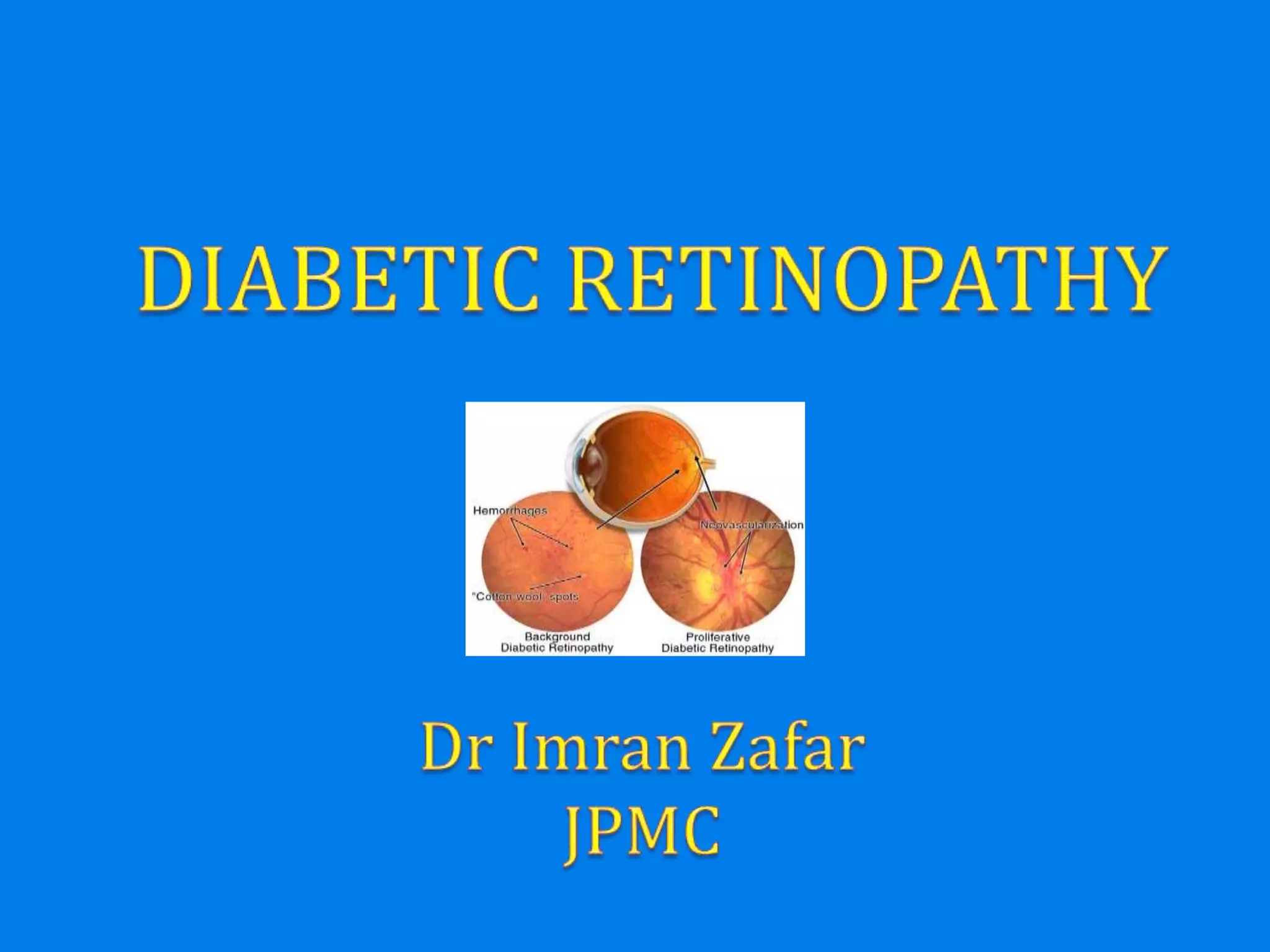
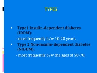
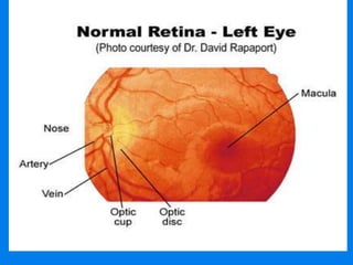
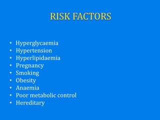
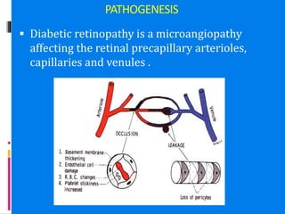
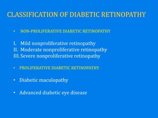
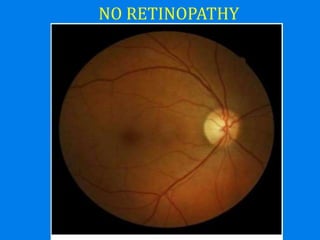
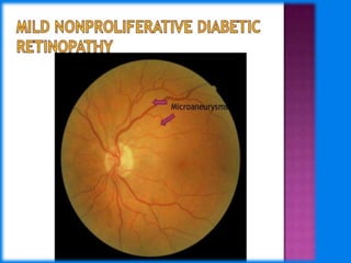
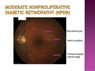
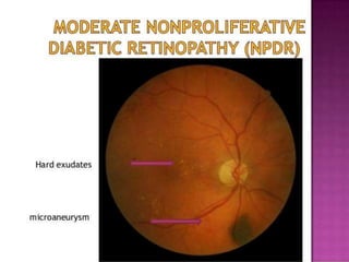
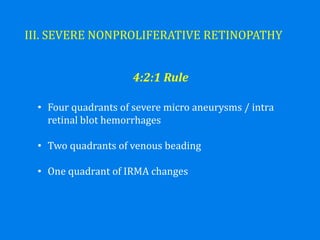
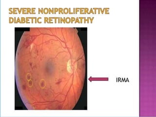
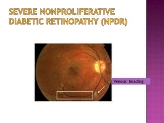
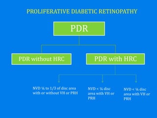
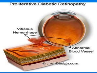
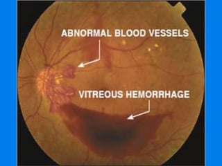
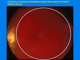
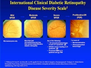
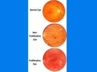
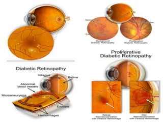
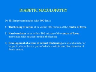
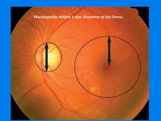
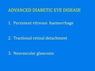
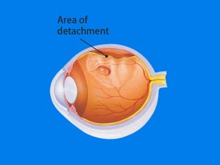
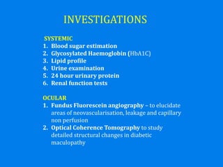
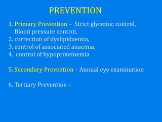
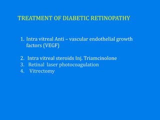
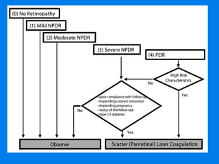
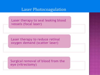
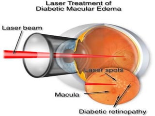
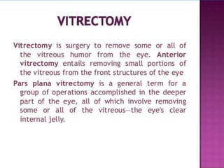
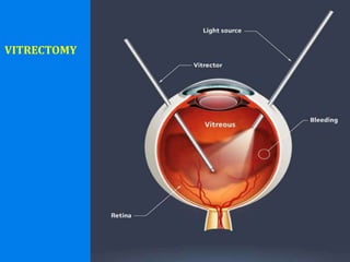
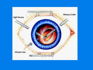
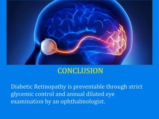
TYPES
 Type1 Insulin-dependent diabetes
(IDDM):
- most frequently b/w 10-20 years.
 Type 2 Non-insulin-dependent diabetes
(NIDDM):
- most frequently b/w the ages of 50-70.
Progressive dysfunction of the retinal
blood vessels caused by chronic
hyperglycaemia.
• Hyperglycaemia
• Hypertension
• Hyperlipidaemia
• Pregnancy
• Smoking
• Obesity
• Anaemia
• Poor metabolic control
• Hereditary
PATHOGENESIS
 Diabetic retinopathy is a microangiopathy
affecting the retinal precapillary arterioles,
capillaries and venules .
• NON-PROLIFERATIVE DIABETIC RETINOPATHY
I. Mild nonproliferative retinopathy
II. Moderate nonproliferative retinopathy
III.Severe nonproliferative retinopathy
• PROLIFERATIVE DIABETIC RETINOPATHY
• Diabetic maculopathy
• Advanced diabetic eye disease
NO RETINOPATHY
I. MILD NONPROLIFERATIVE RETINOPATHY
• At least one micro aneurysm or intraretinal
hemorrhage
• Hard/ Soft exudates may or may not be present
II. MODERATE NONPROLIFERATIVE RETINOPATHY
• Micro aneurysms / intraretinal haemorrhages in 2 or
3 quadrants
• Early mild IRMA Intra retinal Microvascular
abnormalities
• Hard / Soft exudates may or may not be present
III. SEVERE NONPROLIFERATIVE RETINOPATHY
• Four quadrants of severe micro aneurysms / intra
retinal blot hemorrhages
• Two quadrants of venous beading
• One quadrant of IRMA changes
4:2:1 Rule
PROLIFERATIVE DIABETIC RETINOPATHY
PDR
PDR without HRC PDR with HRC
NVD ¼ to 1/3 of disc area
with or without VH or PRH
NVD < ¼ disc
area with VH or
PRH
NVD < ¼ disc
area with VH or
PRH
EXTENSIVE VITREOUS HAEMORRHAGE OBSCURING MOST OF FUNDUS
(WHITE CIRCLE)
DIABETIC MACULOPATHY
On Slit lamp examination with 90D lens :
1. Thickening of retina at or within 500 micron of the centre of fovea
2. Hard exudates at or within 500 micron of the centre of fovea
associated with adjacent retinal thickening
3. Development of a zone of retinal thickening one disc diameter or
larger in size, at least a part of which is within one disc diameter of
foveal centre.
Maculopathy within 1 disc diameter of the fovea.
ADVANCED DIABETIC EYE DISEASE
1. Persistent vitreous haemorrhage
2. Tractional retinal detachment
3. Neovascular glaucoma
INVESTIGATIONS
SYSTEMIC
1. Blood sugar estimation
2. Glycosylated Haemoglobin (HbA1C)
3. Lipid profile
4. Urine examination
5. 24 hour urinary protein
6. Renal function tests
OCULAR
1. Fundus Fluorescein angiography – to elucidate
areas of neovascularisation, leakage and capillary
non perfusion
2. Optical Coherence Tomography to study
detailed structural changes in diabetic
maculopathy
PREVENTION
1. Primary Prevention – Strict glycemic control,
Blood pressure control,
2. correction of dyslipidaemia,
3. control of associated anaemia,
4. control of hypoproteinemia
5. Secondary Prevention – Annual eye examination
6. Tertiary Prevention –
TREATMENT OF DIABETIC RETINOPATHY
1. Intra vitreal Anti – vascular endothelial growth
factors (VEGF)
2. Intra vitreal steroids Inj. Triamcinolone
3. Retinal laser photocoagulation
4. Vitrectomy
Laser Photocoagulation
VITRECTOMY
CONCLUSION
Diabetic Retinopathy is preventable through strict
glycemic control and annual dilated eye
examination by an ophthalmologist.
JAZAKALLAH KHER

DIABETIC RETINOPATHY 2017

  • 2.
    TYPES  Type1 Insulin-dependentdiabetes (IDDM): - most frequently b/w 10-20 years.  Type 2 Non-insulin-dependent diabetes (NIDDM): - most frequently b/w the ages of 50-70.
  • 5.
    Progressive dysfunction ofthe retinal blood vessels caused by chronic hyperglycaemia.
  • 6.
    • Hyperglycaemia • Hypertension •Hyperlipidaemia • Pregnancy • Smoking • Obesity • Anaemia • Poor metabolic control • Hereditary
  • 8.
    PATHOGENESIS  Diabetic retinopathyis a microangiopathy affecting the retinal precapillary arterioles, capillaries and venules .
  • 11.
    • NON-PROLIFERATIVE DIABETICRETINOPATHY I. Mild nonproliferative retinopathy II. Moderate nonproliferative retinopathy III.Severe nonproliferative retinopathy • PROLIFERATIVE DIABETIC RETINOPATHY • Diabetic maculopathy • Advanced diabetic eye disease
  • 12.
  • 13.
    I. MILD NONPROLIFERATIVERETINOPATHY • At least one micro aneurysm or intraretinal hemorrhage • Hard/ Soft exudates may or may not be present
  • 15.
    II. MODERATE NONPROLIFERATIVERETINOPATHY • Micro aneurysms / intraretinal haemorrhages in 2 or 3 quadrants • Early mild IRMA Intra retinal Microvascular abnormalities • Hard / Soft exudates may or may not be present
  • 18.
    III. SEVERE NONPROLIFERATIVERETINOPATHY • Four quadrants of severe micro aneurysms / intra retinal blot hemorrhages • Two quadrants of venous beading • One quadrant of IRMA changes 4:2:1 Rule
  • 21.
    PROLIFERATIVE DIABETIC RETINOPATHY PDR PDRwithout HRC PDR with HRC NVD ¼ to 1/3 of disc area with or without VH or PRH NVD < ¼ disc area with VH or PRH NVD < ¼ disc area with VH or PRH
  • 24.
    EXTENSIVE VITREOUS HAEMORRHAGEOBSCURING MOST OF FUNDUS (WHITE CIRCLE)
  • 28.
    DIABETIC MACULOPATHY On Slitlamp examination with 90D lens : 1. Thickening of retina at or within 500 micron of the centre of fovea 2. Hard exudates at or within 500 micron of the centre of fovea associated with adjacent retinal thickening 3. Development of a zone of retinal thickening one disc diameter or larger in size, at least a part of which is within one disc diameter of foveal centre.
  • 29.
    Maculopathy within 1disc diameter of the fovea.
  • 30.
    ADVANCED DIABETIC EYEDISEASE 1. Persistent vitreous haemorrhage 2. Tractional retinal detachment 3. Neovascular glaucoma
  • 32.
    INVESTIGATIONS SYSTEMIC 1. Blood sugarestimation 2. Glycosylated Haemoglobin (HbA1C) 3. Lipid profile 4. Urine examination 5. 24 hour urinary protein 6. Renal function tests OCULAR 1. Fundus Fluorescein angiography – to elucidate areas of neovascularisation, leakage and capillary non perfusion 2. Optical Coherence Tomography to study detailed structural changes in diabetic maculopathy
  • 33.
    PREVENTION 1. Primary Prevention– Strict glycemic control, Blood pressure control, 2. correction of dyslipidaemia, 3. control of associated anaemia, 4. control of hypoproteinemia 5. Secondary Prevention – Annual eye examination 6. Tertiary Prevention –
  • 34.
    TREATMENT OF DIABETICRETINOPATHY 1. Intra vitreal Anti – vascular endothelial growth factors (VEGF) 2. Intra vitreal steroids Inj. Triamcinolone 3. Retinal laser photocoagulation 4. Vitrectomy
  • 36.
  • 40.
  • 42.
    CONCLUSION Diabetic Retinopathy ispreventable through strict glycemic control and annual dilated eye examination by an ophthalmologist.
  • 43.