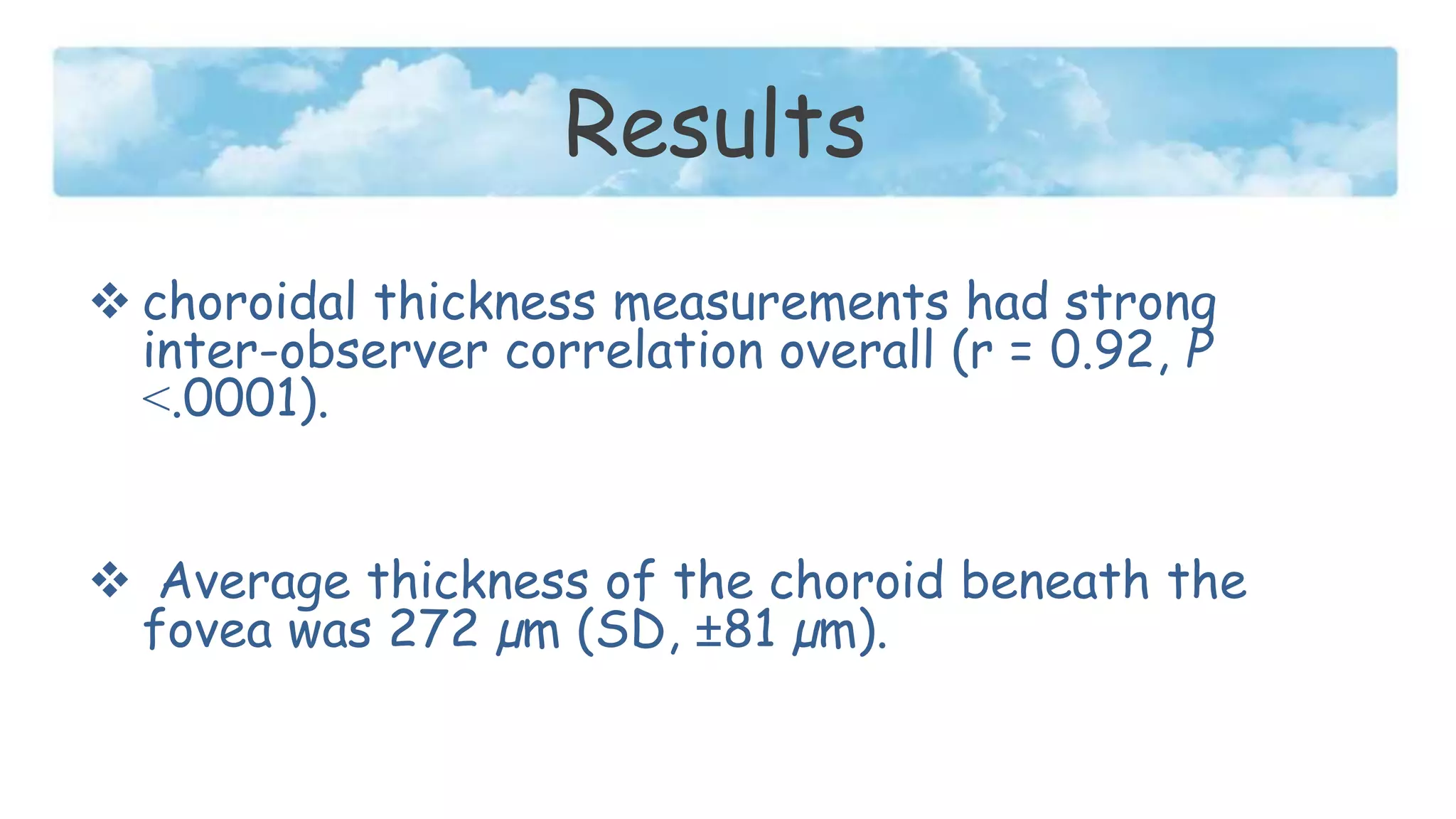
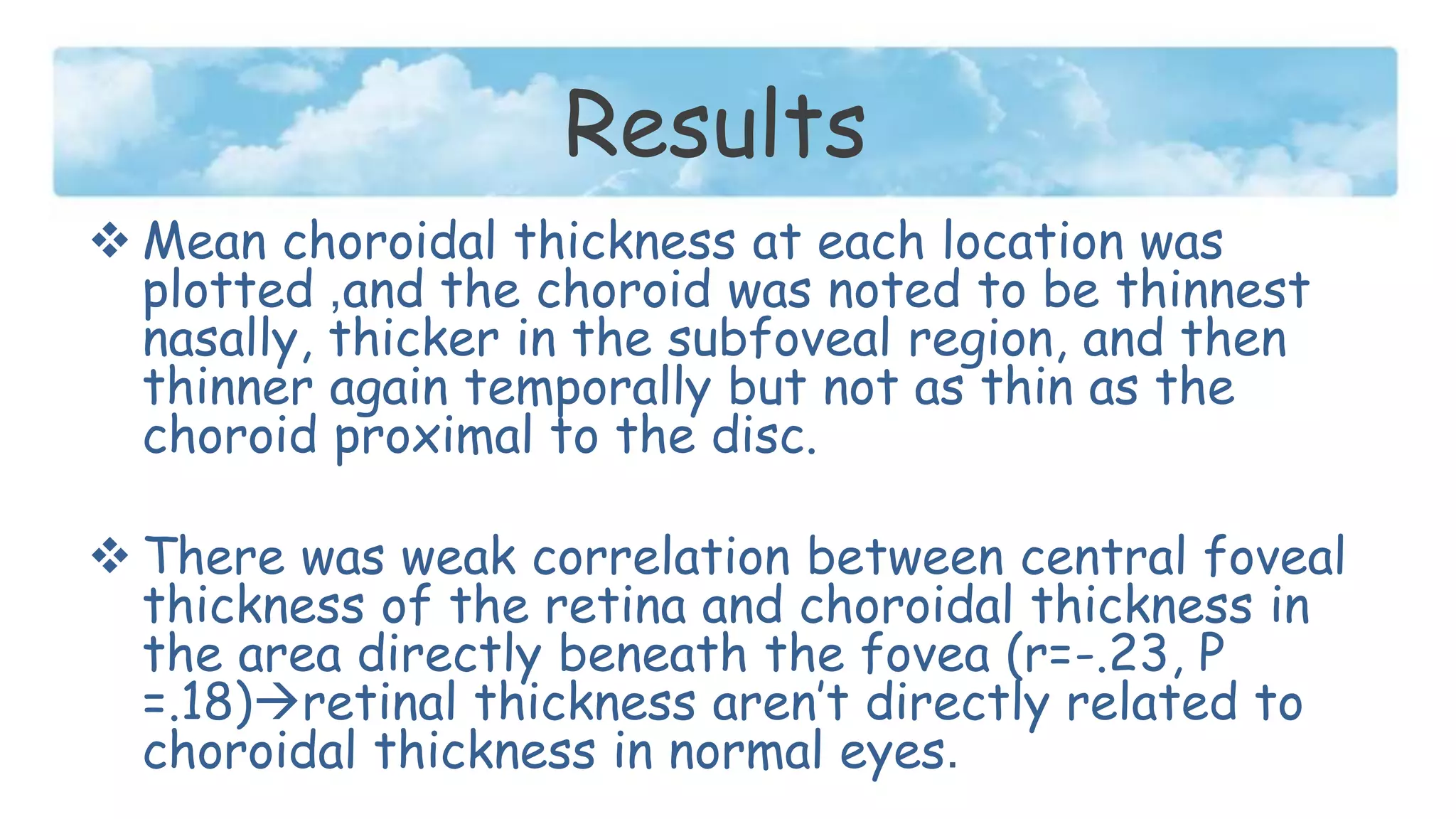
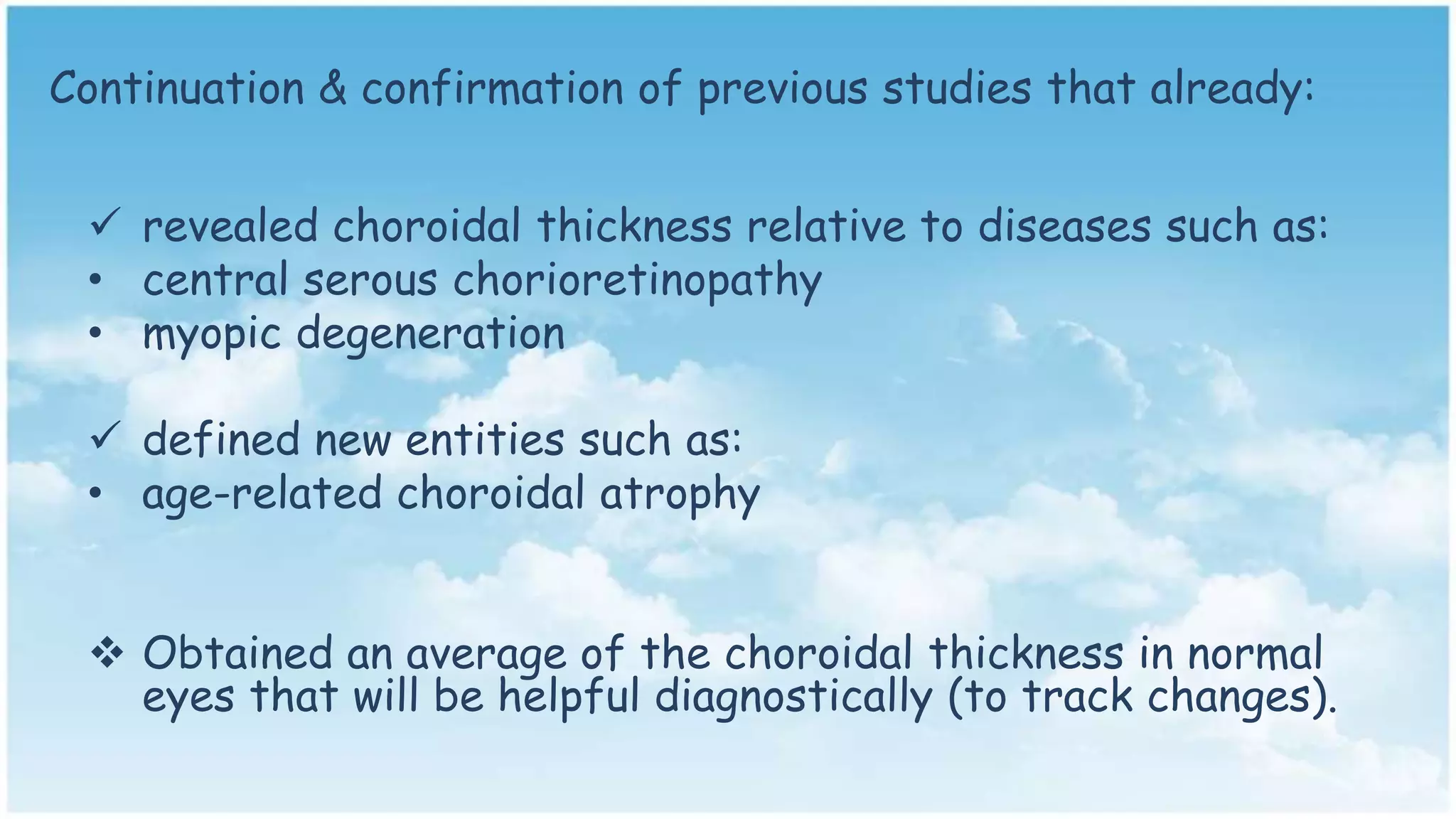
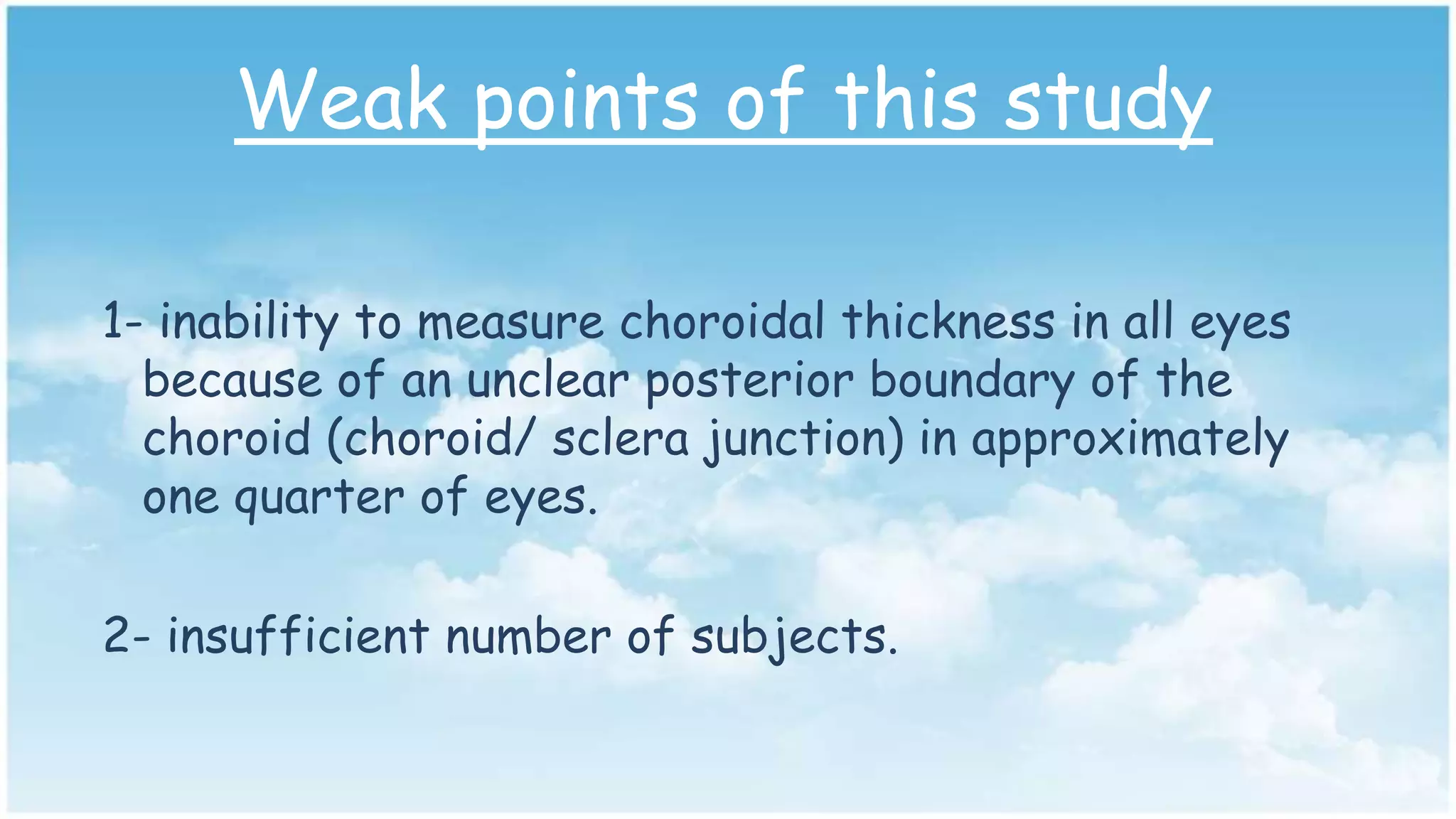
Choroidal Thickness in Normal Eyes
Measured using Cirrus HD
Optical Coherence Tomography
journal critique
By: Manal Al-Romeih
Opto 541
Hypothesis
to evaluate the Cirrus HD-OCT device to
see if its new software would allow accurate
measurement of choroidal thickness and area
in normal eyes.
to determine the correlation between
retinal thickness and choroidal thickness.
Methods
Thirty four eyes of 34 subjects
were measured (one eye per
subject).
With no retinal or choroidal
disease.
Methods
 Subjects with myopia greater than -6.0 D were
excluded.
 Spectral-domain optical coherence tomography
(SD-OCT) with frame enhancement software was
used.
Emit lights into the eye; measure reflectivity of the tissues within a target
cube
Rendering the measurements for visualizing inner-structures
Methods
 Statistical analysis (Pearson correlation)
was used to correlate inter-observer
findings, choroidal thickness and area
measurements with age, and choroidal
thickness with retinal foveal thickness.
2 independent observers measured:
1- choroidal thickness perpendicularly from the posterior
edge of RPE to the choroid/sclera junction at 500 µm
intervals up to 2500 µm temporal and nasal from the
fovea.
2- central foveal thickness.
3- Central 1-mm area of the choroid, 500 µm temporal and
nasal to the fovea.
Histological picture showing the choroid
boundaries
(Top) Demonstration of normal choroidal thickness measured at 500µm intervals, up
to 2500µm temporal and nasal to the fovea. Perpendicular lines were drawn from the
posterior edge of the RPE to the choroid/sclera junction using Cirrus HD-OCT
software.
(Bottom) Central 1-mm cross-sectional area of choroid measured using Image
analysis software; outlined area is 0.32 mm2
Results
 choroidal thickness measurements had strong
inter-observer correlation overall (r = 0.92, P
˂.0001).
 Average thickness of the choroid beneath the
fovea was 272 µm (SD, ±81 µm).
Results
 Mean choroidal thickness at each location was
plotted ,and the choroid was noted to be thinnest
nasally, thicker in the subfoveal region, and then
thinner again temporally but not as thin as the
choroid proximal to the disc.
 There was weak correlation between central foveal
thickness of the retina and choroidal thickness in
the area directly beneath the fovea (r=-.23, P
=.18)retinal thickness aren’t directly related to
choroidal thickness in normal eyes.
Graph of mean macular choroidal thickness in normal eyes. Mean
thickness at each of the 11 locations measured at 500 µm (0.5˜mm)
intervals temporal (T) and nasal (N) to the fovea (F).
This article raised a good topic since:
 The choroid plays a vital role in the patho-physiology of
many diseases affecting the retina.
 Measuring it in vivo was necessary to accurately determine
its true structure and thickness due to its high
vascularization.
 Using OCT imaging techniques is a noninvasive way to
determine optical biopsy of the posterior segment.
Continuation & confirmation of previous studies that already:
 revealed choroidal thickness relative to diseases such as:
• central serous chorioretinopathy
• myopic degeneration
 defined new entities such as:
• age-related choroidal atrophy
 Obtained an average of the choroidal thickness in normal
eyes that will be helpful diagnostically (to track changes).
Obstacle faced measurement of choroidal
thickness
1- Information regarding choroidal thickness in normal eyes
was based primarily on histologic results, which do not
necessarily reflect the true measurements of it, since
histologic fixation produces artifacts because of
processing, tissue deformation, and shrinkage.
2- adequate visualization of the choroid using OCT hasn’t
been possible until recently when Spaide and associates
used spectral domain OCT instrument which offers eye
tracking technology and the capability to capture up to 100
B-scans.
Manjunath et alSpaide et al
34 (34 eyes)30 (54 eyes)Subjects (eyes)
51.1 yrs50.4 yrsMean age
r = −0.62-ve (↓15.6 μm/decade)Age/choroidal
thickness correlation
272 μm287 μmChoroidal thickness
(subfoveal)
New software (raster scan)positioning a SD-OCT device close
enough to the eye to acquire an
inverted image
Technique
Apr 2010Dec 2008Date
Weak points of this study
1- inability to measure choroidal thickness in all eyes
because of an unclear posterior boundary of the
choroid (choroid/ sclera junction) in approximately
one quarter of eyes.
2- insufficient number of subjects.
3- The previous study done by Spaide &
Margolis confirmed the integrity of the
new software used in this study.
One study isn’t enough for good judgment.
Conclusion
• This study led a good research regarding
choroidal thickness in normal eyes since there
is lack in studies in this field.
• On the other hand, evaluating the new cirrus
soft ware had no strong credibility due to
insufficient references in normal choroidal
thickness.
.
Thanks for listening

Choroidal thickness in normal eyes journal critique

  • 1.
    Choroidal Thickness inNormal Eyes Measured using Cirrus HD Optical Coherence Tomography journal critique By: Manal Al-Romeih Opto 541
  • 2.
    Hypothesis to evaluate theCirrus HD-OCT device to see if its new software would allow accurate measurement of choroidal thickness and area in normal eyes. to determine the correlation between retinal thickness and choroidal thickness.
  • 3.
    Methods Thirty four eyesof 34 subjects were measured (one eye per subject). With no retinal or choroidal disease.
  • 4.
    Methods  Subjects withmyopia greater than -6.0 D were excluded.  Spectral-domain optical coherence tomography (SD-OCT) with frame enhancement software was used.
  • 5.
    Emit lights intothe eye; measure reflectivity of the tissues within a target cube Rendering the measurements for visualizing inner-structures
  • 6.
    Methods  Statistical analysis(Pearson correlation) was used to correlate inter-observer findings, choroidal thickness and area measurements with age, and choroidal thickness with retinal foveal thickness.
  • 7.
    2 independent observersmeasured: 1- choroidal thickness perpendicularly from the posterior edge of RPE to the choroid/sclera junction at 500 µm intervals up to 2500 µm temporal and nasal from the fovea. 2- central foveal thickness. 3- Central 1-mm area of the choroid, 500 µm temporal and nasal to the fovea.
  • 8.
    Histological picture showingthe choroid boundaries
  • 9.
    (Top) Demonstration ofnormal choroidal thickness measured at 500µm intervals, up to 2500µm temporal and nasal to the fovea. Perpendicular lines were drawn from the posterior edge of the RPE to the choroid/sclera junction using Cirrus HD-OCT software. (Bottom) Central 1-mm cross-sectional area of choroid measured using Image analysis software; outlined area is 0.32 mm2
  • 10.
    Results  choroidal thicknessmeasurements had strong inter-observer correlation overall (r = 0.92, P ˂.0001).  Average thickness of the choroid beneath the fovea was 272 µm (SD, ±81 µm).
  • 11.
    Results  Mean choroidalthickness at each location was plotted ,and the choroid was noted to be thinnest nasally, thicker in the subfoveal region, and then thinner again temporally but not as thin as the choroid proximal to the disc.  There was weak correlation between central foveal thickness of the retina and choroidal thickness in the area directly beneath the fovea (r=-.23, P =.18)retinal thickness aren’t directly related to choroidal thickness in normal eyes.
  • 12.
    Graph of meanmacular choroidal thickness in normal eyes. Mean thickness at each of the 11 locations measured at 500 µm (0.5˜mm) intervals temporal (T) and nasal (N) to the fovea (F).
  • 13.
    This article raiseda good topic since:  The choroid plays a vital role in the patho-physiology of many diseases affecting the retina.  Measuring it in vivo was necessary to accurately determine its true structure and thickness due to its high vascularization.  Using OCT imaging techniques is a noninvasive way to determine optical biopsy of the posterior segment.
  • 14.
    Continuation & confirmationof previous studies that already:  revealed choroidal thickness relative to diseases such as: • central serous chorioretinopathy • myopic degeneration  defined new entities such as: • age-related choroidal atrophy  Obtained an average of the choroidal thickness in normal eyes that will be helpful diagnostically (to track changes).
  • 15.
    Obstacle faced measurementof choroidal thickness 1- Information regarding choroidal thickness in normal eyes was based primarily on histologic results, which do not necessarily reflect the true measurements of it, since histologic fixation produces artifacts because of processing, tissue deformation, and shrinkage. 2- adequate visualization of the choroid using OCT hasn’t been possible until recently when Spaide and associates used spectral domain OCT instrument which offers eye tracking technology and the capability to capture up to 100 B-scans.
  • 16.
    Manjunath et alSpaideet al 34 (34 eyes)30 (54 eyes)Subjects (eyes) 51.1 yrs50.4 yrsMean age r = −0.62-ve (↓15.6 μm/decade)Age/choroidal thickness correlation 272 μm287 μmChoroidal thickness (subfoveal) New software (raster scan)positioning a SD-OCT device close enough to the eye to acquire an inverted image Technique Apr 2010Dec 2008Date
  • 17.
    Weak points ofthis study 1- inability to measure choroidal thickness in all eyes because of an unclear posterior boundary of the choroid (choroid/ sclera junction) in approximately one quarter of eyes. 2- insufficient number of subjects.
  • 18.
    3- The previousstudy done by Spaide & Margolis confirmed the integrity of the new software used in this study. One study isn’t enough for good judgment.
  • 19.
    Conclusion • This studyled a good research regarding choroidal thickness in normal eyes since there is lack in studies in this field. • On the other hand, evaluating the new cirrus soft ware had no strong credibility due to insufficient references in normal choroidal thickness.
  • 20.