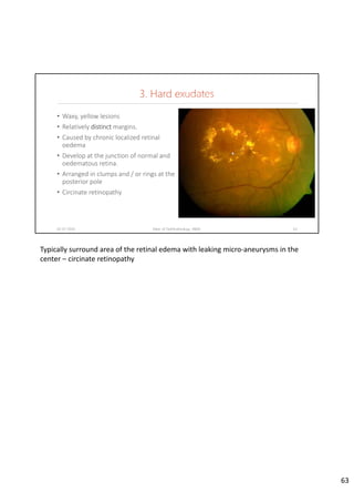
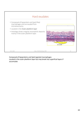
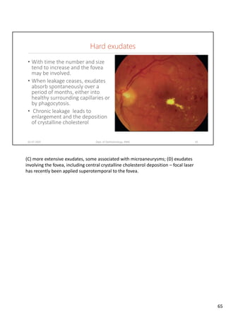
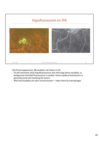
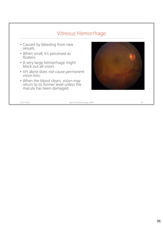
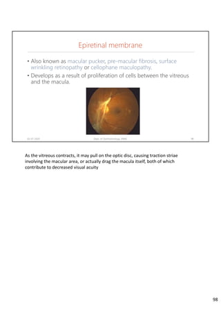
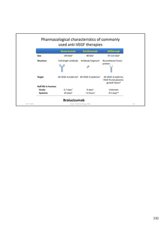
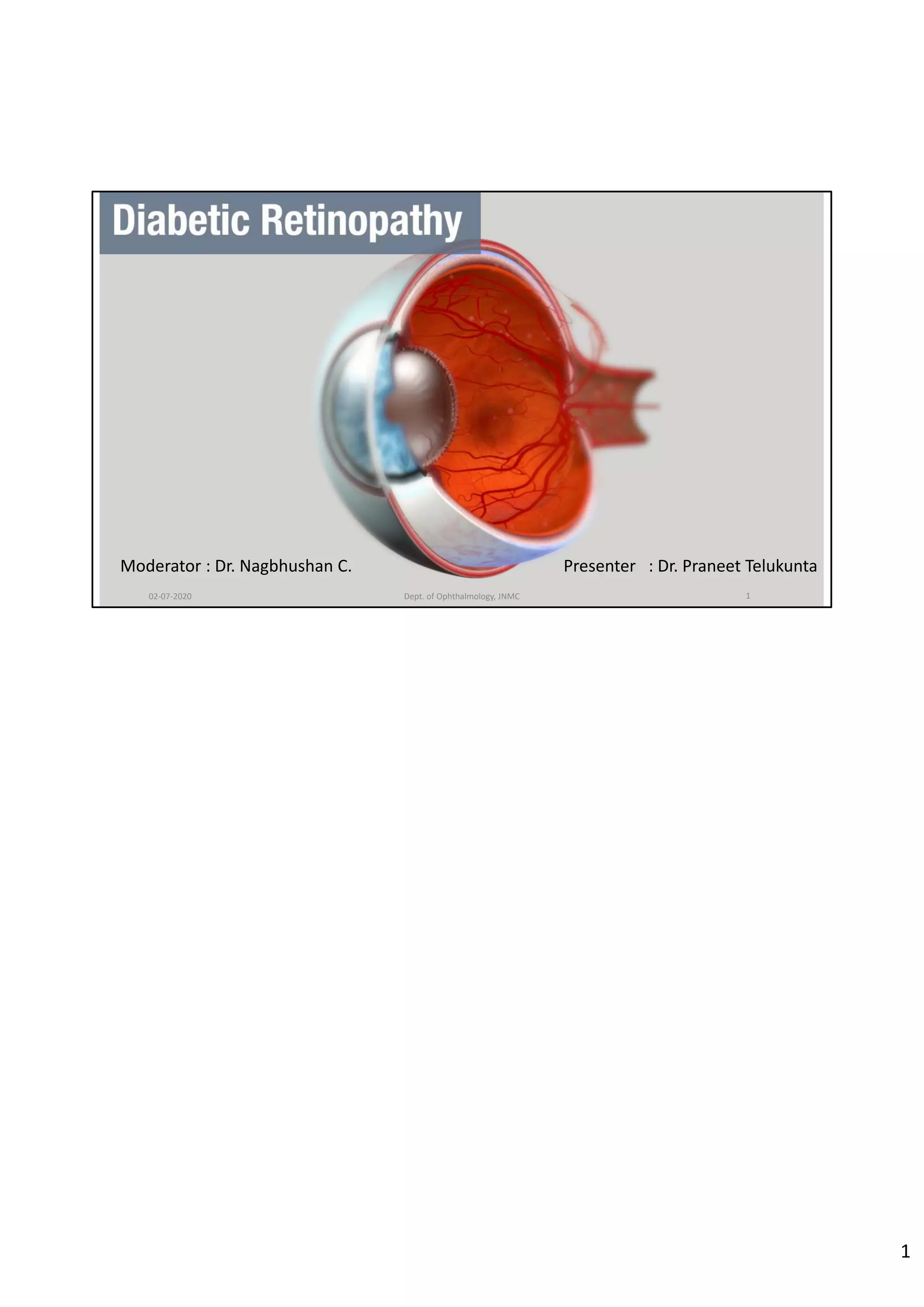
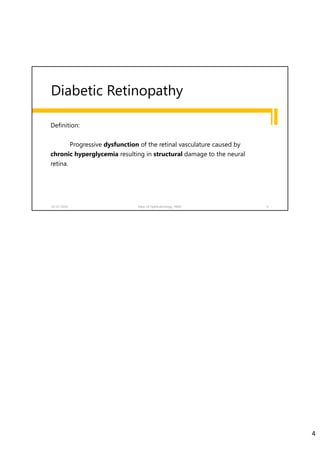
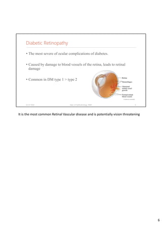
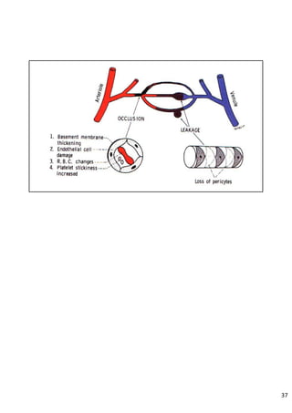
Diabetic retinopathy is caused by damage to the blood vessels in the retina from chronic hyperglycemia. It is the most common retinal vascular disease and a leading cause of vision loss. The risk and severity of diabetic retinopathy increases with longer duration of diabetes, poorer blood glucose control, and presence of other complications like hypertension. The pathogenesis involves microvascular changes like capillary non-perfusion and death of pericytes, as well as increased vascular permeability and neo-vascularization due to elevated levels of vasoactive growth factors. Strict control of blood glucose and other cardiovascular risk factors can help prevent and slow the progression of this vision-threatening condition.
This document summarizes diabetic retinopathy. It defines diabetic retinopathy as a chronic progressive sight-threatening disease caused by prolonged hyperglycemia. Key points include:
- Diabetic retinopathy is a leading cause of blindness and its prevalence is increasing globally.
- Risk factors include duration of diabetes, poor glycemic control, pregnancy, hypertension, nephropathy, and smoking.
- It progresses from non-proliferative to proliferative stages and can lead to vision loss through edema, neovascularization, and retinal detachment.
- Treatments like laser photocoagulation and anti-VEGF drugs aim to prevent vision loss by treating proliferative retinopathy and mac
This document discusses diabetic retinopathy, its causes, stages, treatments, and prevention. It is progressive retinal vessel dysfunction caused by long-term hyperglycemia. Key factors that contribute to its development include hypertension, hyperlipidemia, female sex, pregnancy, smoking, obesity, and poor metabolic control. Stages include non-proliferative and proliferative retinopathy. Treatments include anti-VEGF drugs, laser photocoagulation, vitrectomy, and strict control of blood sugar and blood pressure to prevent its progression.
Hypertensive Retinopathy (HTN-R) for undergraduate MBBS Students.
Covers the basics of Aetiology, pathophysiology, clinical features, Classification and management of HTN-R.
Also encompasses salient points for PGMEE
Central retinal artery occlusion (CRAO) results from obstruction of blood flow through the central retinal artery, causing ischemia of the inner retina. It presents with sudden, painless vision loss. Examination typically reveals a cherry red spot at the macula and whitening of the inner retina. Ancillary testing such as OCT, FFA, and ERG can help evaluate the extent of retinal damage. Prompt diagnosis and treatment of underlying causes is important to prevent further vascular events.
Diabetic retinopathy is a leading cause of blindness worldwide. Prolonged hyperglycemia can damage retinal blood vessels and neurons. Over time, this can lead to vision loss through retinal edema, hemorrhage, fibrosis or neovascularization. Risk factors include duration and control of diabetes, hypertension, and nephropathy. Treatment focuses on laser photocoagulation and intravitreal injections to reduce edema or abnormal blood vessels, along with glycemic control to prevent progression. Regular screening is important to detect diabetic retinopathy early when treatment is most effective.
This document provides information on optic neuritis, including its definition, etiology, pathology, clinical features, diagnosis, and treatment. Some key points:
- Optic neuritis is an inflammatory condition of the optic nerve that can cause visual loss. Common causes include multiple sclerosis, infections, and autoimmune disorders.
- Clinical features include sudden visual loss or blurring in one eye, pain with eye movement, and abnormal pupil response to light. Diagnosis is made based on symptoms and MRI findings.
- Treatment involves corticosteroids via IV or oral administration. The Optic Neuritis Treatment Trial found IV methylprednisolone can help delay conversion to multiple sclerosis.
This document discusses the anatomy and function of the oculomotor nerve (cranial nerve 3). It describes the nucleus and course of the nerve, causes of lesions, clinical features of total oculomotor nerve palsy, treatment options which may include surgery or monitoring, and differential diagnoses. History, examination, and investigations are outlined to evaluate patients presenting with oculomotor nerve palsies.
Diabetic retinopathy is a leading cause of blindness that results from damage to the blood vessels of the retina due to complications of diabetes. It can progress from mild nonproliferative retinopathy, to moderate and severe nonproliferative stages, and finally to the most severe proliferative retinopathy stage. Risk factors include duration of diabetes, blood sugar level, and high blood pressure. Treatment depends on the stage but may include laser photocoagulation surgery or vitrectomy to prevent vision loss. Strict control of blood sugar and blood pressure along with regular eye exams can help prevent and treat diabetic retinopathy.
This document summarizes diabetic retinopathy, including its risk factors, pathogenesis, classification, signs, investigations, and treatments. It notes that diabetic retinopathy is caused by damage to the small blood vessels in the retina from high blood sugar levels over time. The main risk factors are duration of diabetes, poor blood sugar control, pregnancy, and other systemic diseases. The document describes the classification of diabetic retinopathy from mild non-proliferative to more severe proliferative stages. Key signs and investigations are also outlined, along with medical, laser, and surgical therapy options.
The tear film has three layers:
1. A mucin layer secreted by goblet cells that forms a hydrophilic barrier on the cornea.
2. An aqueous layer secreted mainly by the lacrimal gland, containing proteins, electrolytes, and other components.
3. An outer lipid layer secreted by meibomian glands that prevents evaporation and maintains tear film stability.
The tear film is regulated by hormonal and neural pathways and provides nutrients to the cornea, removes waste, lubricates the eye, and protects against infection through its biochemical composition.
Third nerve palsy is a condition that leads to impairment of motor function as the third cranial nerve innervates most eye muscles. It can be congenital or acquired through conditions like diabetes, hypertension, tumors or trauma. Acquired third nerve palsy often involves patients over 45 and can cause ptosis and pupil involvement while congenital palsy is usually unilateral and incomplete without ptosis. The document discusses a case study of a 18-year old female referred for assessment of right eye exotropia secondary to trauma as a child. Her examination findings and diagnosis of right eye exotropia are presented along with a management plan of unilateral eye muscle surgery.
Diabetic retinopathy is progressive dysfunction of retinal blood vessels due to hyperglycemia. It is the leading cause of blindness among working-age adults. The risk increases with duration of diabetes and is also increased by hypertension, smoking, and renal disease. Early stages are asymptomatic but can progress to proliferative diabetic retinopathy with neovascularization, potentially leading to vitreous hemorrhage or retinal detachment. Treatment depends on severity and may include laser photocoagulation, intravitreal injections, or vitrectomy surgery. Regular screening is important to detect diabetic retinopathy early and prevent vision loss.
The cornea is the transparent front part of the eye that transmits and focuses light. It has 3 main layers - an outer epithelial layer, a thick middle stromal layer made of collagen, and an inner single-cell endothelial layer. The cornea derives its strength and curvature from the orderly arrangement of collagen in the stroma. It remains transparent due to its regular structure without blood vessels and the deturgescent properties maintained by the endothelial pump. The cornea has a high metabolic rate powered by glucose and oxygen and is innervated by nerves for vision and protection.
This document discusses the effects of diabetes on the eye. It begins with an introduction to diabetes mellitus and its long-term damaging effects on organs. It then covers the two main types of diabetes and their characteristics. The document discusses the pathogenesis of diabetic retinopathy and how high blood glucose damages blood vessels in the eye. It provides a detailed overview of the stages of diabetic retinopathy from mild non-proliferative to proliferative and potential vision loss outcomes. Management strategies like glucose control, laser therapy, anti-VEGF drugs, and vitrectomy are summarized.
Hypertensive retinopathy is retinal vascular damage caused by systemic hypertension. It can be detected by examining the ocular fundus, where changes to the arterioles and veins may indicate vascular damage from high blood pressure. These changes include arteriolar narrowing, hemorrhages, cotton wool spots, hard exudates, and optic disc swelling. The risk factors include age, family history of hypertension, obesity, smoking, stress, alcohol consumption and lack of exercise. Treatment focuses on lowering blood pressure through lifestyle changes like exercise and diet, as well as medication if needed, to prevent further organ damage.
1) A 20-year-old boy presented with gradual decrease in night vision over 6 months. Examination revealed signs consistent with retinitis pigmentosa including bone-spicule pigmentation and waxy pale optic discs.
2) Retinitis pigmentosa is a hereditary retinal dystrophy where rod photoreceptor cells are initially and predominantly affected, followed by cone degeneration. It typically causes night blindness and progressive loss of peripheral vision.
3) There is no cure for retinitis pigmentosa. Treatment aims to slow progression and improve quality of life through low vision aids, vitamin supplements, and emerging therapies like gene and retinal prosthesis. Prognosis depends on inheritance pattern, with
Over 112,000 people in Indiana have diabetic retinopathy, which is around 20% of diabetics in the state. About 60% of diabetics receive an annual eye exam, but each year between 12,000 to 24,000 new cases of blindness occur from diabetic retinopathy. Diabetic retinopathy is caused by changes in the blood vessels in the eye and can be detected and treated through annual dilated eye exams and procedures like laser surgery. Maintaining good control of blood sugar and receiving regular eye exams can help reduce the risk of vision loss from diabetic eye disease.
Diabetic retinopathy is damage to the blood vessels of the retina due to diabetes. A study compared the standard treatment of pan-retinal photocoagulation (PRP) to initial treatment with intravitreal anti-VEGF injections (ranibizumab) to delay or prevent the need for PRP. At 2 years, vision was maintained or improved in the ranibizumab group while vision remained unchanged in the PRP group. The study suggests initial treatment of proliferative diabetic retinopathy with anti-VEGF injections like ranibizumab may be as effective as immediate PRP treatment and could help delay or reduce the need for destructive PRP therapy.
This document provides information on the anatomy and diseases of the vitreous humor. It discusses that the vitreous humor is a jelly-like structure that fills the back of the eye and provides support. Common diseases include vitreous liquefaction, detachment, hemorrhage, and opacities. Vitreous liquefaction is the most common degenerative change and causes floaters. Posterior vitreous detachment often occurs in older individuals and may lead to retinal tears or breaks. Vitreous opacities can result from inflammatory cells, aggregates, tumors or hemorrhages. Vitreous hemorrhage usually stems from retinal vessels and can cause vision loss.
Diabetic maculopathy is a form of damage to the eye causing by diabetic macular oedema where fluids build up on the macula. It can be cured by laser surgeries.
The document discusses retinal arterial occlusion, including the central retinal artery and its branches. It describes the anatomy and blood supply of the retina. There are several potential mechanisms of retinal arterial occlusion, most commonly atherosclerosis-related thrombosis. Clinical presentations vary depending on the site of occlusion, such as central retinal artery occlusion presenting with a cherry red spot and cilioretinal artery occlusion causing pericentral scotomas. Management includes treating the acute event to restore vision as well as workup and management of any underlying systemic conditions. However, visual recovery is often poor due to retinal infarction.
Retinitis pigmentosa is a genetic disorder that causes the rods and cones in the retina to deteriorate over time. This leads to progressive vision loss starting with night blindness and loss of peripheral vision, which can eventually cause total blindness. There is no cure, but vitamin A supplementation and an omega-3 rich diet have been shown to potentially slow the progression of the disease. It is diagnosed through visual field tests and ERG testing, and patients are referred to low vision specialists who can prescribe aids to help maximize remaining vision.
This document discusses several systemic diseases that can affect the eyes, including:
- Hypertensive retinopathy, which can damage the retina, choroid and optic nerve due to high blood pressure. It is classified based on the severity of retinal vascular changes.
- Dysthyroid eye disease, also known as Graves' ophthalmopathy, which is associated with thyroid disease and causes proptosis, lid retraction, and inflammation of extraocular muscles and orbital fat. It can also lead to intraocular issues like choroidal folds.
- Uveitis, which is inflammation of the uveal tract that can be anterior, intermediate, or posterior based on location. It has various causes and presentations.
GENERAL INFORMATION ABOUT DIABETIC MACULAR EDEMA WITH 2 PATIENT CASES, TREATED WITH 2 DIFFERENT TREATMENT TECHNIQUES.
CLASSIFICATION (CSME)
RISK FACTORS
CAUSES
SIGNS AND SYMPTOMS
MANAGEMENT AND TREATMENT OPTIONS
DIAGNISTIC TESTS, BLOOD AND URINE TEST
SCORING SYSTEM
PATHOLOGY
DIFFERENTIAL DIAGNOSIS
PROGNOSIS
EPIDEMIOLOGY
DESCRIPTION OF 2 CASES, THEIR DIAGNOSTIC RESULT AND DETAILS ABOUT TREATMENTS PERFORMED.
This document provides an overview of sixth nerve palsy, including:
- The sixth cranial nerve innervates the lateral rectus muscle to enable eye abduction. Sixth nerve palsy results in limited ability to turn the eye outward.
- Causes of sixth nerve palsy include idiopathic, vascular issues like hypertension and diabetes, trauma, and tumors. Symptoms include esotropia and diplopia.
- Diagnosis involves assessing eye movement limitations and diplopia. Treatment options include occlusion to control diplopia, botulinum toxin injection, or strabismus surgery if no spontaneous recovery occurs. Prognosis is generally good, with many cases recovering spontaneously in
Retinitis pigmentosa is a group of inherited retinal diseases characterized by progressive degeneration of the photoreceptors. It initially affects rods, resulting in night blindness and peripheral vision loss, and later involves cones leading to tunnel vision. Symptoms include nyctalopia and peripheral field defects. Signs include bone spicule pigmentation, arteriolar attenuation, and disc pallor. It can be inherited in autosomal dominant, recessive or X-linked patterns. Investigations include electroretinography to detect photoreceptor dysfunction and optical coherence tomography. There is currently no cure or treatment to stop progression.
- Diabetic retinopathy is a leading cause of blindness in people aged 20-74 and affects individuals in their most productive years. The risk and severity of retinopathy increases with longer duration of diabetes and poorer blood glucose control.
- The disease involves microvascular changes including microaneurysms, hemorrhages, hard exudates, and new abnormal blood vessel growth. Without treatment, this can lead to vision loss from macular edema, retinal detachment, or vitreous hemorrhage.
- Treatment involves managing blood sugar and blood pressure, as well as laser photocoagulation surgery or intravitreal injections to prevent vision loss from proliferative retinopathy or macular edema.
Diabetic retinopathy is a complication of diabetes that affects the eyes. It is caused by damage to the blood vessels of the light-sensitive tissue at the back of the eye (retina). Over time, diabetes can damage these small blood vessels and cause them to leak blood and other fluids into the retina. This can lead to blurred or decreased vision. The document discusses the causes, risk factors, signs, symptoms, stages, treatments, and importance of screening for diabetic retinopathy. Regular eye exams are emphasized to detect diabetic retinopathy early before vision is affected.
Diabetic retinopathy is a complication of diabetes that affects the small blood vessels in the retina. It is the leading cause of blindness in working age adults. Strict control of blood sugar and blood pressure can help prevent and slow the progression of diabetic retinopathy. Annual eye exams are important for early detection and treatment if needed to prevent vision loss. As diabetic retinopathy progresses, it is classified into mild, moderate, and severe non-proliferative stages and proliferative stage. Treatments include laser therapy and anti-VEGF injections to prevent further vision loss.
This document presents information on diabetic retinopathy including its causes, risk factors, stages, diagnosis, and treatment. It begins with an introduction by Sonali Diwate and objectives of the presentation. It then outlines the topics to be discussed and provides details on the types and pathogenesis of diabetic retinopathy. The stages of diabetic retinopathy from mild nonproliferative to proliferative are explained. Risk factors, diagnostic tests, management approaches like laser therapy and anti-VEGF injections, potential biomarkers and conclusions are summarized. The document aims to raise awareness of diabetic retinopathy as a public health problem and discuss treatments and management of the disease.
microvascular complications of DM 09-12-2023.pptxmanjujanhavi
etiopathogenesis of microvascular complications , pathophysiology of each type of retino, nephropathy ,neuropathy & diabetic foot , prevention , early detection ,patient education
1. Diabetic retinopathy is a microvascular complication of diabetes that affects the small blood vessels in the retina and is a leading cause of blindness.
2. Early changes include loss of retinal pericytes, thickening of capillary basement membranes, and formation of microaneurysms. Later stages involve proliferative retinopathy and macular edema.
3. Risk factors include duration of diabetes, poor blood glucose control, hypertension, and nephropathy. Tight control of blood glucose and blood pressure can help reduce the risk and progression of diabetic retinopathy.
This document provides guidelines for screening, monitoring, classifying severity, and treating diabetic macular edema (DME). It recommends annual screening of diabetic patients aged 15+ for retinopathy and treating any sight-threatening cases found. For DME treatment, it discusses traditional laser photocoagulation as well as newer options like intravitreal corticosteroids and anti-VEGF drugs. Intravitreal injections of anti-VEGF agents are considered first-line therapy for center-involving DME, with laser as an option for non-center cases or if thickening persists after anti-VEGF treatment. Strict control of modifiable risk factors like glycemia, blood pressure, and lipids can also help prevent
DIABETES & PERIODONTAL DISEASES PART 1.pptxDentalYoutube
This document discusses the relationship between diabetes and periodontal disease. It begins with an introduction and overview of diabetes, including definitions, classifications, complications, signs and symptoms, and methods of monitoring glycemic control. It then covers oral manifestations of diabetes, mechanisms by which diabetes can influence the periodontium, and vice versa. The management of diabetic patients and considerations for dental implants are also addressed, with the goal of providing clinicians background knowledge on the implications of diabetes for dental care.
This document provides an overview of diabetic retinopathy. It begins by defining diabetic retinopathy as a microangiopathy affecting the small blood vessels of the retina that is a leading cause of blindness in working-age adults. The document then discusses the pathogenesis of diabetic retinopathy including anatomical lesions like loss of pericytes, capillary basement membrane thickening, and microaneurysms. It also covers biochemical mechanisms involving aldose reductase, advanced glycation end products, and reactive oxygen species. Epidemiology, risk factors like diabetes duration and control, and landmark studies like the Diabetes Control and Complications Trial are summarized.
DIABETES & PERIODONTAL DISEASES PART 2.pptxDentalYoutube
Diabetes is a chronic disease characterized by high blood sugar levels. There are two main types - type 1 caused by lack of insulin production and type 2 related to insulin resistance. Poorly controlled diabetes can lead to oral complications like periodontal disease. Periodontitis is more severe in diabetics due to changes in glucose and bacteria levels in gum tissue, impaired immune response, and effects on collagen metabolism from advanced glycation end products. Dental practitioners must consider a diabetic patient's medication and glycemic control for safe treatment and minimize risks like hypoglycemia.
This document summarizes diabetic retinopathy, including its types, pathogenesis, risk factors, diagnosis, management, and biomarkers. Diabetic retinopathy occurs when diabetes damages the tiny blood vessels inside the retina. It is classified into non-proliferative and proliferative stages. Risk factors include duration of diabetes, poor control, hypertension, and nephropathy. Diagnosis involves eye exams and tests like fluorescein angiography. Management includes laser surgery, injections, and vitrectomy. Novel biomarkers being studied to predict diabetic retinopathy include fibrinogen, apolipoproteins, retinal arteriolar tortuosity, and inflammatory markers like interleukin-6 and C-reactive protein.
This document discusses diabetic retinopathy, which is a complication of diabetes that affects the eyes. It begins by defining diabetes mellitus and its two main types. It then discusses the various stages of diabetic retinopathy from mild non-proliferative diabetic retinopathy to proliferative diabetic retinopathy. Risk factors like duration of diabetes, blood sugar control, hypertension, and pregnancy are covered. Diagnosis, symptoms, and treatment recommendations are provided. The goal is to educate about this chronic eye condition caused by diabetes and its potential to cause vision loss if not properly managed.
Diabetic retinopathy.pptx on diabetic changes in RetinaPrabithaManjeshwar
Diabetic retinopathy is a leading cause of blindness and its incidence is increasing with the rise of diabetes. Poor glycemic control, longer duration of diabetes, and hypertension are major risk factors. The disease progresses from mild non-proliferative retinopathy to more severe proliferative retinopathy, which is driven by hypoxia-induced VEGF expression that causes abnormal blood vessel growth. Intensive glucose control and blood pressure management can delay the onset and progression of diabetic retinopathy.
This document discusses timely insulin initiation and overcoming clinical inertia in the management of type 2 diabetes (T2D). It notes that the global prevalence of diabetes is increasing rapidly and is projected to affect 700 million people by 2045. In Africa, it is estimated that the number of adults with diabetes will rise from 19 million currently to 47 million by 2045. The document summarizes studies from Uganda finding low rates of recommended screening and care processes among diabetic patients. It emphasizes the benefits of early intensive glycemic control, as shown in studies like the UKPDS, and indications for insulin therapy in T2D. The document outlines the physiological insulin secretion pattern and roles of basal and bolus insulin in mimicking this pattern.
Diabetic macular edema Evidence-based management. By Browning,.docxmariona83
Diabetic macular edema: Evidence-based management. By: Browning, David, Stewart, Michael, Lee, Chong, Browning, David J, Stewart, Michael W, Indian Journal of Ophthalmology, 03014738, Dec2018, Vol. 66, Issue 12
Database:
CINAHL with Full Text
Diabetic macular edema: Evidence-based management
Diabetic macular edema (DME) is the most common cause of vision loss in patients with diabetic retinopathy with an increasing prevalence tied to the global epidemic in type 2 diabetes mellitus. Its pathophysiology starts with decreased retinal oxygen tension that manifests as retinal capillary hyperpermeability and increased intravascular pressure mediated by vascular endothelial growth factor (VEGF) upregulation and retinal vascular autoregulation, respectively. Spectral domain optical coherence tomography (SD-OCT) is the cornerstone of clinical assessment of DME. The foundation of treatment is metabolic control of hyperglycemia and blood pressure. Specific ophthalmic treatments include intravitreal anti-VEGF drug injections, intravitreal corticosteroid injections, focal laser photocoagulation, and vitrectomy, but a substantial fraction of eyes respond incompletely to all of these modalities resulting in visual loss and disordered retinal structure and vasculature visible on SD-OCT and OCT angiography. Efforts to close the gap between the results of interventions within randomized clinical trials and in real-world contexts, and to reduce the cost of care increasingly occupy innovation in the social organization of ophthalmic care of DME. Pharmacologic research is exploring other biochemical pathways involved in retinal vascular homeostasis that may provide new points of intervention effective in those cases unresponsive to current treatments.
Keywords: Diabetes; diabetic retinopathy; macular edema
Epidemiology and Risk Factors
Diabetic macular edema (DME) is the most common cause of visual loss in those with diabetic retinopathy and is increasing in prevalence globally.[ 1],[ 2],[ 3] The prevalence of DME in patients with diabetic retinopathy is 2.7%–11%[ 4],[ 5],[ 6],[ 7],[ 8] and it depends on the type of diabetes and the duration of the disease, but for both types 1 and 2 after 25-years duration, it approximates 30%. Systemic factors associated with DME include longer duration of diabetes, higher systolic blood pressure, and higher hemoglobin A1C. The sole ocular factor associated with DME is diabetic retinopathy severity as increasing severity is associated with increasing prevalence of DME.[ 9],[10],[11]
Genetics, Pathoanatomy, and Pathophysiology
The hypothesis that genetic risk and protective alleles exist for development of DME has not been tested with genome wide association studies of adequate size, but studies are underway.[12]
Anatomy
The capillaries in the macula are distributed in four strata within the inner retina with the exception of the single-level arrangement bordering the foveal avascular zone within the ganglion cell l.
This document discusses a drug delivery device to treat diabetic retinopathy, a complication of diabetes that affects the retina. Diabetic retinopathy is classified as either non-proliferative or proliferative and can progress through different stages as the disease worsens over time if not treated. The document outlines the structure and function of the eye, types of diabetes, pathophysiology of diabetic eye disease, and current treatments for diabetic retinopathy which aim to prevent vision loss through early detection and management of risk factors like blood glucose control. A new drug delivery device is proposed to provide an improved treatment option for this condition.
An Amalgamation-Based System for Micro aneurysm Detection and Diabetic Retino...IJMER
We propose an ensemble-based framework to improve microaneurysm detection. Unlike
the well-known approach of considering the output of multiple classifiers, we propose a combination of
internal components of microaneurysm detectors, namely preprocessing methods and candidate
extractors. We have evaluated our approach for microaneurysm detection in an online competition,
where this algorithm is currently ranked as first, and also on two other databases.
Dr. David Greene R3 stem cell Breakthroughs: Stem Cell Therapy in CardiologyR3 Stem Cell
Dr. David Greene, founder and CEO of R3 Stem Cell, is at the forefront of groundbreaking research in the field of cardiology, focusing on the transformative potential of stem cell therapy. His latest work emphasizes innovative approaches to treating heart disease, aiming to repair damaged heart tissue and improve heart function through the use of advanced stem cell techniques. This research promises not only to enhance the quality of life for patients with chronic heart conditions but also to pave the way for new, more effective treatments. Dr. Greene's work is notable for its focus on safety, efficacy, and the potential to significantly reduce the need for invasive surgeries and long-term medication, positioning stem cell therapy as a key player in the future of cardiac care.
Hypertension and it's role of physiotherapy in it.Vishal kr Thakur
This particular slides consist of- what is hypertension,what are it's causes and it's effect on body, risk factors, symptoms,complications, diagnosis and role of physiotherapy in it.
This slide is very helpful for physiotherapy students and also for other medical and healthcare students.
Here is summary of hypertension -
Hypertension, also known as high blood pressure, is a serious medical condition that occurs when blood pressure in the body's arteries is consistently too high. Blood pressure is the force of blood pushing against the walls of blood vessels as the heart pumps it. Hypertension can increase the risk of heart disease, brain disease, kidney disease, and premature death.
TEST BANK For Accounting Information Systems, 3rd Edition by Vernon Richardso...rightmanforbloodline
TEST BANK For Accounting Information Systems, 3rd Edition by Vernon Richardson, Verified Chapters 1 - 18, Complete Newest Version
This particular slides consist of- what is Pneumothorax,what are it's causes and it's effect on body, risk factors, symptoms,complications, diagnosis and role of physiotherapy in it.
This slide is very helpful for physiotherapy students and also for other medical and healthcare students.
Here is a summary of Pneumothorax:
Pneumothorax, also known as a collapsed lung, is a condition that occurs when air leaks into the space between the lung and chest wall. This air buildup puts pressure on the lung, preventing it from expanding fully when you breathe. A pneumothorax can cause a complete or partial collapse of the lung.
Let's Talk About It: Breast Cancer (What is Mindset and Does it Really Matter?)bkling
Your mindset is the way you make sense of the world around you. This lens influences the way you think, the way you feel, and how you might behave in certain situations. Let's talk about mindset myths that can get us into trouble and ways to cultivate a mindset to support your cancer survivorship in authentic ways. Let’s Talk About It!
LGBTQ+ Adults: Unique Opportunities and Inclusive Approaches to CareVITASAuthor
This webinar helps clinicians understand the unique healthcare needs of the LGBTQ+ community, primarily in relation to end-of-life care. Topics include social and cultural background and challenges, healthcare disparities, advanced care planning, and strategies for reaching the community and improving quality of care.
Gemma Wean- Nutritional solution for Artemiasmuskaan0008
GEMMA Wean is a high end larval co-feeding and weaning diet aimed at Artemia optimisation and is fortified with a high level of proteins and phospholipids. GEMMA Wean provides the early weaned juveniles with dedicated fish nutrition and is an ideal follow on from GEMMA Micro or Artemia.
GEMMA Wean has an optimised nutritional balance and physical quality so that it flows more freely and spreads readily on the water surface. The balance of phospholipid classes to- gether with the production technology based on a low temperature extrusion process improve the physical aspect of the pellets while still retaining the high phospholipid content.
GEMMA Wean is available in 0.1mm, 0.2mm and 0.3mm. There is also a 0.5mm micro-pellet, GEMMA Wean Diamond, which covers the early nursery stage from post-weaning to pre-growing.
At Apollo Hospital, Lucknow, U.P., we provide specialized care for children experiencing dehydration and other symptoms. We also offer NICU & PICU Ambulance Facility Services. Consult our expert today for the best pediatric emergency care.
For More Details:
Map: https://cutt.ly/BwCeflYo
Name: Apollo Hospital
Address: Singar Nagar, LDA Colony, Lucknow, Uttar Pradesh 226012
Phone: 08429021957
Opening Hours: 24X7
Feeding plate for a newborn with Cleft Palate.pptxSatvikaPrasad
A feeding plate is a prosthetic device used for newborns with a cleft palate to assist in feeding and improve nutrition intake. From a prosthodontic perspective, this plate acts as a barrier between the oral and nasal cavities, facilitating effective sucking and swallowing by providing a more normal anatomical structure. It helps to prevent milk from entering the nasal passage, thereby reducing the risk of aspiration and enhancing the infant's ability to feed efficiently. The feeding plate also aids in the development of the oral muscles and can contribute to better growth and weight gain. Its custom fabrication and proper fitting by a prosthodontist are crucial for ensuring comfort and functionality, as well as for minimizing potential complications. Early intervention with a feeding plate can significantly improve the quality of life for both the infant and the parents.
Michigan HealthTech Market Map 2024. Includes 7 categories: Policy Makers, Academic Innovation Centers, Digital Health Providers, Healthcare Providers, Payers / Insurance, Device Companies, Life Science Companies, Innovation Accelerators. Developed by the Michigan-Israel Business Accelerator
Deep Leg Vein Thrombosis (DVT): Meaning, Causes, Symptoms, Treatment, and Mor...The Lifesciences Magazine
Deep Leg Vein Thrombosis occurs when a blood clot forms in one or more of the deep veins in the legs. These clots can impede blood flow, leading to severe complications.
Stem Cell Solutions: Dr. David Greene's Path to Non-Surgical Cardiac CareDr. David Greene Arizona
Explore the groundbreaking work of Dr. David Greene, a pioneer in regenerative medicine, who is revolutionizing the field of cardiology through stem cell therapy in Arizona. This ppt delves into how Dr. Greene's innovative approach is providing non-surgical, effective treatments for heart disease, using the body's own cells to repair heart damage and improve patient outcomes. Learn about the science behind stem cell therapy, its benefits over traditional cardiac surgeries, and the promising future it holds for modern medicine. Join us as we uncover how Dr. Greene's commitment to stem cell research and therapy is setting new standards in healthcare and offering new hope to cardiac patients.
Rate Controlled Drug Delivery Systems, Activation Modulated Drug Delivery Systems, Mechanically activated, pH activated, Enzyme activated, Osmotic activated Drug Delivery Systems, Feedback regulated Drug Delivery Systems systems are discussed here.
KEY Points of Leicester travel clinic In London doc.docxNX Healthcare
In order to protect visitors' safety and wellbeing, Travel Clinic Leicester offers a wide range of travel-related health treatments, including individualized counseling and vaccines. Our team of medical experts specializes in getting people ready for international travel, with a particular emphasis on vaccines and health consultations to prevent travel-related illnesses. We provide a range of travel-related services, such as health concerns unique to a trip, prevention of malaria, and travel-related medical supplies. Our clinic is dedicated to providing top-notch care, keeping abreast of the most recent recommendations for vaccinations and travel health precautions. The goal of Travel Clinic Leicester is to keep you safe and well-rested no matter what kind of travel you choose—business, pleasure, or adventure.
International Cancer Survivors Day is celebrated during June, placing the spotlight not only on cancer survivors, but also their caregivers.
CANSA has compiled a list of tips and guidelines of support:
https://cansa.org.za/who-cares-for-cancer-patients-caregivers/
6. • The most severe of ocular complications of diabetes.
• Caused by damage to blood vessels of the retina, leads to retinal
damage
• Common in DM type 1 > type 2
02-07-2020 Dept. of Ophthalmology, JNMC 6
Diabetic Retinopathy
It is the most common Retinal Vascular disease and is potentially vision threatening
6
7. Why should we know how many diabetics are there?
463 million people aged 20-79 years are living with DM as in 2019
02-07-2020 Dept. of Ophthalmology, JNMC 7
https://www.idf.org/aboutdiabetes/what-is-diabetes/facts-figures.html
7
8. Indian scenario
• An estimated 65 million diabetics are present in India
• ICMR survey conducted in 15 states reported that
“7.3% of population aged > 20 years had DM”
02-07-2020 Dept. of Ophthalmology, JNMC 8
https://www.idf.org/aboutdiabetes/what-is-diabetes/facts-figures.html
8
9. Prevalence of DR in Indian Population
• Jotheeshwaran et al.
“ In India, 18.1 % of DM patients aged > 50 years had some DR”
• PK Rani et al.
“Visual impairment due to DR was see in 4 % of 1414 individuals”
02-07-2020 Dept. of Ophthalmology, JNMC 9
2016
9
11. Risk
Factors
Dept.ofOphthalmology,JNMC
Non- modifiable
• Age (increasing age)
• Sex (Females > Males)
• Duration of DM
• Cataract Surgery – worsens
• POAG
• Pregnancy
• Genetic factors
02-07-2020
11
Modifiable
• Control of DM
• Hypertension
• Hyperlipidaemia
• Obesity
• CKD
• Presence of other Diabetic
complications
Presence of other Diabetic complications like
Peripheral neuropathy
Foot ulcers
Amputation
11
12. Duration of
DM
• Most important risk factor
• The best predictor of diabetic retinopathy.
• Pt diagnosed before age 30 years
• 50% DR after 10 yrs
• 90% DR after 30 yrs
12
13. Type I DM and duration of DM
02-07-2020 Dept. of Ophthalmology, JNMC 13
Duration of
Type I DM
Any
DR
PDR DME
5 years 2 % 0 0
> 15 years 97 % 67 % 23 %
Klein et al. 1995
13
14. Type II DM and duration of DM
02-07-2020 Dept. of Ophthalmology, JNMC 14
Klein et al. 1995
Type II DM Insulin OHA
DM > 15 years ( Any DR ) 87% 57%
10 years incidence of PDR 24% 10%
10 years incidence of CSME 18% 9%
5 % of Type II DM will have DR at the time of diagnosis of DM
14
15. 1st examination
• Type I DM : At 3 years after diagnosis of DM
• Type II DM : At the time of diagnosis of DM
02-07-2020 Dept. of Ophthalmology, JNMC 15
15
16. Hyperglycemia – control of DM
• Less important, but relevant to development and
progression of DR
• ↑ HbA1c associated with ↑ risk
• Raised HbA1c is associated with an increased risk of
proliferative disease.
• Target HbA1c level = 6.5 ‐ 7%.
02-07-2020 Dept. of Ophthalmology, JNMC 16
This is because of increased oxygen-binding capacity of HbA1c leading to hypoxia.
16
17. Diabetes Control and Complications DCCT
• Strict control of Type I DM
• Reduce the incidence of new DM by 76 % over a period of 3 years
• Reduce progression of DR by 54%
02-07-2020 Dept. of Ophthalmology, JNMC 17
17
18. United Kingdom Prospective Diabetes Study – T 2 DM
Reduction of Hba1c by 1 % → reduc on of DR risk by 37 %
02-07-2020 Dept. of Ophthalmology, JNMC 18
18
19. Pregnancy
• Associated with rapid progression of DR
• Key factors :
• Greater pre‐pregnancy severity of retinopathy
• Poor pre‐pregnancy control of DM
• Too rapid control during the early stages of pregnancy
• Development of pre‐eclampsia and fluid imbalance
02-07-2020 Dept. of Ophthalmology, JNMC 19
Pregnant diabetic patients should be seen during the first month of pregnancy. If no
retinopathy is found, follow‐up in each trimester is sufficient unless the patient
becomes symptomatic.
19
21. Nephropathy
• Associated with worsening of DR
• Treatment of renal disease:
• ACE inhibitors and Angiotensin II receptor antagonists
• Renal transplantation.
02-07-2020 Dept. of Ophthalmology, JNMC 21
Chase and colleagues found that elevated diastolic blood pressure, even just to “high‐
normal” values, carried an increased risk of retinopathy in young diabetics.
Treatment for renal disease may be associated with improvement of DR and better
response to photocoagulation.
21
22. Other Risk
factors
• Smoking
• Sex : Male > Female
• Hyperlipidemia (TG, LDL)
• Cataract Surgery
• Obesity
• Anemia (leading to hypoxia)
• Carotid artery occlusive disease
• Alcohol
02-07-2020 Dept. of Ophthalmology, JNMC 22
‐Hypomagnesemia
‐Cataract surgery in diabetics is associated with increased incidnece of CME,
Increased risk of progression of retinopathy, Increased risk of anterior segment
neovascularisation.
22
24. Pathogenesis
• The final metabolic pathway which causes diabetic retinopathy is
unknown.
• There are several theories.
02-07-2020 Dept. of Ophthalmology, JNMC 24
24
25. Aldose Reductase pathway
• Aldose reductase converts sugars, when present in high concentration,
into alcohols (glucose to sorbitol).
• Sorbitol cannot easily diffuse out of cells.
• Osmotic forces draw water into the cells resulting in electrolyte imbalance.
• High concentration in Retinal pericytes and Schwann cells.
02-07-2020 Dept. of Ophthalmology, JNMC 25
25
28. Vaso-proliferative factors
• The retina and retinal pigment epithelium release vaso-proliferative
factors, such as VEGF, which induce neo-vascularization.
• VEGF has a direct role in the proliferative retinal vascular abnormalities that
are found in diabetes.
• The concentration of VEGF in aqueous and vitreous directly correlates with
the severity of retinopathy
• VEGF is a potent vaso-permeability factor and is responsible for DME
02-07-2020 Dept. of Ophthalmology, JNMC 28
28
29. Vaso-proliferative factors
• Tissue growth factor-beta (TGFβ) and connective tissue growth factor
(CTGF).
• The inflammatory component results from macrophage and complement
activation.
• It is believed that complement activation results in increased neutrophils,
which then cause endothelial damage.
• Lipids and proteins leak out of the capillaries
• Inflammation thus plays a role in macular edema and diabetic retinopathy.
02-07-2020 Dept. of Ophthalmology, JNMC 29
There are other vasoactive cytokines released in diabetic eyes.
29
30. NEOVASCULARIATION
• Caused by capillary non-perfusion leading to retinal hypoxia.
02-07-2020 Dept. of Ophthalmology, JNMC 30
Angiogenic factors Anti-angiogenic factors
VEGF Endostatin
PDGF Angiostatin
HGF PEDF
FGF
Growth Hormone
New vessel growth is thought to be caused by an imbalance between angiogenic and
anti-angiogenic factors to re-vascularize the hypoxic retina.
IRMA are shunts that run between the retina from arterioles to venules
30
31. Platelets and Blood Viscosity
• Platelets in diabetic patients are “stickier” than platelets of patients without
diabetes
• It is possibly because diabetic patients have elevated factor VIII (von
Willebrand) levels.
• This elevation may be due to elevated growth hormone levels and can be
lowered by strict control of blood glucose.
Factor VIII secreted by vascular endothelial cells.
• Role :
• Erythrocyte coagulation
• Platelet aggregation and adhesion
02-07-2020 Dept. of Ophthalmology, JNMC 31
Platelets in diabetic patients are “stickier” than platelets of patients without diabetes
bcoz diabetic patients have elevated factor VIII (Von Willebrand) levels.
This elevation may be due to elevated growth hormone levels.
31
32. 02-07-2020 Dept. of Ophthalmology, JNMC 32
Platelet Adhesion
Phopholipid
Arachidonic Acid
Thromboxane A2 and ADP
Aggregation
Aspirin
Once the platelets adhere to cell wall, they secrete substances which cause platelets
to adhere to each other(aggregation). When a platelet adhere to anything, a
phospholipid in its cellwall is converted to arachidonic acid. This is converted through
several prostaglandin intermediates to another prostaglandin, thromboxane A2
which is one of the most potent vasoconstrictors and platelet aggregating agents
known.
32
33. • Diabetic patients are especially sensitive to thromboxane and to other
aggregating agents such as epinephrine.
• It has been postulated that abnormal platelet adhesion and aggregation
causes focal capillary occlusions and focal areas of ischaemia in the retina
which in turn are responsible for DR.
• PDR has been reported in patients with severe platelet dysfunction.
02-07-2020 Dept. of Ophthalmology, JNMC 33
33
34. • Other haematological abnormalities seen in diabetics are increased blood
viscosity, erythrocyte aggregation and decreased erythrocyte deformability.
• In diabetics, erythrocytes form clumps instead of the rouleaux seen in non-
diabetics.
• The severity of retinopathy is co-related with severity of clumping and with
elevated fibrinogen and globulin levels.
• These molecules may increase the clumping, which in turn may occlude
capillaries or damage endothelial cells.
02-07-2020 Dept. of Ophthalmology, JNMC 34
34
35. Growth Hormone
• Poulsen noted reversal of diabetic retinopathy in women who had
postpartum hemorrhagic necrosis of the pituitary gland(Sheehan’s
syndrome).
• A 10- to 12-year follow-up study of diabetic dwarfs who lacked
immunoassayable growth hormone found no evidence of retinopathy.
• Growth hormone deficiency was found to be somewhat protective against
retinopathy
02-07-2020 Dept. of Ophthalmology, JNMC 35
Growth hormone acts as an angiogenic factor.
35
36. PATHOGENESIS
• Diabetic retinopathy is a microangiopathy affecting the retinal
pre-capillary arterioles, capillaries and venules.
• Retinopathy has features of both:
• Microvascular leakage (mild- mod NPDR)
• Microvascular occlusion (severe NPDR-PDR)
02-07-2020 Dept. of Ophthalmology, JNMC 36
36
38. Capillaropathy
• Characterized by
• Death of pericytes
• Thickening of capillary basement membrane(sorbitol pathway)
• Loss of vascular smooth muscle cells(capillary acellularity)
• Endothelial proliferation
• Abnormalities of RBCs(leading to defective oxygen transport) and
WBCs.
02-07-2020 Dept. of Ophthalmology, JNMC 38
Histologically, the earliest abnormalities are thickening of capillary basement
membrane and pericyte dropout. Pericytes are mesothelial cells which surround the
endothelial cells. Normally one pericyte for one endothelial cell, but in diabetics
pericytes die off and decrease in number.
38
39. - Increased platelet
stickiness and adhesion.
- Increased plasma viscosity
- Defective fibrinolysis and
prolonged clot lysis time.
Dept. of Ophthalmology, JNMC 02‐07‐2020 39
Capillaropathy
39
40. BREAKDOWN OF BLOOD RETINAL BARRIER
• Blood retinal barrier is composed of two parts –
• Inner BRB - tight junctions of retinal capillaries, endothelial cells
• Outer BRB – tight junctional complexes(zonula occludens and zonula
adherens) located between adjacent RPE cells.
• Breakdown of this BRB leads to leakage of lipoproteins
responsible for formation of exudates.
02-07-2020 Dept. of Ophthalmology, JNMC 40
40
42. Microvascular leakage
Degeneration and loss
of pericytes
Capillary wall weakening
microaneurysm
Intraretinal
hemorrhage
Plasma leakage
Retinal edema
Hard exudate
(Circinate pattern)
Microaneurysms are the first ophthalmoscopically detectable change in DR. They
begin as dilatations in the capillary wall in areas where pericytes are absent. Next
wen the wall of a capillary or microaneurysm is weakened enough, it may rupture
giving rise to an intraretinal haemorrhage. If BRB is sufficiently damaged, fluid begins
to accumulate in the retina (i.e retinal oedema) and there is leakage of lipoprotein
and lipid‐filled macrophages into the retina causing exudates.
42
43. Microvascular occlusion
Endothelial cell damage and proliferation
Capillary basement membrane thickening
Increased plasma viscosity
Deformation of RBC
Increased platelets stickiness
Proliferative
retinopathy
Decreased capillary blood flow
and perfusion
A-V shunt
IRMA
Cotton wool spots
Retinal hypoxia
Neovascularization
and fibrovascular
proliferation
Rubeosis
iridis
VEGF
An important sign of retinal hypoxia is nerve fiber layer infarctions, which are caused
by occlusion of pre‐capillary arterioles. Other signs of retinal hypoxia are venous
beading and IRMA.
43
44. 02-07-2020 Dept. of Ophthalmology, JNMC 44
Increased
VEGF
Neovascularisation
Fibrous
bands
Tractional Retinal
Detachment
Cotton wool spot
Neovascular
Glaucoma
Vitreous
Haemorrhage
Microvascular Occlusions
Ischaemia
Infarction
An important sign of retinal hypoxia is nerve fiber layer infarctions, which are caused
by occlusion of pre‐capillary arterioles.
Other signs of retinal hypoxia are venous beading and IRMA.
44
48. Proliferative Diabetic Retinopathy
02-07-2020 Dept. of Ophthalmology, JNMC 48
Proliferative Diabetic Retinopathy Management
Mild- Moderate PDR
New vessels on the disc (NVD) or New vessels elsewhere
(NVE) but extent insufficient to meet high risk criteria
Treatment considered according to severity of signs,
stability, systemic factors and patient’s personal
circumstances such as reliability of attendance for
review.
If not treated, review in up to 2 months
High – risk PDR
• NVD = 1/3rd disc area
• Any NVD + vitreous haemorrhage (VH)
• NVE > ½ disc area + VH
Laser photocoagulation
Intravitreal anti-VEGF agents
Intravitreal triamcinolone
Pars plana vitrectomy
Lipid lowering drugs
Advanced Diabetic eye disease:
1. Tractional retinal detachment,
2. Persistent vitreous haemorrhage
3. Neovascular glaucoma.
Pars plana vitrectomy
PDR is characterized by neovascularization on or within one disc diameter of the disc
(NVD) and/or new vessels elsewhere (NVE) in the fundus.
PDR requires the presence of Newly formed blood vessels of fibrous tissue or both
arising from retina or optic disc and extending along the inner surface of retina or
optic disc or into vitreous cavity
Should be performed immediately when possible and
certainly same day if symptomatic presentation with good
retinal view
48
49. Descriptive categories
• BACKGROUND DIABETIC RETINOPATHY(BDR)
• It is the earliest phase of DR.
• Microaneurysms, dot and blot haemorrhages and exudates.
• DIABETIC MACULOPATHY
• Refers to the presence of any retinopathy at the macula.
• Vision threatening Oedema and ischaemia
02-07-2020 Dept. of Ophthalmology, JNMC 49
Background diabetic retinopathy ‐ These are generally the earliest signs of DR, and
persist as more advanced lesions appear.
Diabetic maculopathy ‐ but is commonly reserved for significant changes,
particularly vision‐threatening oedema and ischaemia
49
50. Descriptive categories
• PRE-PROLIFERATIVE DIABETIC RETINOPATHY (PPDR)
• Cotton wool spots,
• Venous changes,
• Intraretinal microvascular anomalies (IRMA) and
• Deep retinal haemorrhages.
• Indicated progressive retinal ischemia and heightened progression to retinal
neovascularization.
• DIABETIC PAPILLOPATHY
• It is a form of optic neuropathy seen in young type I diabetics.
• It is unrelated to glycemic control or any other known feature of diabetes.
02-07-2020 Dept. of Ophthalmology, JNMC 50
Preproliferative diabetic retinopathy ‐ . PPDR indicates progressive retinal
ischaemia, with a heightened risk of progression to retinal neovascularization.
50
52. Symptoms
Diabetic Retinopathy is asymptomatic in early stages of the disease.
As the disease progresses symptoms may include
• Blurred vision
• Floaters and flashes
• Fluctuating vision
• Distorted vision
• Dark areas in the vision
• Poor night vision
• Impaired colour vision
• Partial or total loss of vision
02-07-2020 Dept. of Ophthalmology, JNMC 52
52
54. • They are the 1st sign of Diabetic Retinopathy
• How are they formed?
• Loss of pericytes of capillaries due to DM
• Capillary wall weakness
• Weakened wall bulges out or dilates in a saccular
manner to form microaneurysm
02-07-2020 Dept. of Ophthalmology, JNMC 54
1. Microaneurysms
There is one pericyte per endothelial cell in capillaries.
They give structural support to the capillaries
They begin as dilatations in the capillary wall in areas where pericytes are absent,
initially being thin walled.
Seen ophthalmoscopically when size is > 30 micron , better seen on red free filter
Microaneurysms – histopathology. Two arms of a capillary loop that may fuse to
become a microaneurysm – flat preparation of Indian ink‐injected retina
54
56. Microaneurysm
• They are found in the Inner capillary
plexus of the inner nuclear layer
• 10- 125 μ in diameter
• Red dots either singly or in groups
• Found in:
• Center of hard exudate rings
• Areas of retinal edema
• Near area of capillary non-perfusion
• Appearance → fibrosis → disappear(3–4m).
56
Maximally seen in supero-temporal quadrant
Image shows microaneurysms and dot‐blot hemorrhages
56
57. Fundus
Fluorescein
Angiography
• Hyperfluorescent spots -
patent
• Leak dye later
• Hyper cellular MA and
fibrosed MA do not fill up.
‐ FA shows scattered hyperfluorescent spots in the posterior fundus.
‐ Microaneurysms may be indistinguishable clinically from dot haemorrhages.
‐ FA allows differentiation between dot haemorrhages and non‐thrombosed
microaneurysms.
57
58. Significance of Microaneurysm
02-07-2020 Dept. of Ophthalmology, JNMC 58
• They leak plasma and later RBCs
• Retinal edema
• Macular edema
• Hard exudates
• Retinal hemorrhages
• Number of microaneurysms indicates severity of the Diabetic Retinopathy
High microaneurysm turnover rate on CFP taken sequentially is a good biomarker for
progression of DR
58
60. 2. Retinal Hemorrhages
Superficial – flame shaped Deep – dot and blot
02-07-2020 Dept. of Ophthalmology, JNMC 60
When the wall of a capillary or microaneurysm is weakened enough, it may rupture,
giving rise to an intraretinal hemorrhage.
They may be small and round (dot and blot) or flame shaped depending on their depth
within the retinal layers.
Retinal haemorrhage spreads along the line of least resistance, constrained by the
local anatomy of the particular layer from which it arises.
Therefore, a superficial bleed will track parallel to the nerve fibre layer resulting in a
longitudinal spread becoming flame or splinter shaped.
However, deeper in the retina(i.e. in the inner nuclear layer or outer plexiform layer),
since the layers are vertically oriented it results in circumscribed, round
haemorrhages(dot and blot).
60
61. Flame shaped Hemorrhages
02-07-2020 Dept. of Ophthalmology, JNMC 61
• Nerve Fiber Layer (NFL)
• Flame shaped: architecture of
the retinal nerve fibers
• Excessive number:
Measure BP
• Also associated with:
• Hypertension
• Anemia, Leukemias
• Eales disease
• Collagen Vascular disorders
• Vasculitis of any cause
Arise from the larger superficial pre – capillary arterioles
61
62. Dot- blot hemorrhages
02-07-2020 Dept. of Ophthalmology, JNMC 62
• Venous end of capillaries
• Located in the middle layers if
the retina
• Dot hemorrhages may look like
MA but usually larger than 125 µ
• Large dot‐ blot hemorrhages
represent hemorrhagic retinal
infarcts.
125 micron = vein at disc border
Number increases severity
62
63. 3. Hard exudates
• Waxy, yellow lesions
• Relatively distinct margins.
• Caused by chronic localized retinal
oedema
• Develop at the junction of normal and
oedematous retina.
• Arranged in clumps and / or rings at the
posterior pole
• Circinate retinopathy
02-07-2020 Dept. of Ophthalmology, JNMC 63
Typically surround area of the retinal edema with leaking micro‐aneurysms in the
center – circinate retinopathy
63
65. Hard exudates
• With time the number and size
tend to increase and the fovea
may be involved.
• When leakage ceases, exudates
absorb spontaneously over a
period of months, either into
healthy surrounding capillaries or
by phagocytosis.
• Chronic leakage leads to
enlargement and the deposition
of crystalline cholesterol
02-07-2020 Dept. of Ophthalmology, JNMC 65
(C) more extensive exudates, some associated with microaneurysms; (D) exudates
involving the fovea, including central crystalline cholesterol deposition – focal laser
has recently been applied superotemporal to the fovea.
65
66. Hypofluorescent on FFA
02-07-2020 Dept. of Ophthalmology, JNMC 66
‐ (A) Clinical appearance; (B) exudates not shown on FA.
‐ FA will commonly show hypofluorescence only with large dense exudates, as
background choroidal fluorescence is masked, retinal capillary fluorescence is
generally preserved overlying the lesions
‐ Why hard exudates are seen around vessels? – taken there by macrophages
66
67. Hard exudates
: Significance
02-07-2020 67
• They are part of the CSME and there
is risk of visual loss.
• Extensive hard exudates can go to
settle under NS retina and damage
photoreceptors and cause irreversible
loss of vision
• Take 3 – 6 months to disappear
• Cholesterol profile should be paid
attention to.
Dept. of Ophthalmology, JNMC
67
68. 4. Retinal Edema
02-07-2020 Dept. of Ophthalmology, JNMC 68
• Retinal edema is retinal thickening seen with stereoscopic fundus
examination by slit lamp bio-microscopy with 90D / 78D lens.
• This is usually in the posterior pole seen as elevation .
• The choroidal pattern will be less visible through the edematous retina.
• If it involves the fovea, foveal reflex would be lost.
• Best detected with OCT.
FFA – leaking microaneurysms and dilated capillaries as cause of retinal edema
68
69. • White or greyish white lesions
• Feathery white borders as they are in the
superficial nerve fibre layer.
• Commonly seen along the arcades or near
the disc
• Also called soft exudates or nerve fibre
infarcts, are an accumulation of intracellular
fluid and organelles as a result of local
ischemia and not exudation.
02-07-2020 Dept. of Ophthalmology, JNMC 69
5. Cotton Wool Spots
Described by Mcleod D in 1975.
Local ischemia causes effective obstruction of axoplasmic flow in the normally
transparent nerve fiber layer; the subsequent swelling of the nerve fibers gives
cotton‐wool spots their characteristic white fluffy appearance.
Common in HTN but can occur in non HTN diabetic patients
Fluorescein angiography shows lack of capillary perfusion in the area corresponding
to a cotton‐wool spot.
69
70. Cotton wool spots
• Result from interruption of
axoplasmic flow in the nerve
fibre layer thereby causing a
gross, localized axonal
distension known as ‘cytoid
bodies’
• May obscure the underlying
blood vessels
02-07-2020 Dept. of Ophthalmology, JNMC 70
Disruption of axoplasmic flow in axons results in the swollen ends known as cytoid
bodies (globular structures in the NFL) on HPE
Cotton wool spots. (A) Histology shows cytoid bodies in the retinal nerve fibre layer;
(B) clinical appearance;
As cotton wool spots heal, debris is removed by autolysis and phagocytosis
70
71. Cotton wool spots
• Once the cotton wool spot resolves,
the nerve fiber and ganglion cells at
that spot atrophy, giving rise to
‘depression sign’.
• FA shows focal hypofluorescence due
to local ischaemia and blockage of
background choroidal fluorescence
02-07-2020 Dept. of Ophthalmology, JNMC 71
As cotton wool spots heal, debris is removed by autolysis and phagocytosis; (C) red‐
free photography showing differing appearance of cotton wool spots and
haemorrhages, the hemorrhages appearing black – the smaller well‐defined white
lesions are exudates
OCT – hyperreflectivity at NFL
71
72. Cotton wool spots : significance
02-07-2020 Dept. of Ophthalmology, JNMC 72
• Indicate worsening of Diabetic Retinopathy
• Less significance than Hemorrhages, venous beading and IRMAs.
72
74. • Fine, reddish, irregular blood
vesses that run from retinal
arterioles to venules – shunt
vessels.
• They bypass the capillary bed.
• Intra-retinal location.
• They don’t cross major vessels,
• They do not leak on FFA when
compared to NVE.
02-07-2020 Dept. of Ophthalmology, JNMC 74
6. Intraretinal Microvascular abnormality (IRMA)
They signify progressive retinal ischemia and are therefore often seen adjacent to
areas of marked capillary hypoperfusion.
FA shows focal hyperfluorescence associated with adjacent areas of capillary closure
(‘dropout’) but without leakage.
74
75. IRMA - significance
02-07-2020 Dept. of Ophthalmology, JNMC 75
• Signify progressive retinal ischemia
• May be present in moderate NPDR.
75
76. • Venous dilatation
• Venous beading
• Venous looping
• Venous doubling
02-07-2020 Dept. of Ophthalmology, JNMC 76
7. Venous changes
Another early finding in DR is Venous anomalies seen in ischaemia consist of
generalized dilatation and tortuosity, looping(seen adjacent to areas of capillary non‐
perfusion), beading (focal narrowing and dilatation) and sausage‐like segmentation.
76
77. Venous dilatation
02-07-2020 Dept. of Ophthalmology, JNMC 77
• Indicates loss of autoregulation with increased blood flow initially
• Not a specific sign
Can be present from beginning
77
78. Venous beading
02-07-2020 Dept. of Ophthalmology, JNMC 78
• Major vessels have beaded appearance
• It may be seen in moderate NPDR, but
severity will be less or present only in
one quadrant
• They are very important signs of
worsening Diabetic Retinopathy
78
84. Neovascularization
02-07-2020 Dept. of Ophthalmology, JNMC 84
• New vessels are a hallmark of Proliferative Diabetic Retinopathy (PDR)
• DR progression →Hypoxia→ upregula on of VEGF
• VEGF → endothelial proliferation and their migration → Neovascularization.
• VEGF levels in vitreous of patients with PDR are higher than those without PDR
84
85. New vessels indicate
02-07-2020 Dept. of Ophthalmology, JNMC 85
• Worsening condition
• Possibility of severe visual loss
• Require immediate treatment
• Points to probability of other systemic complication of DM like
• IHD, CKD, stroke, neuropathy.
85
86. Neovascularization on the disc (NVD)
02-07-2020 Dept. of Ophthalmology, JNMC 86
Grow on the disc or within 1 disc diameter of the disc
Disc new vessels. (A) Moderate; (B) severe with cotton‐wool spots; (C) FA showing
leaking disc vessels, with extensive peripheral capillary dropout and a small focus of
leaking vessels elsewhere
86
87. Neovascularization elsewhere (NVE)
02-07-2020 Dept. of Ophthalmology, JNMC 87
Grow on the surface of the retina outside one disc diameter of the disc
New vessels are seen as bright red or fibrovascular membranes on the retinal surface
Sometimes they may be seen protruding in to the vitreous
87
88. Neovascularization
02-07-2020 Dept. of Ophthalmology, JNMC 88
New vessels grow from retinal vessels on the retinal surface.
They pierce the ILM of the retina
Once they pierce the ILM they grow on the post hyaloid surface.
88
89. Fate of new vessels
02-07-2020 Dept. of Ophthalmology, JNMC 89
• They are fragile and may break
• Vitreous hemorrhage
• Sub-hyaloid hemorrhage
• They leak proteins into the vitreous and cause changes into it
• Posterior hyaloid face starts slowly to detach from the retina and the new
vessels attached may start bleeding.
• New vessels regress slowly replaced by fibrous tissue
• Posterior hyaloid face may pull the FVM – tractional RD
When the bleeder breaks the posterior hyaloid and enters the vitreous gel‐ Vitreous
hemorrhage
When the blood collects between ILM and the posterior hyaloid facem then it is
subhyaloid hemorrhage
Fibrosis may cause thickening of the posterior hyaloid
89
90. New vessels on the Iris (NVI)
• Also known as ‘Rubeosis iridis’
• High likelihood of progression to neovascular glaucoma
02-07-2020 Dept. of Ophthalmology, JNMC 90
90
91. • Once the stimulus for growth of new vessels is present, the path of
subsequent growth taken by neovascularization is along the route of least
resistance.
• For example, the absence of a true internal limiting membrane on the disc
could explain the prevalence of new vessels at that location.
• Also, neovascularization seems to grow more easily on a preformed
connective tissue framework.
• Thus, a shallow detached posterior vitreous face is a frequent site of
growth of new vessels.
02-07-2020 Dept. of Ophthalmology, JNMC 91
91
92. Proliferative Diabetic Retinopathy
Developmental stages of proliferative retinopathy –
“Dobree”.
Stage 1:
• Proliferative vessels usually arise from veins and often begin as a collection
of fine naked vessels, the IRMA.
• Later extra-retinal new vessels develop on the retina or the optic nerve
head.
• NVD & NVE
02-07-2020 Dept. of Ophthalmology, JNMC 92
Once new vessels appear on the retina then it is PDR
92
93. • Stage 2 - The new vessels, initially naked, usually progress through a
stage of further proliferation, with associated connective tissue formation.
• Stage 3 - Terminating stage
- Regression of the vascular systems with contracture of the connective
tissue components.
- Development of retinovitreal bands.
- Thickening of the posterior vitreous face.
- The appearance of retinoschisis, retinal detachment and retinal break
formation.
02-07-2020 Dept. of Ophthalmology, JNMC 93
93
94. • As PDR progresses, the fibrous component becomes more prominent, with
the fibrotic tissue being either vascular or avascular.
• The fibrovascular variety usually is found in association with vessels that
extend into the vitreous cavity or with abnormal new vessels on the surface
of the retina or disc.
• The avascular variety usually results from organization or thickening of the
posterior hyaloid face.
02-07-2020 Dept. of Ophthalmology, JNMC 94
94
95. • Scar tissue from neovascularization shrinks leading to tractional RD.
02-07-2020 Dept. of Ophthalmology, JNMC 95
Retinal detachment
Two types of diabetic retinal detachments occur, those that are caused by traction
alone (nonrhegmatogenous) and those caused by retinal break formation
(rhegmatogenous). Vitreous traction is transmitted to the retina along these
proliferations and may lead to traction retinal detachment.
95
96. • Caused by bleeding from new
vessels.
• When small, it’s perceived as
floaters.
• A very large hemorrhage might
block out all vision.
• VH alone does not cause permanent
vision loss.
• When the blood clears, vision may
return to its former level unless the
macula has been damaged.
02-07-2020 Dept. of Ophthalmology, JNMC 96
Vitreous Hemorrhage
96
97. • Davis et al. have stressed the role of the contracting vitreous in the
production of vitreous hemorrhage, retinal breaks, and retinal detachment.
• Neovascular vessels do not ‘grow’ forward into the vitreous cavity but rather
are pulled into the vitreous by the contracting vitreous to which they adhere.
• When a haemorrhage occurs, if the erythrocytes are behind the posterior
vitreous face, they usually quickly settle to the bottom of the eye and are
absorbed. However, when erythrocytes break into the vitreous body, they
adhere to the gel and clearing may take months or years.
02-07-2020 Dept. of Ophthalmology, JNMC 97
It has long been assumed that sudden vitreous contractions tear the fragile new
vessels, causing vitreous hemorrhage. However, the majority of diabetic vitreous
hemorrhages occur during sleep, possibly because of an increase in blood pressure
secondary to early morning hypoglycemia or to rapid eye movement sleep.
97
98. • Also known as macular pucker, pre-macular fibrosis, surface
wrinkling retinopathy or cellophane maculopathy.
• Develops as a result of proliferation of cells between the vitreous
and the macula.
02-07-2020 Dept. of Ophthalmology, JNMC 98
Epiretinal membrane
As the vitreous contracts, it may pull on the optic disc, causing traction striae
involving the macular area, or actually drag the macula itself, both of which
contribute to decreased visual acuity
98
99. • When vitreous contraction has reached completion (i.e. when the vitreous
has detached from all areas of the retina except those where vitreo-retinal
adhesions associated with new vessels prevent such detachment),
proliferative retinopathy tends to enter the burned-out or ‘involutional’ stage.
• Vitreous hemorrhages decrease in frequency and severity.
• Some degree of RD is present.
02-07-2020 Dept. of Ophthalmology, JNMC 99
Burned out PDR
99
100. • A marked reduction in the calibre of retinal vessels is characteristic of this
stage. Previously dilated or beaded veins return to normal calibre.
• Arterioles decrease in calibre and also the number of visible branches are
reduced.
• Only occasional retinal haemorrhages and microaneurysms are present.
• Fibrous tissue may become thinner and more transparent, allowing the retina
to be seen more clearly.
• Marked loss of vision at this stage is explained by severe retinal ischaemia.
02-07-2020 Dept. of Ophthalmology, JNMC 100
100
102. Diabetic Macular Edema
• It is defined as retinal thickening or hard exudates (or both) at or within 1 disc
diameter of the center of the macula
02-07-2020 Dept. of Ophthalmology, JNMC 102
Diabetic maculopathy is the most common cause of visual impairment in
diabetic patients, particularly type II.
If the BRB is sufficiently damaged, fluid begins to accumulate in the retina, especially
in the macula.
ETDRS
It can be present in both NPDR and PDR
According to the Wisconsin Epidemiologic Study of Diabetic
Retinopathy(WESDR), the prevalence rate of macular edema is 10% in the
diabetic population as a whole.
102
103. • Oedema is due to capillary leakage and the fluid is initially located between
the outer plexiform and inner nuclear layers.
• Later it may also involve the inner plexiform and nerve fibre layers.
02-07-2020 Dept. of Ophthalmology, JNMC 103
The fluid accumulates in all layers of the retina but the largest quantities are found in
the outer plexiform layer. Other layers have extensive intertwinning of axons which
bind the axons and cells together thereby resisting any intercellular accumulation of
fluid. Diffuse retinal oedema is caused by extensive capillary leakage, and localized
oedema by focal leakage from microaneurysms and dilated capillary segments.
103
104. Clinically detected by
• Slit lamp biomicroscopic fundus examination
• Look for
• Loss of foveal reflex
• Retinal thickening and extent
• Cystoid changes
• Subretinal fluid – gentle elevation of retina over some area with loss of
choroidal pattern under it
02-07-2020 Dept. of Ophthalmology, JNMC 104
Retinal edema as recognized by thickening of retina is best detected by slit-
lamp biomicroscopy with a contact macular lens or Hruby lens.
104
105. • With further accumulation of fluid the fovea assumes a cystoid
appearance - cystoid macular oedema (CMO) that is readily detectable on
OCT and assumes a central flower petal pattern on FA.
02-07-2020 Dept. of Ophthalmology, JNMC 105
Oedema causes scattering of light by multiple interfaces it creates in retina which
decreases retina’s translucency so that the normal RPE and choroidal background
pattern is blurred. Finally the pockets of fluid in the outer plexiform layer, if large
enough can be seen as cystoid macular edema.
105
107. 02-07-2020 Dept. of Ophthalmology, JNMC 107
Retinal thickening
within 500 μm of the
centre of the fovea
Exudates within 500 μm of the
centre of the fovea, if
associated with retinal
thickening; the thickening itself
may be outside the 500 μm
Retinal thickening one disc area (1500 μm) or larger, any part
of which is within one disc diameter of the centre of the macula
Clinically Significant Macular Edema(CSME)
As defined by ETDRS
107
108. Diabetic Macular Edema
DME is of 4 types: (FFA classification)
• Focal
• Diffuse
• Ischemic
• Mixed
02-07-2020 Dept. of Ophthalmology, JNMC 108
108
109. Focal DME
• Well-circumscribed retinal thickening associated with complete or
incomplete rings of exudates.
• FA shows late, focal hyperfluorescence due to leakage, usually with good
macular perfusion.
02-07-2020 Dept. of Ophthalmology, JNMC 109
Focal diabetic maculopathy. (A) A ring of hard exudates temporal to the macula; (B)
FA late phase shows focal area of hyperfluorescence due to leakage corresponding to
the centre of the exudate ring.
Focal photocoagulation – good prognosis.
109
110. Diffuse DME
• Diffuse retinal thickening, which may be associated with cystoid changes.
• There are typically also scattered microaneurysms and small haemorrhages.
• FA shows mid- and late-phase diffuse hyperfluorescence.
02-07-2020 Dept. of Ophthalmology, JNMC 110
Landmarks may be obscured by oedema, which may render localization of the fovea
impossible. (A) Dot and blot haemorrhages – diffuse retinal thickening is present; (B)
late‐phase FA shows extensive hyperfluorescence at the posterior pole due to leakage
Grid photocoagulation – guarded prognosis
Flower petal appearance
110
111. Ischemic DME
• Signs are variable
• Macula - relatively normal
• Reduced visual acuity.
• FA shows capillary non-perfusion at the fovea (an enlarged FAZ) and
frequently other areas of capillary non-perfusion at the posterior pole and
periphery.
02-07-2020 Dept. of Ophthalmology, JNMC 111
FA venous phase shows hypofluorescence due to capillary non‐perfusion at the
central macula and elsewhere
111
112. Mixed DME
• Diabetic Maculopathies rarely exist isolated and most commonly have two or
more of the component.
02-07-2020 Dept. of Ophthalmology, JNMC 112
Focal
Ischemic
Diffuse
112
114. Investigations in DR
• General Investigations
• FBS, HbA1c – to assess the control
• Blood Pressure
• Specific Ocular Investigations
• FFA
• OCT
• OCTA
• Ultra – wide field FFA
• USG B-scan
• Microperimetry, Visual fields
02-07-2020 Dept. of Ophthalmology, JNMC 114
114
115. FFA
• When Should we do FFA?
• Routine use has reduced with introduction of OCT
• Focal / grid LASER for treatment of CSME- to identify leakage points
• Ischemic maculopathy (VA is poor, but fundus findings do not match)
02-07-2020 Dept. of Ophthalmology, JNMC 115
115
117. Enlarged FAZ – ischemic maculopathy
02-07-2020 Dept. of Ophthalmology, JNMC 117
117
118. OCT in DME
• Advantages
• Non- invasive, quick (2-5 minutes)
• Follow up OCTs to see regression
• In vivo- optical biopsy
• Types of DME
• Diffuse
• Cystoid
• Subretinal fluid
• Mixed
• Tractional & ERM
02-07-2020 Dept. of Ophthalmology, JNMC 118
OCT plays a great role in diagnosis, decision of treatment, follow up and
prognositication of DME
118
120. Characteristics of lesions in OCT
• Hard exudates and Hemorrhages – hyperreflective- white
• Intra retinal edema , cystoid spaces, subretinal fluid – hyporeflective – dark
• Shadow effect – hard exudates, vessels, hemorrhages
02-07-2020 Dept. of Ophthalmology, JNMC 120
Disturbed ELM and Ellipsoid zone
Cystoid spaces increased number and size
DRIL
Retinal thickness‐ weak sign
RPE atrophy
120
121. OCT Angiography
• Non-invasive
• Detects only 67 % microaneurysms
• Detects FAZ and capillary drop out areas
• Limited to central 3 – 6 mm only
02-07-2020 Dept. of Ophthalmology, JNMC 121
Swept source to visualize vasculature
121
122. Ultra wide field FFA
• Detects peripheral capillary drop out areas
02-07-2020 Dept. of Ophthalmology, JNMC 122
122
123. USG B-scan
• To assess retina when media is not clear
• Cataract
• NVI / NVA with cataract
• Vitreous hemorrhage
• Helps to diagnose Vitreous Hemorrhage, Retinal detachment, TRD
or PVD
02-07-2020 Dept. of Ophthalmology, JNMC 123
123
125. Treatment of DME
• General measures
• Control of DM, HTN, other risk factors
• History of medications:
• Oral Hypoglycemic agents : Pioglitazone / Rosiglitazone
• Statins
• Modalities of treatment of DME
• Oral medications
• LASER photocoagulation
• Anti-VEGF injections intravitreally
• Intravitreal steroids
02-07-2020 Dept. of Ophthalmology, JNMC 125
Medications worsen DME
125
126. LASER
• Focal
• When localized edema is treated by treating leaking MA
• Modified Grid
• Diffuse area of edema is treated by mainly applying burns in edematous
area along with treatment of MA where visible
02-07-2020 Dept. of Ophthalmology, JNMC 126
126
128. PRPC - Procedure
• Take written, informed consent
• Instill topical / peribulbar anesthesia
• Usually slit lamp mounted laser / indirect laser ophthalmoscopy is used.
• Mainster wide field lens is used (165 D)
• Spot size 200 – 500 micron
• Duration 0.1 sec
• Power start from 200 mW and titrate it to get mild white burns
02-07-2020 Dept. of Ophthalmology, JNMC 128
128
129. Disadvantages of laser
• Only 3 % had VA gain of ≥ 3 lines
• Microperimetry – field defects in near fixation
• Contrast sensitivity – decreases
• Accidental burn to fovea
02-07-2020 Dept. of Ophthalmology, JNMC 129
Risk of visual loss reduced but
CNV ccassionally due to laser causing Bruch’s membrane rupture
129
130. Anti- VEGF use in DME – EBM guidelines
• If eyes have DME on OCT with CSFT > 250 μm
• VA 6/9 – 6/96 (20/32 to 20/320)
• Anti- VEGF is better than laser treatment
• 8 letter gain in anti-VEGF eyes v/s
• 1 letter gain in LASER treated eyes
• 7-9 injections are needed in the first year to get this benefit
02-07-2020 Dept. of Ophthalmology, JNMC 130
130
132. Anti VEGF in DME
Advantages
• Improvement in VA
• Reduction in central foveal
thickness
• Severity of DR ↓
• Development of PDR ↓
02-07-2020 Dept. of Ophthalmology, JNMC 132
Disadvantages
• Repeated injections
• Frequent visits
• Increased cost
• Risk of endophthalmitis, vitreous
hemorrhage, cataract, RD.
• Systemic considerations
132
136. Three major randomized clinical trials have largely determined
the strategies for appropriate clinical management of patients
with DR –
1) Diabetic Retinopathy Study (DRS)
2) Early Treatment Diabetic Retinopathy Study (ETDRS)
3) Diabetic Retinopathy Vitrectomy Study (DRVS)
02-07-2020 Dept. of Ophthalmology, JNMC 136
Clinical trials
DRS ‐ proved the use of photocoagulation in the treatment
ETDRS – gave data regarding when to do photocoagulation
These are the studies which measured the efficacy of photocoagulation
DRVS – proved the advantage of early vitrectomy in VH complicating PDR. This study
measured the efficacy of vitrectomy.
136
137. MAJOR ELIGIBILITY CRITERIA
1) Visual acuity >/= 20/100 (6/36) in each eye.
2) PDR in at least one eye or severe NPDR in both
3) Both eyes suitable for photocoagulation
MAJOR DESIGN FEATURES
One eye of each patient was assigned randomly to photocoagulation –
scatter (panretinal), local (for surface vessels) and focal (for macular
oedema). The other eye was assigned to follow-up without
photocoagulation.
02-07-2020 Dept. of Ophthalmology, JNMC 137
Diabetic Retinopathy study (DRS)
137
138. MAJOR CONCLUSIONS
1) Photocoagulation reduced the risk of severe visual loss by 50% or more.
2) Modest risks of decrease in visual acuity and visual field.
3) Treatment benefit outweighs risks for eye with high-risk PDR.
02-07-2020 Dept. of Ophthalmology, JNMC 138
138
139. MAJOR ELIGIBILITY CRITERIA
1) VISUAL ACUITY >/= 20/40 (6/12) {20/400(60/120) if reduction caused by
macular oedema}.
2) Mild NPDR to non high-risk PDR, with or without macular oedema.
3) Both eyes suitable for photocoagulation.
MAJOR DESIGN FEATURES
1) One eye of each patient was assigned randomly to early photocoagulation
and the other eye to deferral (careful follow up and photocoagulation if high
risk PDR develops).
2) Patients assigned randomly to asprin or placebo.
02-07-2020 Dept. of Ophthalmology, JNMC 139
ETDRS
139
140. MAJOR CONCLUSIONS
1) Focal photocoagulation (direct laser for focal leaks and grid laser for diffuse
leaks ) reduced the risk of moderate visual loss by 50% or more and
increased the chance of a small improvement in visual acuity
2) Both early scatter with or without focal photocoagulation and deferral were
followed by low rates of severe visual loss
3) Focal photocoagulation should be considered for eyes with CSME
4) Scatter photocoagulation is not indicated for mild to moderate NPDR but
should be considered as retinopathy approaches the high risk stage and
usually should not be delayed when the high risk stage is present
02-07-2020 Dept. of Ophthalmology, JNMC 140
140
141. DIABETIC RETINOPATHY VITRECTOMY STUDY (DRVS)
GROUP H – Recent severe vitreous hemorrhage
MAJOR ELIGIBILITY CRITERIA
1) Visual acuity </= 5/200 (5/60)
2) Vitreous hemorrhage consistent with visual acuity ,duration 1-6 months
3) Macula attached
MAJOR DESIGN FEATURES
1) In most patients, only one eye was eligible
2) Eligible eye or eyes assigned randomly to early vitrectomy or conventional
management (vitrectomy if centre of macula detaches or if vitreous hemorrhage
persists for 1 year, photocoagulation as needed and as possible)
02-07-2020 Dept. of Ophthalmology, JNMC 141
141
142. MAJOR CONCLUSIONS
• Chances of recovery of VA >/= 10/20 (3/6) increased by early vitrectomy at
least in patients with type I diabetes, who were younger and had more
severe PDR
02-07-2020 Dept. of Ophthalmology, JNMC 142
142
143. GROUP NR- Very severe PDR with useful vision
MAJOR ELIGIBILITY CRITERIA
1) Visual acuity >/= 10/200 (3/60)
2) Centre of macula attached
3) Extensive, active, neovascular or fibrovascular proliferations
MAJOR DESIGN FEATURES
• Same as Group H (except conventional management included vitrectomy
after a 6 months waiting period in eyes that developed severe VH)
02-07-2020 Dept. of Ophthalmology, JNMC 143
143
144. MAJOR CONCLUSIONS
- Chances of VA >/= 10/20 (3/6) increased by early vitrectomy at least for eyes
with severe new vessels.
- Early vitrectomy for eyes with recent severe VH and VA < 5/200 (5/60) was
beneficial, especially for patients with type 1 DM.
- Furthermore ,the chances of achieving VA of 10/20(3/6) or better increased
when early vitrectomy was performed in eyes with severe new vessels, again
especially for patients with type I DM.
02-07-2020 Dept. of Ophthalmology, JNMC 144
144
145. References
• Kanski’s Clinical Ophthalmology 9/e
• Ophthalmology by Myron Yanoff & Jay S.Duker
• Principles and Practice of Ophthalmology by Albert & Jakobiec’s
02-07-2020 Dept. of Ophthalmology, JNMC 145
145