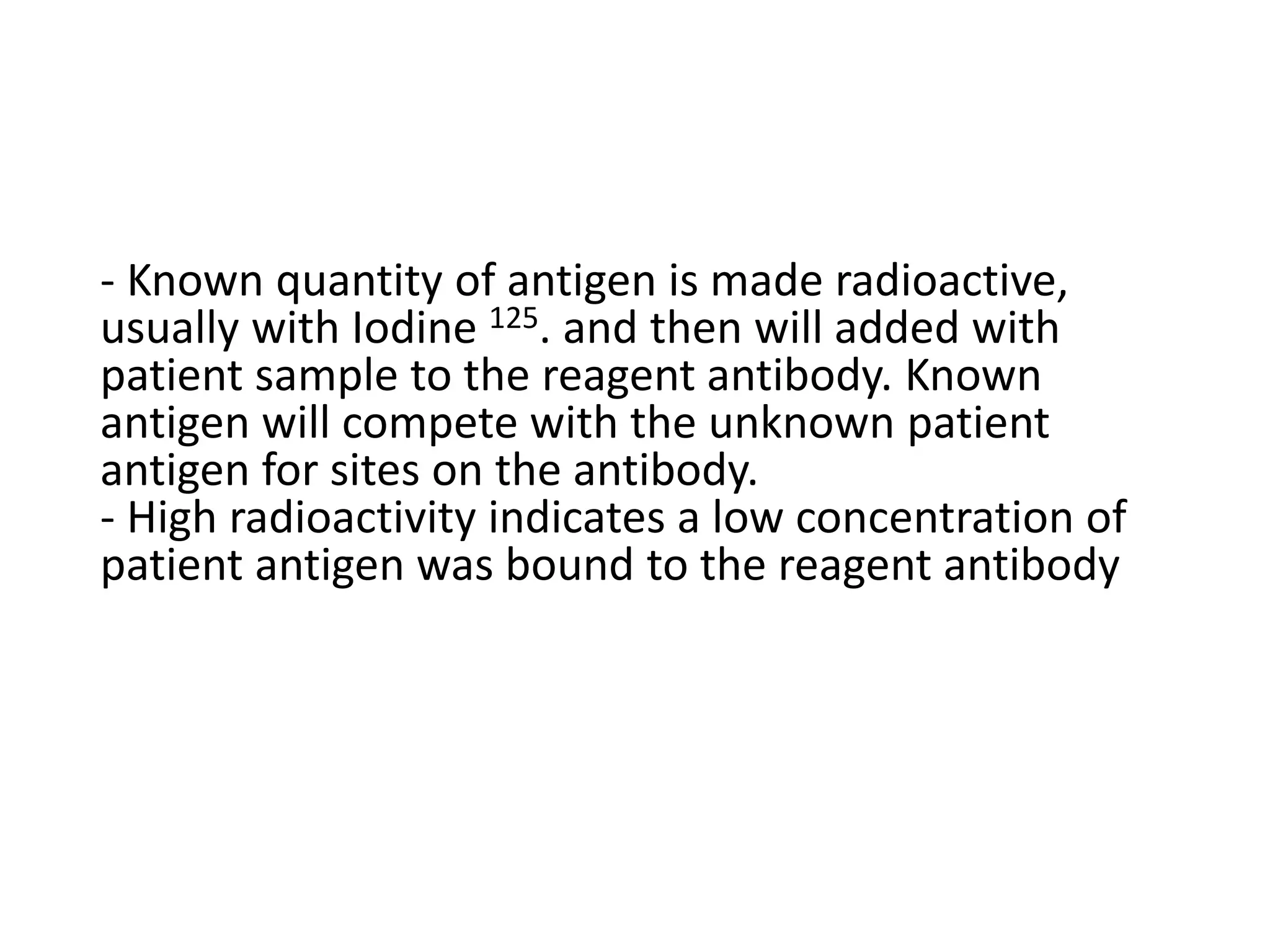
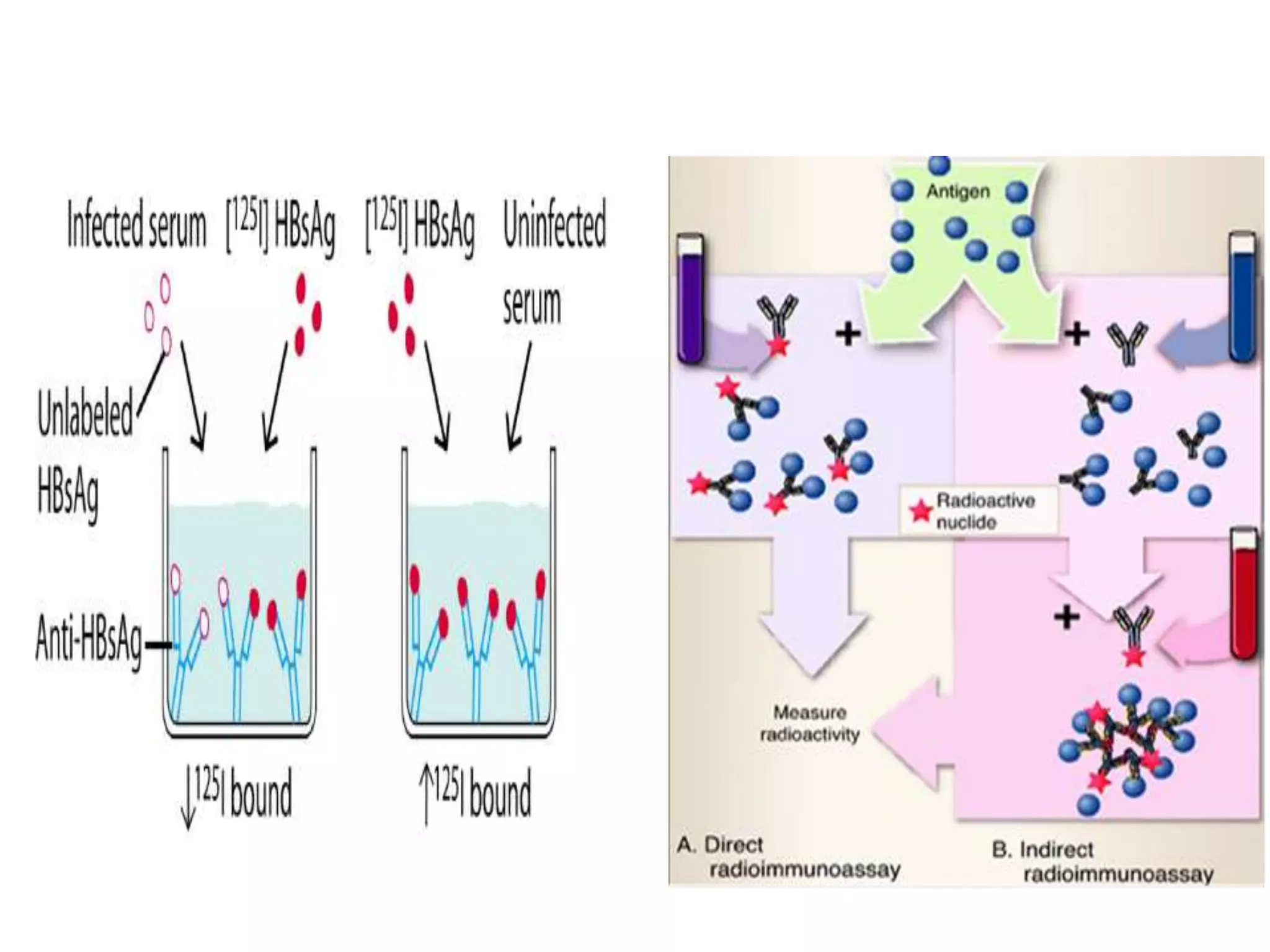
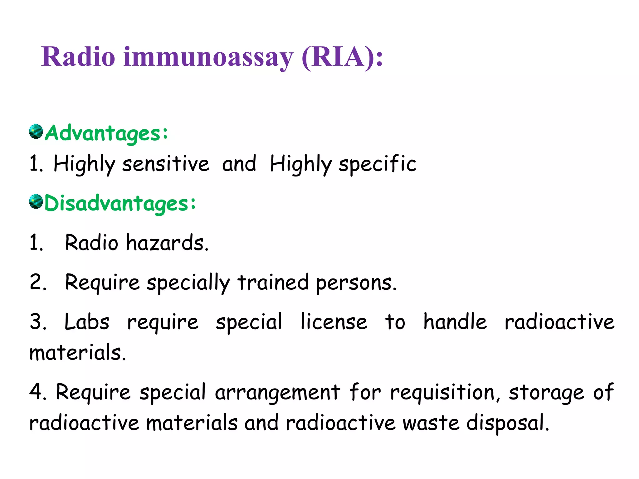
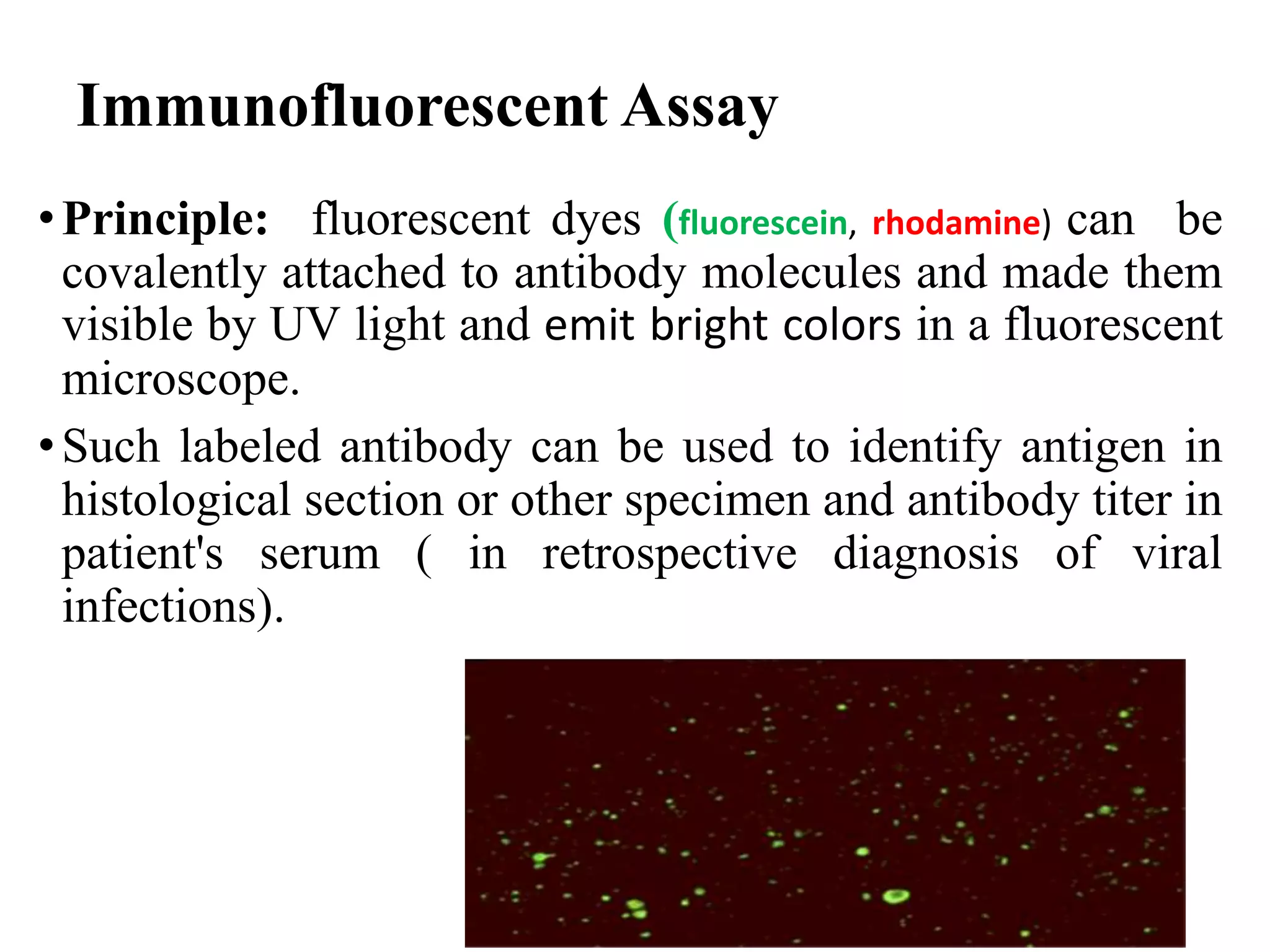
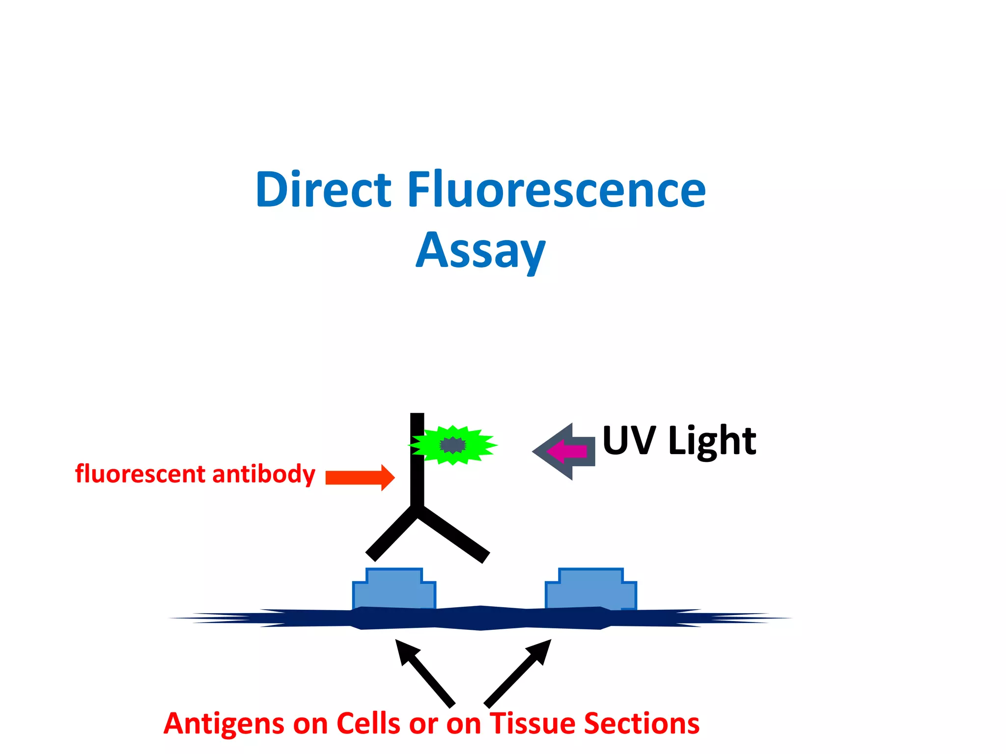
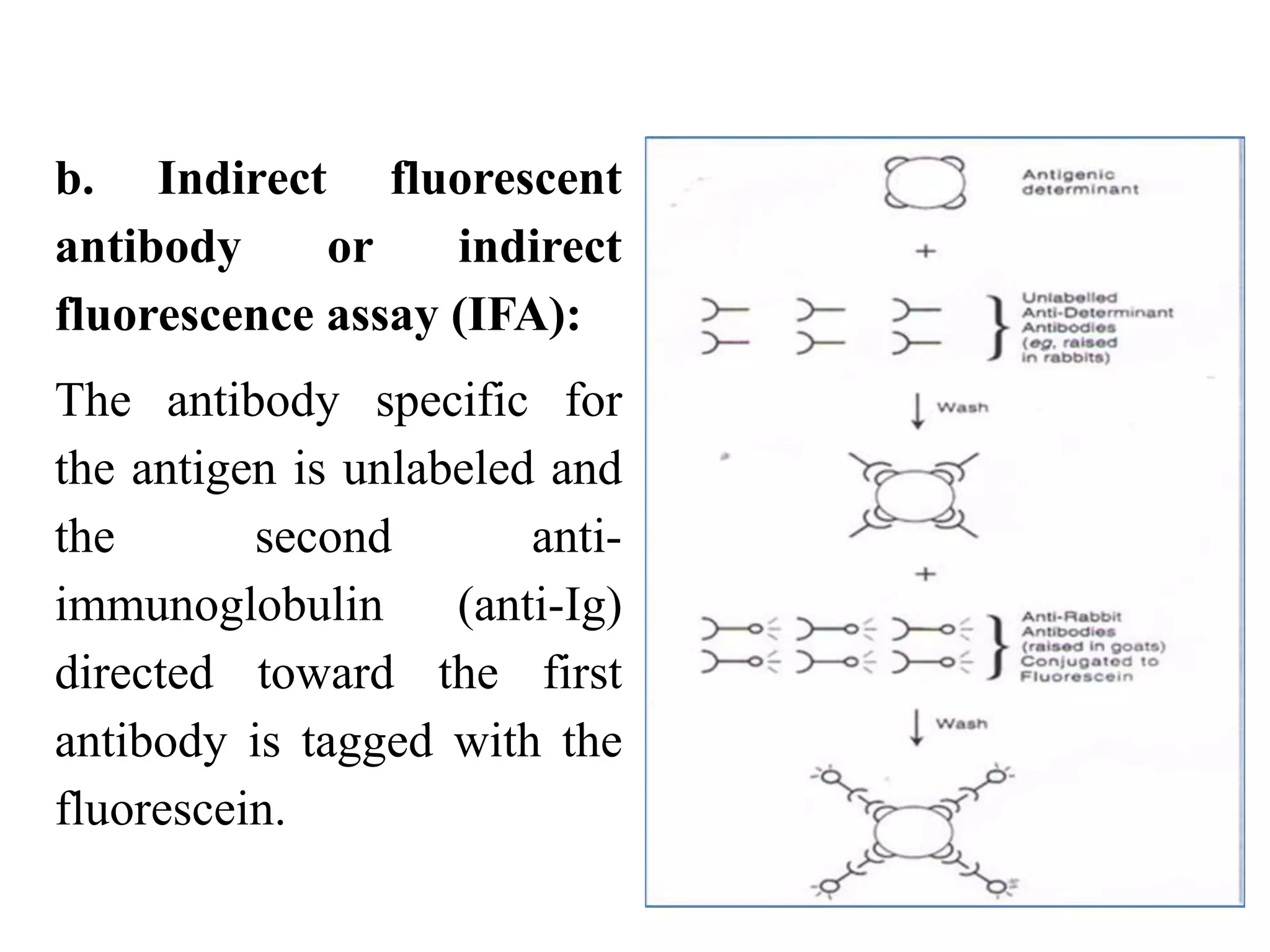
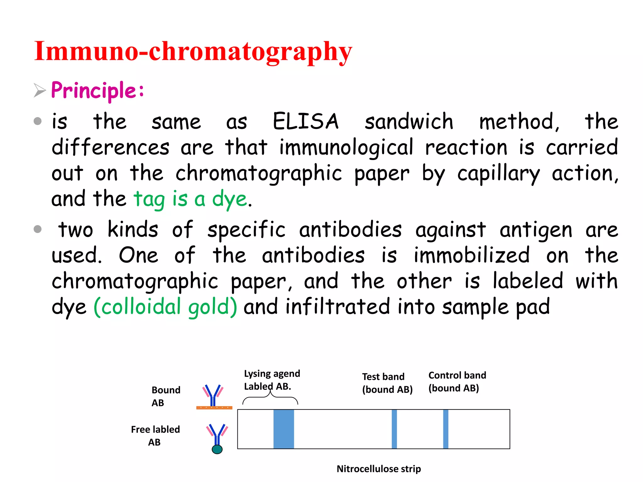
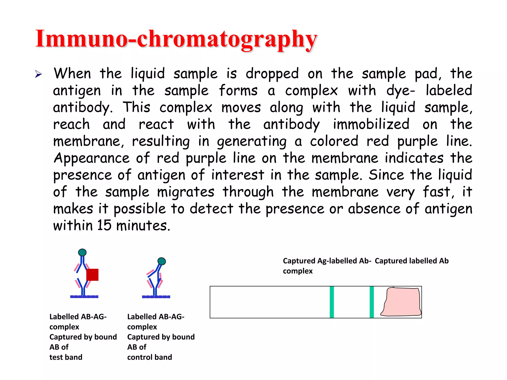
A RIA is a very sensitive in vitro assay technique used to measure concentrations of substances, usually measuring antigen concentrations (for example, hormone .
Bangalore Call Girls Majestic 📞 9907093804 High Profile Service 100% Safe
Radio Immuno Assay, Immuno Fluorescent Test, Lab 4.pptx
1. Practical Immunology
Lab 4
Radio Immuno Assay, Immuno Fluorescent Test, and immunochromatography Tests
By:
Lecturer Shaima’a Al-Salihy
2. Radio immunoassay (RIA):
Principle: competitive binding
Radioactive labeled Ag (or Ab) competes with
the patient’s unlabeled Ag (or Ab) for binding
sites of known amount of Ab (or Ag). The tag is
a radioactive isotope like 32P or 125I.
High radioactivity, small amount of patient substance and low
radioactivity high amount of patient substance
There is two types of (RIA): direct and indirect
3. - Known quantity of antigen is made radioactive,
usually with Iodine 125. and then will added with
patient sample to the reagent antibody. Known
antigen will compete with the unknown patient
antigen for sites on the antibody.
- High radioactivity indicates a low concentration of
patient antigen was bound to the reagent antibody
4.
5. Radio immunoassay (RIA):
Advantages:
1. Highly sensitive and Highly specific
Disadvantages:
1. Radio hazards.
2. Require specially trained persons.
3. Labs require special license to handle radioactive
materials.
4. Require special arrangement for requisition, storage of
radioactive materials and radioactive waste disposal.
7. Immunofluorescent Assay
•Principle: fluorescent dyes (fluorescein, rhodamine) can be
covalently attached to antibody molecules and made them
visible by UV light and emit bright colors in a fluorescent
microscope.
•Such labeled antibody can be used to identify antigen in
histological section or other specimen and antibody titer in
patient's serum ( in retrospective diagnosis of viral
infections).
8. a. Direct fluorescent
antibody (DFA): known
labelled Ab interacts
directly with unknown
antigen
For example detection of
RSV from Respiratory
specimen.
9. Antigens on Cells or on Tissue Sections
UV Light
Direct Fluorescence
Assay
fluorescent antibody
10. b. Indirect fluorescent
antibody or indirect
fluorescence assay (IFA):
The antibody specific for
the antigen is unlabeled and
the second anti-
immunoglobulin (anti-Ig)
directed toward the first
antibody is tagged with the
fluorescein.
13. Immuno-chromatography
Principle:
is the same as ELISA sandwich method, the
differences are that immunological reaction is carried
out on the chromatographic paper by capillary action,
and the tag is a dye.
two kinds of specific antibodies against antigen are
used. One of the antibodies is immobilized on the
chromatographic paper, and the other is labeled with
dye (colloidal gold) and infiltrated into sample pad
Lysing agend
Labled AB.
Test band
(bound AB)
Control band
(bound AB)
Nitrocellulose strip
Bound
AB
Free labled
AB
14. Immuno-chromatography
When the liquid sample is dropped on the sample pad, the
antigen in the sample forms a complex with dye- labeled
antibody. This complex moves along with the liquid sample,
reach and react with the antibody immobilized on the
membrane, resulting in generating a colored red purple line.
Appearance of red purple line on the membrane indicates the
presence of antigen of interest in the sample. Since the liquid
of the sample migrates through the membrane very fast, it
makes it possible to detect the presence or absence of antigen
within 15 minutes.
Captured Ag-labelled Ab-
complex
Captured labelled Ab
Labelled AB-AG-
complex
Captured by bound
AB of
test band
Labelled AB-AG-
complex
Captured by bound
AB of
control band
15. • Dye-labelled antibody, specific for target
antigen, is present on the lower end of
nitrocellulose strip or in a plastic well
provided with the strip.
• Antibody, also specific for the target
antigen, is bound to the strip in a thin (test)
line
• Either antibody specific for the labelled
antibody, or antigen, is bound at the control
line
16. Immuno-chromatography
Advantages:
1. Commercially available
2. Single use, rapid test
3. Easy to perform
4. Can detect antigen or antibody
5. Inexpensive
Limitations:
1. Results are qualitative.
2. less sensitive or less accurate compared to
existing tests.
- Known quantity of antigen is made radioactive, usually with Iodine 125. and then will added with patient sample to the reagent antibody. Known antigen will compete with the unknown patient antigen for sites on the antibody.
- High radioactivity indicates a low concentration of patient antigen was bound to the reagent antibody
Dye-labelled antibody, specific for target antigen, is present on the lower end of nitrocellulose strip or in a plastic well provided with the strip.
Antibody, also specific for the target antigen, is bound to the strip in a thin (test) line
Either antibody specific for the labelled antibody, or antigen, is bound at the control line