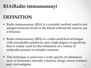
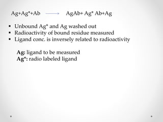
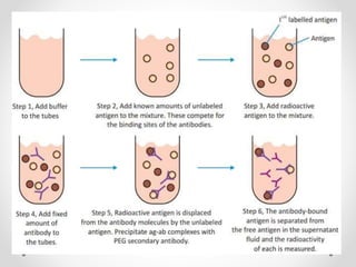
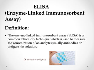
MODERN PHARMACEUTICAL
ANALYTICAL TECHNIQUES
(MPC 101T)
SANA SAIFI
M.PHARM
Topic: Immunological Assays: RIA (Radio immune
assay), ELISA.
IMMUNOASSAY
Biochemical test that measures the concentration of a substance in a
biological liquid, typically serum or urine, using the reaction of an
antibody or antibodies to its antigen.
PURPOSE:
 The purpose of immunoassay is to measure (or, in a qualitative assay to
detect) an analyte.
 Immunoassay is a method of choice for measuring analytes normally
present at very low concentrations that cannot be determined
accurately by other expensive tests.
Common uses include measurement of drugs, hormones, specific proteins, tumor
markers and markers of cardiac injury.
CONTENTS
Definition
Introduction
Principle and theory
Requirements
Applications
RIA(Radio immunoassay)
DEFINITION
 Radio immunoassay (RIA) is a scientific method used to test
antigen hormone levels in the blood without the need to use
a bioassay.
 Radio immunoassay (RIA) is a radio-analytical technique
with remarkable sensitivity and a high degree of specificity
that is widely used for the estimation of a variety of
molecules present in complex matrices.
 This technique is used over a wide spectra of substances
such as hormones, steroids, vitamins, drugs, tumor markers
and viral antigens.
RADIO IMMUNO ASSAY
(RIA)
HISTORY
 The technique was introduced in 1960 by BERSON and
YALOW as an assay for the concentration of insulin in
plasma.
 It represented the first time that hormone levels in the
blood could be detected by an invitro assay.
INTRODUCTION
 This isotopic measuring method was developed in 1959 by
two Americans, biophysicist ROSALYN YALOW and
physician SOLOMON A. BERSON.
 RIA combines the specificity of an antigen antibody reaction
with sensitivity of radioactive measurements.
 This technique used for detection of micro quantities of
protein, viral antigens, antibodies, structural proteins,
vitamins and drug and their metabolites.
 It can also be used for detection of pictogram quantities (10
g) of biological constituents present in biological fluid.
 RIA is used in place of bioassay in various branches of
science like biochemistry. microbiology, hematology and
clinical pharmacology.
PRINCIPLE AND THEORY
 RIA involves the separation of a protein (from a mixture)
using the specificity of antibody –antigen binding and
quantitation using radioactivity. It uses Antigens and
Antibodies for an immune reaction.
 RIA works on basic principle of biochemistry that
competitive binding between antigens for same antibody
binding site.
 The competition of an analyte with its radio isotopically
labeled counterpart for a limited amount of antibody, the
specific reagent, is the underlying principle of this
technique.
 Increasing the analyte concentration inhibits the binding
of the labeled analyte to the antibody.
Ag+Ag*+Ab AgAb+ Ag* Ab+Ag
 Unbound Ag* and Ag washed out
 Radioactivity of bound residue measured
 Ligand conc. is inversely related to radioactivity
Ag: ligand to be measured
Ag*: radio labeled ligand
REQUIREMENTS
 Micro titer plate / Test tubes
 Pure antigen
 Radio labelled antigen
 Antibodies
 Standard
 Centrifuge
 Radioactive counter
MICRO TITRE PLATE
 A microtitre plate is mostly used for this assay.
 A microtitre plate could have 60,24,96,384, or even
sometimes 1536 wells arranged in rows.
 Each well of a microtitre can only hold very small
amounts of liquid.
ANTIGEN
Pure Antigen:
 Antigen may be obtained from biological sample or by
synthetic form.
 It should be pure.
 It is used as standard or calibrator.
Radio labelling of antigen :
 The most commonly used radio labelles in RIA are tritium
and iodine.
 They have adequate activity and have long
enough half lifes.
ANTIBODY
 Specific antibodies are obtained by injecting the Ag to
animals.
 Ag I.e. Drug molecule + bovine serum albumin.
STANDARD
 Draw a standard curve by taking conc. Of Ag in x-axis and
radio activity in y-axis.
 It will help to determine the unknown conc.by
using radio activity.
CENTRIFUGE
 Range: 1200-2500 rpm
 Uses for the separation of precipitated form and
supernatant liquid form.
Radio active counters :
 Two types of counters are used
They are:
a) Gamma counter.
b) Scintillation counters.
a) Gamma counter :
 These are used for the gamma energy emitting isotopes.
e.g, common iodine isotopes.
b) Scintillation counter :
 These are used for counting beta energy emitting isotopes
e.g., tritium, carbon -14 isotopes
 PROCEDURE:
 These test tubes are incubated untill the reaction is
completed.
 A competition occur between Ag & Ag* for the binding
sites of Ab.
 This leads to displacement of Ag* by Ag.
 Centrifugation is done for separation of
bound and free form
SEPARATION TECHNIQUES
 After completion of reaction of reaction free form and
bound forms are determined by separation techniques.
 Various techniques include gel filtration,
Electrophoresis, solid phase adsorption of Ag, Ab &
fractional precipitate.
Applications:
Radio immuno assay of morphine :
This involves:
 Synthesis of immunogen
 Antiserum production.
 Procedure.
 Radio immuno assay of clonazepam.
 Radio immuno assay of barbiturates.
 RIA of flurazepam in human plasma.
 RIA of hydrocodan in human plasma.
 Determination of Ag concentration.
 Estimation of hormones like LH, FSH, ACTH.
 To detect hepatitis and HIV antigens.
 Estimation of vitamins like folic acid, riboflavin etc.
CONCLUSION:
 RIA is invitro assay technique used for the
determination of Ag concentration present in given
sample.
 In advance to RIA many other techniques are
introduced like RAST, EIA, RIST, IRMA,
competitive RIA.
ELISA
(Enzyme-Linked Immunosorbent
Assay)
Definition:
 The enzyme-linked immunosorbent assay (ELISA) is a
common laboratory technique which is used to measure
the concentration of an analyte (usually antibodies or
antigens) in solution.
 Microtiter well plate
Principle of ELISA:
 Based on Basic Immunology Response
 Lock and Key Concept:
1) Antigen (key) 2) Antibody (lock):
-Key fits into the lock
 Enzyme conjugate substrates.
• Bound to a secondary antibody that binds with the
antibody-antigen complex.
 ELISA use an enzyme to detect the binding of antigen (Ag)
antibody (Ab).
 The enzyme converts a colorless substrate (chromogen) to
a colored product, indicating the presence of Ag: Ab
binding.
 An ELISA can be used to detect either the presence of
antigens or antibodies in a sample depending how the
test is designed.
 A number of enzymes have been employed ELISA,
including
1. Alkaline phosphatase
2. Horseradish peroxidase
3. Galactosidase
 These assays approach the sensitivity of RIAS and have the
advantage of being safer and less costly.
INDIRECT ELISA
 Antibody can be detected or quantitatively determined
with an indirect ELISA.
 Serum or some other sample containing primary antibody
(Ab1) is added to an antigen-coated microtiter well and
allowed to react with the antigen attached to the well.
 After any free antibody (Ab1) is washed away, the presence
of antibody bound to the antigen is detected by adding an
enzyme-conjugated secondary anti-isotype antibody (Ab2),
which binds to the primary antibody.
 Any free Ab₂ then is washed away and a substrate for the
enzyme is added.
 The amount of colored reaction product that forms is
measured by specialized spectrophotometric plate
readers, which can measure the absorbance of all of the
wells of a 96-well plate in seconds.
.
SANDWICH ELISA
 Antigen can be detected or measured by a sandwich
ELISA.
 In this technique, the antibody (rather than the antigen)
is immobilized on a microtiter well.
 A sample containing antigen is added and allowed to
react with the immobilized antibody.
 After the well is washed, a second enzyme- linked
antibody specific for a different epitope on the antigen is
added and allowed to react with the bound antigen.
 After any free second antibody is removed washing,
substrate is added, and the colored reaction product is
measured.
COMPETITIVE ELISA
 Another variation for measuring amounts of antigen is
competitive ELISA.
 In this technique, antibody is first incubated in solution
with a sample containing antigen.
 The antigen-antibody mixture is then added to an antigen-
coated microtiter well.
 The more antigen present in the sample, the less free
antibody will be available to bind to the antigen-coated
well.
 Addition of an enzyme-conjugated secondary antibody
(Ab₂) specific for the isotype of the primary antibody
can be used to determine the amount of primary
antibody bound to the well as in an indirect ELISA.
 In the competitive assay however, higher the
concentration of antigen in the original sample, the
lower will be the absorbance.
APPLICATION
 Serum antibody concentration.
 Detecting potential food allergens.(milk, peanut, walnut,
almonds and eggs)
 Tracking the spread of disease in disease outbreaks. (e.g.
HIV, bird flu, common COLD, STDs.)
 Detection of antigen. (e.g. pregnancy hormones, drug
allergens, GMO, mad cow disease)
 Detection of antibodies to past exposure to disease. (e.g.
Lyme disease, trichinosis, HIV, bird flu)
THANK YOU

Immunological Assays;RIA and ELISA.pjsjsptx

  • 1.
    MODERN PHARMACEUTICAL ANALYTICAL TECHNIQUES (MPC101T) SANA SAIFI M.PHARM Topic: Immunological Assays: RIA (Radio immune assay), ELISA.
  • 2.
    IMMUNOASSAY Biochemical test thatmeasures the concentration of a substance in a biological liquid, typically serum or urine, using the reaction of an antibody or antibodies to its antigen. PURPOSE:  The purpose of immunoassay is to measure (or, in a qualitative assay to detect) an analyte.  Immunoassay is a method of choice for measuring analytes normally present at very low concentrations that cannot be determined accurately by other expensive tests. Common uses include measurement of drugs, hormones, specific proteins, tumor markers and markers of cardiac injury.
  • 3.
  • 4.
    RIA(Radio immunoassay) DEFINITION  Radioimmunoassay (RIA) is a scientific method used to test antigen hormone levels in the blood without the need to use a bioassay.  Radio immunoassay (RIA) is a radio-analytical technique with remarkable sensitivity and a high degree of specificity that is widely used for the estimation of a variety of molecules present in complex matrices.  This technique is used over a wide spectra of substances such as hormones, steroids, vitamins, drugs, tumor markers and viral antigens.
  • 5.
  • 6.
    HISTORY  The techniquewas introduced in 1960 by BERSON and YALOW as an assay for the concentration of insulin in plasma.  It represented the first time that hormone levels in the blood could be detected by an invitro assay.
  • 7.
    INTRODUCTION  This isotopicmeasuring method was developed in 1959 by two Americans, biophysicist ROSALYN YALOW and physician SOLOMON A. BERSON.  RIA combines the specificity of an antigen antibody reaction with sensitivity of radioactive measurements.  This technique used for detection of micro quantities of protein, viral antigens, antibodies, structural proteins, vitamins and drug and their metabolites.  It can also be used for detection of pictogram quantities (10 g) of biological constituents present in biological fluid.
  • 8.
     RIA isused in place of bioassay in various branches of science like biochemistry. microbiology, hematology and clinical pharmacology.
  • 9.
    PRINCIPLE AND THEORY RIA involves the separation of a protein (from a mixture) using the specificity of antibody –antigen binding and quantitation using radioactivity. It uses Antigens and Antibodies for an immune reaction.  RIA works on basic principle of biochemistry that competitive binding between antigens for same antibody binding site.  The competition of an analyte with its radio isotopically labeled counterpart for a limited amount of antibody, the specific reagent, is the underlying principle of this technique.  Increasing the analyte concentration inhibits the binding of the labeled analyte to the antibody.
  • 10.
    Ag+Ag*+Ab AgAb+ Ag*Ab+Ag  Unbound Ag* and Ag washed out  Radioactivity of bound residue measured  Ligand conc. is inversely related to radioactivity Ag: ligand to be measured Ag*: radio labeled ligand
  • 11.
    REQUIREMENTS  Micro titerplate / Test tubes  Pure antigen  Radio labelled antigen  Antibodies  Standard  Centrifuge  Radioactive counter
  • 12.
    MICRO TITRE PLATE A microtitre plate is mostly used for this assay.  A microtitre plate could have 60,24,96,384, or even sometimes 1536 wells arranged in rows.  Each well of a microtitre can only hold very small amounts of liquid.
  • 13.
    ANTIGEN Pure Antigen:  Antigenmay be obtained from biological sample or by synthetic form.  It should be pure.  It is used as standard or calibrator. Radio labelling of antigen :  The most commonly used radio labelles in RIA are tritium and iodine.  They have adequate activity and have long enough half lifes.
  • 14.
    ANTIBODY  Specific antibodiesare obtained by injecting the Ag to animals.  Ag I.e. Drug molecule + bovine serum albumin.
  • 15.
    STANDARD  Draw astandard curve by taking conc. Of Ag in x-axis and radio activity in y-axis.  It will help to determine the unknown conc.by using radio activity.
  • 16.
    CENTRIFUGE  Range: 1200-2500rpm  Uses for the separation of precipitated form and supernatant liquid form.
  • 17.
    Radio active counters:  Two types of counters are used They are: a) Gamma counter. b) Scintillation counters.
  • 18.
    a) Gamma counter:  These are used for the gamma energy emitting isotopes. e.g, common iodine isotopes. b) Scintillation counter :  These are used for counting beta energy emitting isotopes e.g., tritium, carbon -14 isotopes
  • 19.
     PROCEDURE:  Thesetest tubes are incubated untill the reaction is completed.  A competition occur between Ag & Ag* for the binding sites of Ab.  This leads to displacement of Ag* by Ag.  Centrifugation is done for separation of bound and free form
  • 21.
    SEPARATION TECHNIQUES  Aftercompletion of reaction of reaction free form and bound forms are determined by separation techniques.  Various techniques include gel filtration, Electrophoresis, solid phase adsorption of Ag, Ab & fractional precipitate.
  • 22.
    Applications: Radio immuno assayof morphine : This involves:  Synthesis of immunogen  Antiserum production.  Procedure.
  • 23.
     Radio immunoassay of clonazepam.  Radio immuno assay of barbiturates.  RIA of flurazepam in human plasma.  RIA of hydrocodan in human plasma.  Determination of Ag concentration.  Estimation of hormones like LH, FSH, ACTH.  To detect hepatitis and HIV antigens.  Estimation of vitamins like folic acid, riboflavin etc.
  • 24.
    CONCLUSION:  RIA isinvitro assay technique used for the determination of Ag concentration present in given sample.  In advance to RIA many other techniques are introduced like RAST, EIA, RIST, IRMA, competitive RIA.
  • 25.
    ELISA (Enzyme-Linked Immunosorbent Assay) Definition:  Theenzyme-linked immunosorbent assay (ELISA) is a common laboratory technique which is used to measure the concentration of an analyte (usually antibodies or antigens) in solution.  Microtiter well plate
  • 26.
    Principle of ELISA: Based on Basic Immunology Response  Lock and Key Concept: 1) Antigen (key) 2) Antibody (lock): -Key fits into the lock  Enzyme conjugate substrates. • Bound to a secondary antibody that binds with the antibody-antigen complex.
  • 27.
     ELISA usean enzyme to detect the binding of antigen (Ag) antibody (Ab).  The enzyme converts a colorless substrate (chromogen) to a colored product, indicating the presence of Ag: Ab binding.  An ELISA can be used to detect either the presence of antigens or antibodies in a sample depending how the test is designed.
  • 28.
     A numberof enzymes have been employed ELISA, including 1. Alkaline phosphatase 2. Horseradish peroxidase 3. Galactosidase  These assays approach the sensitivity of RIAS and have the advantage of being safer and less costly.
  • 29.
    INDIRECT ELISA  Antibodycan be detected or quantitatively determined with an indirect ELISA.  Serum or some other sample containing primary antibody (Ab1) is added to an antigen-coated microtiter well and allowed to react with the antigen attached to the well.  After any free antibody (Ab1) is washed away, the presence of antibody bound to the antigen is detected by adding an enzyme-conjugated secondary anti-isotype antibody (Ab2), which binds to the primary antibody.
  • 30.
     Any freeAb₂ then is washed away and a substrate for the enzyme is added.  The amount of colored reaction product that forms is measured by specialized spectrophotometric plate readers, which can measure the absorbance of all of the wells of a 96-well plate in seconds. .
  • 31.
    SANDWICH ELISA  Antigencan be detected or measured by a sandwich ELISA.  In this technique, the antibody (rather than the antigen) is immobilized on a microtiter well.  A sample containing antigen is added and allowed to react with the immobilized antibody.  After the well is washed, a second enzyme- linked antibody specific for a different epitope on the antigen is added and allowed to react with the bound antigen.  After any free second antibody is removed washing, substrate is added, and the colored reaction product is measured.
  • 33.
    COMPETITIVE ELISA  Anothervariation for measuring amounts of antigen is competitive ELISA.  In this technique, antibody is first incubated in solution with a sample containing antigen.  The antigen-antibody mixture is then added to an antigen- coated microtiter well.  The more antigen present in the sample, the less free antibody will be available to bind to the antigen-coated well.
  • 34.
     Addition ofan enzyme-conjugated secondary antibody (Ab₂) specific for the isotype of the primary antibody can be used to determine the amount of primary antibody bound to the well as in an indirect ELISA.  In the competitive assay however, higher the concentration of antigen in the original sample, the lower will be the absorbance.
  • 35.
    APPLICATION  Serum antibodyconcentration.  Detecting potential food allergens.(milk, peanut, walnut, almonds and eggs)  Tracking the spread of disease in disease outbreaks. (e.g. HIV, bird flu, common COLD, STDs.)  Detection of antigen. (e.g. pregnancy hormones, drug allergens, GMO, mad cow disease)  Detection of antibodies to past exposure to disease. (e.g. Lyme disease, trichinosis, HIV, bird flu)
  • 36.