Parasitology is the scientific discipline concerned with the study of the biology of parasites and parasitic diseases, including the distribution, biochemistry, physiology, molecular biology, ecology, evolution and clinical aspects of parasites, including the host response to these agents.
Entamoeba histolytica was first discovered by Losch in 1875.
It is worldwide distribution.
It is prevalent in tropical and subtropical countries where sanitary conditions are poor.
In india, it is prevalent in Chandigarh, Tamil Nadu & Maharashtra.
It is found in the colon of man.
It is monogenetic because the whole life cycle completed within a single host, i.e. man.
This is a series of lectures on microbiology useful for undergraduate medical and paramedical students.. This lecture is a comprehensive coverage of all parasites, protozoa and helminths...
This is a series of lectures on microbiology useful for undergraduate medical and paramedical students.. This lecture is a comprehensive coverage of all parasites, protozoa and helminths...
Morphology, Life cycle, Clinical manifestations and laboratory diagnosis of E. histolytica from Clinical and Microbiological point of view for UG and PG Students.
Virology is the scientific study of biological viruses. It is a subfield of microbiology that focuses on their detection, structure, classification and evolution, their methods of infection and exploitation of host cells for reproduction, their interaction with host organism physiology and immunity,
Strategies Novartis can use to GROW from a Billion Dollar Company to a Trillion Dollar Company like Alphabet Inc
Novartis is a leading healthcare company which is situated in Switzerland and uses digital technologies and innovative science to come up with transformative ways of treatment in areas of great medicinal needs. This article explains what Novartis strategies and what they should employ so that they can rise from a billion dollar company to a trillion dollar company like the Google Alphabet Inc.
Novartis was formed in March 1996 by the merging of pharmaceutical and agrochemical divisions of Ciba-Geigy and Sandoz companies. Thanks to the merging of the two companies, Novartis is one of the biggest pharmaceutical companies in the world. Novartis is one of the largest companies which achieved a great milestone within a few decades. Novartis as a whole is divided into three major divisions: Sandoz (generics), Innovative Medicines and Alcon (eyecare). Novartis is also involved in collaborative research projects that are publicly funded.
Below are some of Novartis best selling drugs and their revenue
1.Cosenty – This is the top selling drug with a revenue of 4.788 billion dollars
2.Enfresto – This has a revenue of 4.644 billions dollars
3.Promacta – This has a revenue 0f 2.088 billion dollars
Medicine manufactured by Novartis and their uses
Medicine Medicine use
Cosentyx Used to treat psoriatic arthritis
Entresto Used to treat heart failure
Lucentis Used to block abnormal vessel growth in the back of the eye
Tasigna Used to treat chronic myelogenous leukemia which has the Philadelphia chromosome
Jakavi Used to treat myelofibrosis, polycythemia vera and graft-versus-host disease
Promacta Used to treat patients with abnormal low platelet count
Sandostatin Used to treat patients with tumor experiencing symptoms like flushing and diarrhea
Xolair Used to treat moderate and severe asthma
Gilenya Used to treat multiple sclerosis
How Novartis became one of the biggest pharmaceutical companies in the world
1.Market control through partnership
Geigy, Sandoz and Ciba combined their power so that they can compete with strong foreign firms and formed a cartel called the Basal Syndicate or Basal IG. Basal IG secured most of the manufacturing facilities all over the US and across Europe. It later joined with IG Farben and other chemical companies to form a big cartel called the Quadrapartite Cartel which dominated all of the European market and enjoyed the profits made from the joint manufacturing.
2.Growth acceleration through mergers
Since competition was very rampant in the pharmaceutical industry, Ciba and Geigy decided to merge with Sandoz AG to form Novartis. With this merge, Novartis became one of the growing giants in the pharmaceutical industry. This made Novartis gain a lot of fame and build a strong reputation over other companies. Novartis majored on agrochemical and pharmaceutical industries which made it easy to focus on a specific mar
توثيق مراجع البحث العلمي على أنو: "إثبات ادلصادر البيانات وادلعلومات ونسبها إىلnedalalazzwy
عترب البحث العلمي ادلمنهج ذاك الذي يستويف يف مجيع مراحلو مراعاة معايري البحث العلمي ادلنهجي
خاصة فيما خيص األمانة يف اعتماد ادلراجع سواء كانت دراسات سابقة او مراجع لبعض االقتباسلت والعبارات
وزبتلف عملية التوثيق للمراجع باختالف مصدرىا ونوعها واختالف رلال زبصصها فتوثيق التت ملال خيتلف
عنو يف توثيق ادلقاالت الصحفية وخيتلف عن توثيق ادلواد االلتًتونية وىذه األخرية خيتلف يف توثيقها تبعا ألنواعها
ىي األخرى واذلدف من ذلك ىو حفاظ الباحث على سهولة العودة اىل ادلصادر وادلراجع ادلستخدمة بالنسبة
لقراء حبقو العلمي وىو أيضا من باب األمانة العلمية
A single nucleotide polymorphism (abbreviated SNP, pronounced snip) is a genomic variant at a single base position in the DNA. Scientists study if and how SNPs in a genome influence health, disease, drug response and other traits.
Mycology is the branch of biology concerned with the study of fungi, including their genetic and biochemical properties, their taxonomy and their use to humans, including as a source for tinder, traditional medicine, food, and entheogens, as well as their dangers, such as toxicity or infection.
Rabies virus, scientific name Rabies lyssavirus, is a neurotropic virus that causes rabies in humans and animals. Rabies transmission can occur through the saliva of animals and less commonly through contact with human saliva. Rabies lyssavirus, like many rhabdoviruses, has an extremely wide host range.
Immunofluorescence (IF) is a technique that permits visualization of virtually many components in any given tissue or cell type. This broad capability is achieved through combinations of specific antibodies tagged with fluorophores. Consequently, the pos
fastidious organism is any organism that has complex or particular nutritional requirements. In other words, a fastidious organism will only grow when specific nutrients are included in its medium.
An antigen is any substance that causes your immune system to produce antibodies against it. This means your immune system does not recognize the substance, and is trying to fight it off. An antigen may be a substance from th
Multiplex PCR is a technique whereby PCR is used to amplify several different DNA sequences simultaneously. It is a type of target enrichment approach. It was first described in 1988 as a method to detect deletion mutations in the dystrophin gene – the largest known human gene
Radio Immuno Assay, Immuno Fluorescent Test, Lab 4.pptxnedalalazzwy
A RIA is a very sensitive in vitro assay technique used to measure concentrations of substances, usually measuring antigen concentrations (for example, hormone .
What is enzyme-linked immunosorbent assay?
A laboratory technique that uses antibodies linked to enzymes to detect and measure the amount of a substance in a solution, such as serum. The test is done using a solid surface to which the antibodies and other molecules stick.
Infectious diseases can be viral, bacterial, parasitic or fungal infections. There's also a rare group of infectious diseases known as transmissible spongiform encephalopathies (TSEs).
What is toxoplasmosis? Toxoplasmosis is an infection caused by a single-celled parasite called Toxoplasma gondii. While the parasite is found throughout the world, more than 40 million people in the United States may be infected with the Toxoplasma parasite.
Integrons are genetic elements that contain a site-specific recombination system able to integrate, express and exchange specific DNA elements, called gene cassettes. 5. The complete integron is not considered to be a mobile element as such as it lacks functions for self-mobility.
Mycoplasma pneumoniae are bacteria that can cause illness by damaging the lining of the respiratory system (throat, lungs, windpipe). People can have the bacteria in their nose or throat at one time or another without being ill. People spread Mycoplasma pneumoniae bacteria to others by coughing or sneezing.
A microarray is a laboratory tool used to detect the expression of thousands of genes at the same time. DNA microarrays are microscope slides that are printed with thousands of tiny spots in defined positions, with each spot containing a known DNA sequence or gene.
Cloning is a technique scientists use to make exact genetic copies of living things. Genes, cells, tissues, and even whole animals can all be cloned. Some clones already exist in nature. Single-celled organisms like bacteria make exact copies of themselves each time they reproduce.
A cell cycle is a series of events that takes place in a cell as it grows and divides. A cell spends most of its time in what is called interphase, and during this time it grows, replicates its chromosomes, and prepares for cell division. The cell then leaves interphase, undergoes mitosis, and completes its division.
Polymerase chain reaction (abbreviated PCR) is a laboratory technique for rapidly producing (amplifying) millions to billions of copies of a specific segment of DNA, which can then be studied in greater detail.
Assay of sodium hydroxide solution.pptxnedalalazzwy
sodium hydroxide is useful for its ability to alter fats. It is used to make soap and as a main ingredient in household products such as liquid drain cleaners. Sodium hydroxide is usually sold in pure form as white pellets or as a solution in water.
TEST BANK for Operations Management, 14th Edition by William J. Stevenson, Ve...kevinkariuki227
TEST BANK for Operations Management, 14th Edition by William J. Stevenson, Verified Chapters 1 - 19, Complete Newest Version.pdf
TEST BANK for Operations Management, 14th Edition by William J. Stevenson, Verified Chapters 1 - 19, Complete Newest Version.pdf
ARTIFICIAL INTELLIGENCE IN HEALTHCARE.pdfAnujkumaranit
Artificial intelligence (AI) refers to the simulation of human intelligence processes by machines, especially computer systems. It encompasses tasks such as learning, reasoning, problem-solving, perception, and language understanding. AI technologies are revolutionizing various fields, from healthcare to finance, by enabling machines to perform tasks that typically require human intelligence.
New Directions in Targeted Therapeutic Approaches for Older Adults With Mantl...i3 Health
i3 Health is pleased to make the speaker slides from this activity available for use as a non-accredited self-study or teaching resource.
This slide deck presented by Dr. Kami Maddocks, Professor-Clinical in the Division of Hematology and
Associate Division Director for Ambulatory Operations
The Ohio State University Comprehensive Cancer Center, will provide insight into new directions in targeted therapeutic approaches for older adults with mantle cell lymphoma.
STATEMENT OF NEED
Mantle cell lymphoma (MCL) is a rare, aggressive B-cell non-Hodgkin lymphoma (NHL) accounting for 5% to 7% of all lymphomas. Its prognosis ranges from indolent disease that does not require treatment for years to very aggressive disease, which is associated with poor survival (Silkenstedt et al, 2021). Typically, MCL is diagnosed at advanced stage and in older patients who cannot tolerate intensive therapy (NCCN, 2022). Although recent advances have slightly increased remission rates, recurrence and relapse remain very common, leading to a median overall survival between 3 and 6 years (LLS, 2021). Though there are several effective options, progress is still needed towards establishing an accepted frontline approach for MCL (Castellino et al, 2022). Treatment selection and management of MCL are complicated by the heterogeneity of prognosis, advanced age and comorbidities of patients, and lack of an established standard approach for treatment, making it vital that clinicians be familiar with the latest research and advances in this area. In this activity chaired by Michael Wang, MD, Professor in the Department of Lymphoma & Myeloma at MD Anderson Cancer Center, expert faculty will discuss prognostic factors informing treatment, the promising results of recent trials in new therapeutic approaches, and the implications of treatment resistance in therapeutic selection for MCL.
Target Audience
Hematology/oncology fellows, attending faculty, and other health care professionals involved in the treatment of patients with mantle cell lymphoma (MCL).
Learning Objectives
1.) Identify clinical and biological prognostic factors that can guide treatment decision making for older adults with MCL
2.) Evaluate emerging data on targeted therapeutic approaches for treatment-naive and relapsed/refractory MCL and their applicability to older adults
3.) Assess mechanisms of resistance to targeted therapies for MCL and their implications for treatment selection
Title: Sense of Taste
Presenter: Dr. Faiza, Assistant Professor of Physiology
Qualifications:
MBBS (Best Graduate, AIMC Lahore)
FCPS Physiology
ICMT, CHPE, DHPE (STMU)
MPH (GC University, Faisalabad)
MBA (Virtual University of Pakistan)
Learning Objectives:
Describe the structure and function of taste buds.
Describe the relationship between the taste threshold and taste index of common substances.
Explain the chemical basis and signal transduction of taste perception for each type of primary taste sensation.
Recognize different abnormalities of taste perception and their causes.
Key Topics:
Significance of Taste Sensation:
Differentiation between pleasant and harmful food
Influence on behavior
Selection of food based on metabolic needs
Receptors of Taste:
Taste buds on the tongue
Influence of sense of smell, texture of food, and pain stimulation (e.g., by pepper)
Primary and Secondary Taste Sensations:
Primary taste sensations: Sweet, Sour, Salty, Bitter, Umami
Chemical basis and signal transduction mechanisms for each taste
Taste Threshold and Index:
Taste threshold values for Sweet (sucrose), Salty (NaCl), Sour (HCl), and Bitter (Quinine)
Taste index relationship: Inversely proportional to taste threshold
Taste Blindness:
Inability to taste certain substances, particularly thiourea compounds
Example: Phenylthiocarbamide
Structure and Function of Taste Buds:
Composition: Epithelial cells, Sustentacular/Supporting cells, Taste cells, Basal cells
Features: Taste pores, Taste hairs/microvilli, and Taste nerve fibers
Location of Taste Buds:
Found in papillae of the tongue (Fungiform, Circumvallate, Foliate)
Also present on the palate, tonsillar pillars, epiglottis, and proximal esophagus
Mechanism of Taste Stimulation:
Interaction of taste substances with receptors on microvilli
Signal transduction pathways for Umami, Sweet, Bitter, Sour, and Salty tastes
Taste Sensitivity and Adaptation:
Decrease in sensitivity with age
Rapid adaptation of taste sensation
Role of Saliva in Taste:
Dissolution of tastants to reach receptors
Washing away the stimulus
Taste Preferences and Aversions:
Mechanisms behind taste preference and aversion
Influence of receptors and neural pathways
Impact of Sensory Nerve Damage:
Degeneration of taste buds if the sensory nerve fiber is cut
Abnormalities of Taste Detection:
Conditions: Ageusia, Hypogeusia, Dysgeusia (parageusia)
Causes: Nerve damage, neurological disorders, infections, poor oral hygiene, adverse drug effects, deficiencies, aging, tobacco use, altered neurotransmitter levels
Neurotransmitters and Taste Threshold:
Effects of serotonin (5-HT) and norepinephrine (NE) on taste sensitivity
Supertasters:
25% of the population with heightened sensitivity to taste, especially bitterness
Increased number of fungiform papillae
HOT NEW PRODUCT! BIG SALES FAST SHIPPING NOW FROM CHINA!! EU KU DB BK substit...GL Anaacs
Contact us if you are interested:
Email / Skype : kefaya1771@gmail.com
Threema: PXHY5PDH
New BATCH Ku !!! MUCH IN DEMAND FAST SALE EVERY BATCH HAPPY GOOD EFFECT BIG BATCH !
Contact me on Threema or skype to start big business!!
Hot-sale products:
NEW HOT EUTYLONE WHITE CRYSTAL!!
5cl-adba precursor (semi finished )
5cl-adba raw materials
ADBB precursor (semi finished )
ADBB raw materials
APVP powder
5fadb/4f-adb
Jwh018 / Jwh210
Eutylone crystal
Protonitazene (hydrochloride) CAS: 119276-01-6
Flubrotizolam CAS: 57801-95-3
Metonitazene CAS: 14680-51-4
Payment terms: Western Union,MoneyGram,Bitcoin or USDT.
Deliver Time: Usually 7-15days
Shipping method: FedEx, TNT, DHL,UPS etc.Our deliveries are 100% safe, fast, reliable and discreet.
Samples will be sent for your evaluation!If you are interested in, please contact me, let's talk details.
We specializes in exporting high quality Research chemical, medical intermediate, Pharmaceutical chemicals and so on. Products are exported to USA, Canada, France, Korea, Japan,Russia, Southeast Asia and other countries.
Ethanol (CH3CH2OH), or beverage alcohol, is a two-carbon alcohol
that is rapidly distributed in the body and brain. Ethanol alters many
neurochemical systems and has rewarding and addictive properties. It
is the oldest recreational drug and likely contributes to more morbidity,
mortality, and public health costs than all illicit drugs combined. The
5th edition of the Diagnostic and Statistical Manual of Mental Disorders
(DSM-5) integrates alcohol abuse and alcohol dependence into a single
disorder called alcohol use disorder (AUD), with mild, moderate,
and severe subclassifications (American Psychiatric Association, 2013).
In the DSM-5, all types of substance abuse and dependence have been
combined into a single substance use disorder (SUD) on a continuum
from mild to severe. A diagnosis of AUD requires that at least two of
the 11 DSM-5 behaviors be present within a 12-month period (mild
AUD: 2–3 criteria; moderate AUD: 4–5 criteria; severe AUD: 6–11 criteria).
The four main behavioral effects of AUD are impaired control over
drinking, negative social consequences, risky use, and altered physiological
effects (tolerance, withdrawal). This chapter presents an overview
of the prevalence and harmful consequences of AUD in the U.S.,
the systemic nature of the disease, neurocircuitry and stages of AUD,
comorbidities, fetal alcohol spectrum disorders, genetic risk factors, and
pharmacotherapies for AUD.
The prostate is an exocrine gland of the male mammalian reproductive system
It is a walnut-sized gland that forms part of the male reproductive system and is located in front of the rectum and just below the urinary bladder
Function is to store and secrete a clear, slightly alkaline fluid that constitutes 10-30% of the volume of the seminal fluid that along with the spermatozoa, constitutes semen
A healthy human prostate measures (4cm-vertical, by 3cm-horizontal, 2cm ant-post ).
It surrounds the urethra just below the urinary bladder. It has anterior, median, posterior and two lateral lobes
It’s work is regulated by androgens which are responsible for male sex characteristics
Generalised disease of the prostate due to hormonal derangement which leads to non malignant enlargement of the gland (increase in the number of epithelial cells and stromal tissue)to cause compression of the urethra leading to symptoms (LUTS
Acute scrotum is a general term referring to an emergency condition affecting the contents or the wall of the scrotum.
There are a number of conditions that present acutely, predominantly with pain and/or swelling
A careful and detailed history and examination, and in some cases, investigations allow differentiation between these diagnoses. A prompt diagnosis is essential as the patient may require urgent surgical intervention
Testicular torsion refers to twisting of the spermatic cord, causing ischaemia of the testicle.
Testicular torsion results from inadequate fixation of the testis to the tunica vaginalis producing ischemia from reduced arterial inflow and venous outflow obstruction.
The prevalence of testicular torsion in adult patients hospitalized with acute scrotal pain is approximately 25 to 50 percent
Tom Selleck Health: A Comprehensive Look at the Iconic Actor’s Wellness Journeygreendigital
Tom Selleck, an enduring figure in Hollywood. has captivated audiences for decades with his rugged charm, iconic moustache. and memorable roles in television and film. From his breakout role as Thomas Magnum in Magnum P.I. to his current portrayal of Frank Reagan in Blue Bloods. Selleck's career has spanned over 50 years. But beyond his professional achievements. fans have often been curious about Tom Selleck Health. especially as he has aged in the public eye.
Follow us on: Pinterest
Introduction
Many have been interested in Tom Selleck health. not only because of his enduring presence on screen but also because of the challenges. and lifestyle choices he has faced and made over the years. This article delves into the various aspects of Tom Selleck health. exploring his fitness regimen, diet, mental health. and the challenges he has encountered as he ages. We'll look at how he maintains his well-being. the health issues he has faced, and his approach to ageing .
Early Life and Career
Childhood and Athletic Beginnings
Tom Selleck was born on January 29, 1945, in Detroit, Michigan, and grew up in Sherman Oaks, California. From an early age, he was involved in sports, particularly basketball. which played a significant role in his physical development. His athletic pursuits continued into college. where he attended the University of Southern California (USC) on a basketball scholarship. This early involvement in sports laid a strong foundation for his physical health and disciplined lifestyle.
Transition to Acting
Selleck's transition from an athlete to an actor came with its physical demands. His first significant role in "Magnum P.I." required him to perform various stunts and maintain a fit appearance. This role, which he played from 1980 to 1988. necessitated a rigorous fitness routine to meet the show's demands. setting the stage for his long-term commitment to health and wellness.
Fitness Regimen
Workout Routine
Tom Selleck health and fitness regimen has evolved. adapting to his changing roles and age. During his "Magnum, P.I." days. Selleck's workouts were intense and focused on building and maintaining muscle mass. His routine included weightlifting, cardiovascular exercises. and specific training for the stunts he performed on the show.
Selleck adjusted his fitness routine as he aged to suit his body's needs. Today, his workouts focus on maintaining flexibility, strength, and cardiovascular health. He incorporates low-impact exercises such as swimming, walking, and light weightlifting. This balanced approach helps him stay fit without putting undue strain on his joints and muscles.
Importance of Flexibility and Mobility
In recent years, Selleck has emphasized the importance of flexibility and mobility in his fitness regimen. Understanding the natural decline in muscle mass and joint flexibility with age. he includes stretching and yoga in his routine. These practices help prevent injuries, improve posture, and maintain mobilit
Knee anatomy and clinical tests 2024.pdfvimalpl1234
This includes all relevant anatomy and clinical tests compiled from standard textbooks, Campbell,netter etc..It is comprehensive and best suited for orthopaedicians and orthopaedic residents.
How STIs Influence the Development of Pelvic Inflammatory Disease.pptx
Classification of medical parasitology Lec.2.pptx
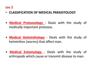
1. Lec 2
• CLASSIFICATION OF MEDICAL PARASITOLOGY
• Medical Protozoology - Deals with the study of
medically important protozoa.
• Medical Helminthology - Deals with the study of
helminthes (worms) that affect man.
• Medical Entomology - Deals with the study of
arthropods which cause or transmit disease to man.
2. Classification of Medically important
Parasites
The parasite divide into three main groups
Arthropoda(Medical
Entomology) include
Insecta (Butter fly)
Arachnida (Mite)
Crustacea (Cyclops)
Metazoa
Parasite consist of
multicellular cells,
Bilaterally symmetrical
animals, having well-
differentiated tissues
and complex organ
Protozoa
Parasite consist of a
single celled organism
which is morphologically
and functionally
complete and can
perform all function of
life ,reproduction by
asexual or sexual
3. Taxonomic classification of Protozoa
Species-
examples
Genus-
examples
Class
Phylum
Sub kingdom
E. hstolytica
E.nana
I.butchlii
D.fragilis
Entamoeba
Endolimax
Iodameba
Dientameba
Sarcodina-
(Amoeba)
move by
pseudopodia
Sarcomastig-
ophora
further divided into
Protozoa
G. Lamblia
T.vaginalis
T.brucci
L.donovani
Giardia
Trichmonas
Trypanosoma
Leishmania
Mastigophora
(Flagellates)
move by flagella
P. falciparum
T.gonidi
C.parvum
I.beli
Plasmodium
Toxoplasma
Cryptosporidium
Isospora
Apicomplexa
(Sporozoa)
no organelle of
Locomotion
B. coli
Balantidium
Ciliophora
move by cillia
4. Amebas: Move by extending cytoplasmic projections
(pseudopodia)
Ciliates: Move by synchronous beating of hair- like cilia
Sporozoa:(also called apicomplexa) are obligate,
intracellular parasites. They generally have non motile
adult forms.
Flagellates: Move by rotating whip-like flagella
5. Protozoa are diverse groups of unicellular, eukaryotic
organisms:
There are about 45,000 protozoan species; around 8000 are
parasitic, and around 250 species are important to humans.
Many have evolved structural features (organelles) that mimic the
organs of multicellular organisms.
Reproduction is generally by mitotic binary fission, through in
some protozoal species ,sexual ( meiotic) reproduction with several
variations occurs as well. Protozoal infections are common in
developing tropical and subtropical regions where sanitary
conditions and control of the vectors of transmission are poor .
However, with increased world travel and immigration, protozoal
diseases are no longer confined to specific geographic locales.
6. • Shape
• There is no one shape or morphology which would include a majority of
the protozoa.
• Shape range from the amorphous and ever changing forms of amoeba,
to relatively rigid forms.
• All protozoa have certain morphologic features, like nucleus,
cytoplasm(endoplasm &ectoplasm)
• Nuclear structure - important in species differentiation.
• Size - helpful in identifying organisms; must have calibrated objectives
on the microscope in order to measure accurately.
• Cytoplasmic inclusions - chromatoid bars; red blood cells; food vacuoles
containing bacteria, yeast, etc.
• Appearance of cytoplasm - smooth & clean or vacuolated.(endoplasm &
ectoplasm)
• Endoplasm : the nucleus consist of moderately dense, finally granular
protoplasm that function in the digestion of ingested food and other
process.
• Ectoplasm : serves for locomotion, for obtaining and ingesting food, and
for respiration and excretion.
• Type of motility - directional or non-directional; sluggish or fast.
7. Nuclear Structure:
• Chromatin - nuclear DNA. Present as “peripheral”
chromatin and the karyosome.
• Karyosome - a small mass of chromatin within nuclear
space. Also called “endosome” or “centrosome.”
• Peripheral Chromatin - chromatin adhering to the nuclear
membrane.
• Nuclear membrane - membrane surrounding all nuclear
material.
Feeding
• Protozoa may absorb food via their cell membranes
• Amoebae & other intestinal forms surround food and
engulf it into food vacuoles.
• Others Like Balantidium have opening or mouth pores
they sweep foods into food vacuoles and contractile
vacuoles
8. • Protozoa Reproduction
• A sexual Binary fission
Multiple Fission
• Sexual Fusion of gametes
Conjugation
Some Protozoa use a combination, of sexual and asexual reproduction
• Protozoa Motility
• Mechanism : Flagella , Cilia, Ameboid motility and Gliding motility
• Protozoa generally have two Stage:
• Trophozoite - the motile vegetative ,quite stage; multiply via binary
fission; colonizes host.
• Cyst - the inactive, non-motile, infective stage; survives the
environment due to the presence of a cyst wall. Cysts do not multiply,
however, some organisms divide within the cyst wall.
9. IMPORTANT PROTOZOA
Amebas are unicellular organism belong to the: Sarcodina, common
in the environment, found different species of amoebae naturally
parasitize the human mouth and intestines.
They are three groups of Amoeba
Free Living
Neagleria fowleria
Nonpathogenic ;-
Entamoeba coli
E.gingivalis
Endolimax nana
Iodameba butschili
Pathogenic :-
Entamoeba histolytica
10. • Entamoeba histolytica
• Disease : Amoebiases
• E. histolytica associated with intestinal & extra
intestinal infection.
• E. histolytica inhabits large intestine.
• The other Species are important because the
may be confused with E. histolytica
• They are transmitted by Feco-orally route.
• It occurs in three stages:
• Trophozoite, precyst and cyst
3/27/2023
11. Trophozoite :-
Viable trophozoites vary in size from
about 12-60μm in diameter.
Motility is rapid, progressive, and
unidirectional, through pseudopods.
The nucleus is characterized by
evenly arranged chromatin on the
nuclear membrane and the presence
of centrally located karyosome.
The cytoplasm is usually described as
finely granular with few ingested
bacteria or debris in vacuoles.
In the case of dysentery, however,
RBCs may be visible in the cytoplasm,
and this feature is diagnostic for
E.histolytica.
Lecture One
11
12. • Pre cyst
• It is colourless, Round or oval, Range between
10 – 20 μm in size smaller than Trophozoite &
larger Than Cyst, Sluggish movement, No RBCs
3/27/2023
Lecture two
13. Cyst : (infective stage)
• Found in the lumen of large
intestine.
• Cysts range in size from 10-
20μm. contains four nuclei
when mature, has inclusions
namely; glycogen
• As the cyst matures, the
glycogen completely
disappears.
• The structure of the nucleus is
same as of trophozoite.
3/27/2023
Lecture two
15. Life cycle:
It passes its life cycle in only one host. Man acquires the
infection by ingestion of water and food contaminated with
mature cysts( infective dose usually 1000 cysts ).Infection
may be acquired by anal-oral sexual practices among male
homosexuals. In the small intestine the cyst wall is lysed by
trypsin and a single tetranucleate amoeba is liberated. Each
nucleus divides by binary fission giving rise to eight nuclei,
thus from each mature cyst eight small amoebulae
( Metacystic trophozoites)are produced. This process is
known as excystation . Metacystic trophozoites are carried in
the faecal stream into the caecum. They invade the mucosa
and ultimately lodge in the sub mucous tissue of large
intestine
.
16. Life cycle:
During growth, E. histolytica secretes a proteolytic enzyme of the
nature of histolysin which brings about destruction and necrosis of
tissue and produces flask-shaped ulcers. The amoebae are mostly
present at the periphery of the lesion .At this stage, a large
numberof trophozoites are excreted alone with blood and mucus
in the stool leading to amoebic dysentery. In a few cases, erosion of
the large intestine may be so extensive that trophozoites gian
entrance into the radicles of portal vein and are carried away to
the
liver where they multiply leading to amoebic hepatitis and
amoebic liver abscess.
The trophpzoites , in the lumen of the large intestine, discharge
undigested food particles and transform into precysts and then
into mature cyst . These are the infective forms of the parasite
.This process is Known as encystation.
Lab.Three
17. • Encystation
• Trophozoite round up
• Secretion of cyst wall
• Aggregation of ribosomes (Chromatoid Bodies)
• Two round of nuclear division(1 4) nuclei
• Excystation
• Occurs in small intestine
• Cyst wall disruption
• Nuclear division (4 8)
• Cytoplasmic divisions (8 amebula)
• Trophozoite Migrate to large intestine.
3/27/2023
Lecture two
18. Pathogenesis
E. histolytica causes intestinal and extraintestinal amoebiasis .
Infection with E. histolytica may be totally a symptomatic
(90%) or life threatening event.
E. histolytica, although not strictly an apportunistic pathogen
in that it can cause disease in immunocompetent individuals,
is more common in patients with HIV infection.
Amoebiasis tends to be more sever in pregnant and lactating
mothers , and in children especially in neonats.
19. Pathogenesis
Some of the mechanisms that have been proposed for causation
of disease are:
-Secretory enzyme : trypsin, pepsin, amylase and hyaluronidase
have been isolated from trophozoite , which resulting tissue
destruction.
-Soluble or trophozoite –free products: these are called as
enterotoxins or cytotoxine , their role in mediating damage to
the tissue.
-Contact-dependent cytolysis: E. histolytica can also cause tissue
injury by direct contact with target cells, lectin mediated
adherence of trophozoite, amebapore forming large
membrane holes. Cytolysis, which appears to require both
intact microfilament function and amoebic phospholipase.
The lysis of neutrophils, which are attracted to trophozoites,
may amplify tissue damage. Dissolution of the extracellular
matrix by cysteine proteases
.
20. -
Other factor influencing pathogenesis
Strain variation
Role of bacteria
Infective dose
Nutritional status
Associated disease
Pregnancy
Drugs
Immunity
Intestinal mucus
Dietary iron
-
21. Pathogenesis
• Non invasive (asymptomatic)
• Caused by E. dispar, less Frequently by E.hisolytica
• E. dispar adheres to cell in vary much the same as E. histolytica.
• asymptomatic cyst passer
• Non-dysentric diarrhea, abdominal cramp, other GI symptoms
Invasive (symptomatic) E. histolytica
• Necrosis of mucosa ulcer, dysentery
• Ulcer enlargement severe dysentery, colitis, peritonitis
• Metastasis extraintestinal amoebiases.
A- liver amoebiasis
B-Pulmonary amoebiasis
C- cerabral amoebiasis
D- other extraintestinal foci
3/27/2023
Lecture two
22.
Intestinal amoebiasis
Develop early as two to four weeks after infection with E.
histolytica or after asymptomatic periods of months or even
years.
• the amoebae invade the colonic mucosa, producing
characteristic ulcerative lesions and a profuse bloody diarrhea
(amoebic dysentery). the ulcers may be generalized involving
the whole length of the large intestine or may be localized in
the ileo-caecal or sigmoido-rectal region .
• . The size vary from pin-head size to more than 2.5 cm in
diameter .They may be deep or superficial.
• Abdominal discomfort and episodes of diarrhea of varying duration
including blood-mixed.
• Dysentery which ameba can detected, including Trophozoite
containing RBCs
• Fever ,dehydration and toxemia can also present
• In this cases ,antibodies are usually present in serum.
23.
E. histolytica may also cause appendicitis and amoebomas.
The latter are pseudotumoural lesions, whose formation is
associated with necrosis, inflammation and oedema of the
mucosa and submucosa of the colon. Amoebomas are
generally single, but occasionally multiple.
The condition is usually acute with dysentery, abdominal
pain and a palpable mass in the corresponding area of the
abdomen.
.
24. • Extra intestinal amoebiasis
About 5% individuals with intestinal amoebiasis, 1-3 months
after the disappearance of the dysentric attack, develop hepatic
amoebiasis. E. histolytica are carried as emboli by the radicles of
the portal vein from the base of the ulcer in the large intestine.
They multiply in the liver and lead to cytolytic action. The
amoebae cause obstruction of the portal venules resulting in
anaemic necrosis of hepatic cells.
Amoebic liver abscess varies in size. It may occur in any part
of the liver. Atypical liver abscess include an acute illness with
fever, right upper abdominal tenderness and pain, or sub acutely
with prominent weight loss, fever and abdominal pain.
Laboratory abnormalities include leukocytosis and an elevated
alkaline phosphatase level. .
Pus of the Liver abscess:
The center of an amoebic liver abscess contains a viscous red-
brown or grey-yellow fluid consisting of cytolysed liver cells ,
red blood cells and leucocytes. It is referred as pus but contains
very few pus cells .
25. Complications of amoebic liver abscess
With the continued lysis of liver tissue, the abscess may grow in
various directions coming in contact with neighbouring organs
through which its contents may be discharged .
A right-sided liver abscess may rupture externally .In such cases
amoebae may cause infection of the skin leading to granuloma
cutis.
It may rupture into the lungs and pus containing the trophozoites
may be expectorated . It may also rupture into right pleural cavity
leading to empyema thoracis ,below the diaphragm causing
subphrenic abscess and into the peritoneal cavity producing
generalized peritonitis.
26. A left-sided liver abscess may rupture into the
stomach leading to haematemesis and in the
pericardial cavity leading to pericarditis .
From the liver, E. histolytica may inter into
general circulation involving lungs, brain,
spleen, skin,etc
27. • Pulmonry Amoebiasis
• Primary:- rare condition even without hepatic amoebiasis,
trophozoite can reach the pulmonary capillaries, via the portal
circulation.
• Secondary :- arise as a complication of liver abscess from the
liver to the base of right lung, resulting in pneumonia.
Cerebral amoebiasis
• is single and of small size located mostly in one of the cerebral
hemisphere.
• Splenic amoebiasis
• Found in association with hepatic abscess
• Cutaneous amoebiasis
• May develop when the skin is in prolonged contact with
amoeba from any cause, such as liver abscess, or colostomy
wound in the site of ruptured appendicular and peri-colic
abscess.
• Mucosa bathed in fluids contain Trophozoite
• Perianal ulcers
28. Epidemiology
1-The infection is due to transmission of mature
cysts with contaminated foods (Fruit,
Vegetables), drinking water or fecally
contaminated hands of infected persons or
carriers.
2-A symptomatic patient are important in the
transmission of the disease.
3-contamination of water is prime source of
infection in many areas.
4- flies and cockroaches can function as
mechanical transmitters by carrying cysts from
the feces to foods. 3/27/2023
Lecture two
29. 5- E.histolytica has a worldwide
distribution. Although it is found in cold
areas, the incidence is highest in tropical
and subtropical regions that have poor
sanitation and contaminated water
6-Super chlorination or addition of iodine
to drinking water are insufficient to kill
cyst.
7-More common un children over 5 years
and in adult males rather than females.
3/27/2023
Lecture two
30. Lecture two
Diagnosis
In intestinal amoebiasis:
•
Examination of a fresh dysenteric faecal specimen or
rectal scraping for trophozoite stage. (Motile amoebae
containing red cells are diagnostic of amoebic
dysentery).
• Examination of formed or semiformed faeces for
cyst stage. (Cysts indicate infection with either a
pathogenic E.histolytica or non-pathogenic E.dispar.)
3/27/2023
31. Extraintestinal amoebiasis
• Hepatic amoebiasis : based on aspirate & liver biopsy to
identify trophozoite.
• Pulmonary ; based on identify trophzoites in sputum
sample.
• Serlogical tests :
• IHA,IFAT
• ELISA,PCR (distinguishes E.histolytica from E. dispar).
32. Treatment
• Treatment of amoebiasis is based on the use of
amoebicides and replacement of fluid, electrolytes
and blood.
• Amoebicides with luminalaction:
Diiodohydroxyquin,Diloxanide furoate, Paromomycin.
• Amoebicides effective in the liver, intestinal wall and
other tissues: Emetine, Dehydroemetine.
• Amoebicides effective only in the liver : Chloroquine
• Amoebicides effective in both the tissue and the
intestinal lumen: Metronidazole, nitroinidazole.
3/27/2023
Lecture two
33. Prevention:
• - Avoiding faecal contamination of food and water.
• - There should be proper disposal of human faces through proper
drainage system. Contamination may result from discharge of sewage into
rivers.
• - Purified water should be distributed through pipelines to avoid
contamination.
• - Boiled water is safe, the amount of chlorine normally used to purify
water is insufficient to kill cyst.
• - Asymptomatic carriers passing large numbers of cysts in their stools
are important source of infection, they should be removed from food-
handing occupations and treated properly.
• - Using human excreta as fertilizer may lead to contamination of
vegetables .Vegetables that are usually eaten raw should be cleaned with
uncontaminated running water and treated with 5% acetic acid before
consuming .
• - Houseflies and cockroaches ingest cysts and can pass them after
periods as long as 24 hours .They can also carry cysts mechanically on their
body .therefore, food exposed to flies and cockroaches should not be
consumed .
• For symptomatic intestinal disease, or extra intestinal infections, the drugs
of choice are metronidazole.
34. Control
• Personal hygiene
• Group hygiene
• Protection of water supply from being contaminated
with feces
3/27/2023
Lecture two