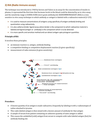
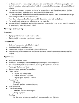
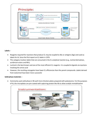
Immunological assays use antibodies or antigens to detect the presence or concentration of a molecule in a solution. There are several types of immunoassays including radioimmunoassays (RIA), enzyme-linked immunosorbent assays (ELISA), and bioluminescence assays. RIA uses radioactive labels on antigens or antibodies for highly sensitive detection, but requires special safety precautions. ELISA is a common plate-based assay that uses enzyme labels for detection and has advantages like sensitivity, reproducibility, and flexibility. Bioluminescence assays convert chemical energy from reactions involving luciferins, luciferases, and oxygen into detectable light for applications like cell proliferation analysis.
Mass spectroscopy
1.Introduction
2. Principle
3. Theory
4. Instrumentation
5. Different types of Ionization
6. FAB
7. MALDI
8. APCI
9. ESI
10.Quardapole
Mass spectroscopy
1.Introduction
2. Principle
3. Theory
4. Instrumentation
5. Different types of Ionization
6. FAB
7. MALDI
8. APCI
9. ESI
10.Quardapole
PRINCIPLES of FT-NMR & 13C NMR
Fourier Transform
FOURIER TRANSFORM NMR SPECTROSCOPY
THEORY OF FT-NMR
13C NMR SPECTROSCOPY
Principle
Why C13-NMR is required though we have H1-NMR?
CHARACTERISTIC FEATURES OF 13 C NMR
Chemical Shifts
NUCLEAR OVERHAUSER ENHANCEMENT
Short-Comings of 13C-NMR Spectra
spectrofluorometer is the instrument for recording fluorescence emission and absorption spectra When a beam of light is incident on certain substances they emit visible light or radiations. This is known as fluorescence. Fluorescence starts immediately after the absorption of light and stops as soon as the incident light is cut off. The substances showing this phenomenon are known as flourescent substances.
Types of crystals & Application of x raykajal pradhan
some basic information:-
A crystal lattice is a 3-D arrangement of unit cells.
Unit cell is the smallest unit of a crystal, By stacking identical unit cells, the entire lattice can be constructed
A crystal’s unit cell dimensions are defined by six numbers, the lengths of the 3 axes, a, b, and c, and the three interaxial angles, α, β and γ.
If a unit cell has the same type of atom at the corners of the unit cell but not also in the middle of the faces nor in the centre of the cell, it is called primitive and given by symbol P
7 types of crystal system details
14 bravis lattice
APPLICATION X-RAY CRYSTALLOGRAPHY
1. Structure of crystals
2. Polymer characterisation
3. State of anneal in metals
4. Particle size determination
a) Spot counting method
b) Broadening of diffraction lines
c) Low-angle scattering
5.Applications of diffraction methods to complexes
a) Determination of cis- trans isomerism
b) Determination of linkage isomerism
6.Miscellaneous applications
ICH and WHO Guideline for Validation and Calibration.pptxRAHUL PAL
Validation: Action of proving and documenting that any process, procedure or method actually and consistently leads to the expected results.
Calibration: The set of operations that establish, under specified conditions, the relationship between values indicated by an instrument or system for measuring (for example, weight, temperature and pH), recording, and controlling, or the values represented by a material measure, and the corresponding known values of a reference standard.
Quadrupole and Time of Flight Mass analysers.Gagangowda58
Description about important mass analysers Quadrupole and TOF: Principle, Construction and Working, Advantages and Disadvantages and their Applications.
Assignment on General principles of ImmunoassayDeepak Kumar
Assignment on General principles of immunoassay: theoretical basis and optimization of immunoassay, heterogeneous and homogenous immunoassay systems. Immunoassay methods evaluation; protocol outline, objectives and preparation. Immunoassay for digoxin and insulin
COMPRESSION AND COMPACTION , physics of tablet compression, compression, consolidation, effects of friction, distribution of forces, compaction profiles, solubility.
Paper electrophoresis
gel electrophoresis
capillary electrophoresis
zone electrophoresis
moving boundary electrophoresis
iso electric focusing electrophoresis
GASTRO RETENTIVE DRUG DELIVERY SYSTEM (GRDDS)JayeshRajput7
Gastro retentive drug delivery system which includes, principles concepts, advantages and disadvantages of GRDDS, Modulation of GI transit time, Approaches to extend GI transit, Buccal drug delivery systems, Principle of muco adhesion, advantages and disadvantages of GRDDS, Mechanism of drug permeation, Methods of formulation and its evaluations.
Documentation in pharaceutical industryJayeshRajput7
documentation in pharmaceutical industry, master formula record (MFR), DMF (drug master file), distribution records, generic drugs product development, hatch waxman act, CFR (code of federal regulation), drug product performance, in vitro ANDA regulatory approval process, NDA approval process, BE and drug product assessment, in-vivo scale up process approval changes, post marketing surveillance, outsourcing BA and BE to CRO (contract research organisation), Regulatory requireents for product approval, API, Biologics, Novel therapies obtaining NDA, ANDA for generic drug ways and means of US registration for foreign drugs.
CMC, post approval regulatory affairs, etcJayeshRajput7
this document covers points such as CMC, post approval regulatory affairs, regulation for combination products, and medical devices, common technical document (CTD) and electronic common technical document (eCTD) format, industry and FDA liasion, ICH guidelines of ICH Q,S,E,M, regulatory requirements of EU, MHRA, TGA and ROW countries.
Welcome to TechSoup New Member Orientation and Q&A (May 2024).pdfTechSoup
In this webinar you will learn how your organization can access TechSoup's wide variety of product discount and donation programs. From hardware to software, we'll give you a tour of the tools available to help your nonprofit with productivity, collaboration, financial management, donor tracking, security, and more.
This is a presentation by Dada Robert in a Your Skill Boost masterclass organised by the Excellence Foundation for South Sudan (EFSS) on Saturday, the 25th and Sunday, the 26th of May 2024.
He discussed the concept of quality improvement, emphasizing its applicability to various aspects of life, including personal, project, and program improvements. He defined quality as doing the right thing at the right time in the right way to achieve the best possible results and discussed the concept of the "gap" between what we know and what we do, and how this gap represents the areas we need to improve. He explained the scientific approach to quality improvement, which involves systematic performance analysis, testing and learning, and implementing change ideas. He also highlighted the importance of client focus and a team approach to quality improvement.
The Indian economy is classified into different sectors to simplify the analysis and understanding of economic activities. For Class 10, it's essential to grasp the sectors of the Indian economy, understand their characteristics, and recognize their importance. This guide will provide detailed notes on the Sectors of the Indian Economy Class 10, using specific long-tail keywords to enhance comprehension.
For more information, visit-www.vavaclasses.com
Instructions for Submissions thorugh G- Classroom.pptxJheel Barad
This presentation provides a briefing on how to upload submissions and documents in Google Classroom. It was prepared as part of an orientation for new Sainik School in-service teacher trainees. As a training officer, my goal is to ensure that you are comfortable and proficient with this essential tool for managing assignments and fostering student engagement.
The French Revolution, which began in 1789, was a period of radical social and political upheaval in France. It marked the decline of absolute monarchies, the rise of secular and democratic republics, and the eventual rise of Napoleon Bonaparte. This revolutionary period is crucial in understanding the transition from feudalism to modernity in Europe.
For more information, visit-www.vavaclasses.com
Palestine last event orientationfvgnh .pptxRaedMohamed3
An EFL lesson about the current events in Palestine. It is intended to be for intermediate students who wish to increase their listening skills through a short lesson in power point.
Synthetic Fiber Construction in lab .pptxPavel ( NSTU)
Synthetic fiber production is a fascinating and complex field that blends chemistry, engineering, and environmental science. By understanding these aspects, students can gain a comprehensive view of synthetic fiber production, its impact on society and the environment, and the potential for future innovations. Synthetic fibers play a crucial role in modern society, impacting various aspects of daily life, industry, and the environment. ynthetic fibers are integral to modern life, offering a range of benefits from cost-effectiveness and versatility to innovative applications and performance characteristics. While they pose environmental challenges, ongoing research and development aim to create more sustainable and eco-friendly alternatives. Understanding the importance of synthetic fibers helps in appreciating their role in the economy, industry, and daily life, while also emphasizing the need for sustainable practices and innovation.
The Art Pastor's Guide to Sabbath | Steve ThomasonSteve Thomason
What is the purpose of the Sabbath Law in the Torah. It is interesting to compare how the context of the law shifts from Exodus to Deuteronomy. Who gets to rest, and why?
Ethnobotany and Ethnopharmacology:
Ethnobotany in herbal drug evaluation,
Impact of Ethnobotany in traditional medicine,
New development in herbals,
Bio-prospecting tools for drug discovery,
Role of Ethnopharmacology in drug evaluation,
Reverse Pharmacology.
1. Masters of pharmacy, Pharmaceutical technology (Pharmaceutics)
Subject- Modern pharmaceutical analytical techniques (Mpt-101T)
Lesion no- 6 IMMUNOLOGICAL ASSAYS By- Drx JAYESH M RAJPUT
Points:-
1) Immunological assays (Immunoassay)
An immunoassay is a biochemical test that measures the presence or concentration of a macromolecule or a
small molecule or a small molecule in a solution through the use of an antibody (usually) or an antigen
(sometimes). The molecule detected by the immunoassay is often referred to as an “analyte” and is in many
cases a protein although it may be other kinds of molecules, of different size and types, as long as the proper
antibodies that have the adequate properties for the assay are developed. Analytes in biological liquids such
as serum or urine are frequently measured using immunoassays for medical and research purposes.
Immunoassays come in many different formats and variations. Immunoassays may be run in multiple steps
with reagents being added and washed away or separated at different points in the assay. Multiple-step
assays are often called separation immunoassays or heterogeneous immunoassays. Some immunoassays
can be carried out simply by mixing the reagents and sample and making a physical measurement. Such
assays are called homogeneous immunoassays, or less frequently non-separation immunoassays.
The use of a calibrator is often employed in immunoassays. Calibrators are solutions that are known to
contain the analyte in questions, and the concentration of that analyte is generally known. Comparison of an
assay’s response to a real sample against the assay’s response produced by the calibrator makes it possible
to interpret the signal in terms of the presence or concentration of analyte in the sample.
Immunoassay relies on the ability of an antibody to recognize and bind a specific macromolecule in what
might be a complex mixture of macromolecules. In immunology the particular macromolecule bound by an
antibody is referred to as an antigen and the area on an antigen to which the antibody binds is called an
epitope, in some cases, an immunoassay may use an antigen to detect for the presence of antibodies, which
recognize that antigen, in a solution. In other words, in some immunoassays, the analyte may be an antibody
rather than an antigen. In addition to the binding of an antibody to its antigen, the other key feature of all
immunoassays is a means to produce a measurable signal in response to the binding. Most though not all,
immunoassays involve chemically linking antibodies or antigens with some kind of detectable label. A large
number of labels exist in modern immunoassays, and they allow for detection through different means
many labels are detectable because they emit radiation, produce a color change in a solution, fluorescence
under light, or can be induced to emit light.
immunoassay means a method to measure any particular substance in a mixture using its specific-
binding antibody
one of the merits of immunoassay is that we can measure a substance that is present in a mixture of
various contaminants
immunoassay have become very popular in view of their high sensitivity, safety, economy and simple
instrument requirements
2. 2) RIA (Radio Immuno assays)
The technique was introduced in 1960 by berson and Yalow as an assay for the concentration of insulin in
plasma, it represented the first time that hormone levels in the blood could be detected by an in vitro assay,
and the sensitivity range is 0.0006-0.006 micro gram antibody/ml. RADIOIMMUNOASSAY (RIA) is a very
sensitive in vitro assay technique in which antibody or antigen is labeled with a radioactive material (I-125)
it is used to measure concentrations of antigens using specificity of antigen-antibody binding and
quantization using radioactivity
it is also called as binder-ligand assay where binder is the component to which radioactive material is
labeled and ligand (antigen or antibody) is the component which is to be detected
it is more specific and sensitive method and can detect antigen upto pictogram quantities
Principle of RIA
It involves three principles:
an immune reaction i.e. antigen, antibody binding
a competitive binding or competitive displacement reaction (it gives specificity)
measurement of radio emission (it gives sensitivity)
Procedure:
A known quantity of an antigen is made radioactive, frequently by labeling it with γ-radioisotopes of
iodine attached to tyrosine
This radio labeled antigen is then mixed with a known amount of antibody for that antigen
A sample of serum from patient containing an unknown quantity of same antigen is added
This causes the unlabelled (cold) antigen from serum to compete with radio labeled antigen (hot) for
antibody binding site
3. As the concentration of cold antigen is increased, more of it binds to antibody, displacing the radio
labeled variant and reducing the ratio of antibody bound radio labeled antigen to free radio labeled
antigen
The bound antigens are then separated from the unbound ones, and the radioactivity of the free
antigen remaining in the supernatant is measured using a γ-counter
The concentration of the test antigen can be calculated from the ratio of the bound and total antigen
labels, using a standard dose response curve
From these data, a standard binding curve, like the one shown in red, can be drawn
The samples to be assayed (the unknowns) are run in parallel
After determining the ratio of bound to free antigen in each unknown, the antigen concentrations can
be read directly from the standard curve
Advantages & disadvantages
Advantages
Highly specific: immune reactions are specific
Highly sensitivity: immune reactions are sensitive
Disadvantages
Radiation hazards: uses radiolabelled reagents
Requires specially trained persons
Labs require special license to handle radioactive material
Requires special arrangements for requisition, storage of radioactive material, radioactive waste
disposal
Applications
Detection of narcotic drugs
Blood bank screening for the hepatitis (a highly contagious condition) virus
Measurement of growth hormone levels, immunoglobulin’s tumor markers
Tracking of the leukemia virus
Diagnosis and treatment of peptic ulcers
Endocrinology
- Insulin, HCG, vasopressin
- Detects endocrine disorders
- Physiology of endocrine function
Pharmacology
- Morphine
- Detect drug abuse or drug poisoning
Study drug kinetics
Clinical immunology
- Antibodies for inhalant allergens
- Allergy diagnosis
Oncology
- Carcino-embryonic antigen, and early cancer detection and diagnosis
4. 3) ELISA (Enzyme linked Immuno-sorbent assay)
Elisa is a biochemical technique used mainly in immunology to detect the presence of an antibody or
an antigen in a sample
In this method the antigen or antibody is conjugated to an enzyme
It is a plate based assays designed for detecting and quantifying substances such as peptides,
proteins, antibodies, antigens and hormones
It involves detection of “analyte” in a liquid sample using liquid reagent (wet lab) or dry strips (dry
lab)
The test can be done in polystyrene tubes (macro-ELISA) or polyvinyl microtitre plates (micro-
ELISA)
Basic principles (based on immunology response)
Lock and key concept
- Antigen (key): substance when introduced into the body produces antibodies
- Antibody (lock): protein in the body that is used by immune system to identify and neutralize
foreign targets (referred to as antigens)
- Key fits into the lock
Enzyme conjugate substrates
- Enzyme that converts colorless substrates to a colored product
- Bound to the antibody that is part of the antibody-antigen complex OR
- Bound to a secondary antibody that binds with the antibody-antigen complex
5. Advantages of ELISA test
Sensitive: nanogram levels or lower
Reproducible
Minimal reagents
Qualitative and quantitative
Qualitative- Eg: HIV testing
Quantitative assays- Eg: TDM
Greater scope: wells can be coated with antigens OR antibodies
No radiation hazards
Fast-90 samples tested in 2-3 hr
Sensitivity (up to 10 pg/ml)
Specificity (sample with high concentration contaminants)
Four common ELISA tests- based on the binding structure between the antibody and antigen
1) Direct- ELISA
It uses the directly labeling the antibody itself, microwell plates are coated with a sample containing the
target antigen, and the binding of labeled antibody is quantitated by a colourimetric, chemiluminescent, of
fluorescent end-point
Apply a sample of known antigen to a surface, often in the well of a microtitre plate. The antigen is
fixed to the surface to render it immobile
The plate wells or other surface are then coated with blocking buffer
Detecting antibody (labeled by an enzyme), usually diluted in blocking buffer, is applied to the plate
for binding to the antigen coated on the plate
The plate is washed, so that unbound antibody is removed after this wash, only the antibody- antigen
complexes remain attached to the well
Apply a substrate which is converted by the enzyme to elicit a chromogenic or fluorescent signal
6. View/quantify the result using a spectrophotometer or other optical device
Positive and negative controls should always be included in the test
At every step, incubation and washing is done to wash off unbound reagents
Advantages of direct ELISA:
Quick methodology since only one antibody is used
Cross-reactivity of secondary antibody is eliminated
Disadvantages of direct ELISA:
Immuno- reactivity of the primary antibody may be reduced as a result of labeling
Labeling of energy primary antibody label from one experiment to another
Little signal amplification
2) Indirect- ELISA
For antibody detection, the wells of microtitre plates are coated with antigen
Sera to be tested are added in these coated wells
If antibody is present, antigen-antibody reaction takes place
To detect this reaction, a goat antihuman immunoglobulin antibody conjugated with an enzyme is
added
A substrate is added and enzyme acts on substrate to produce a colour
Advantages of indirect ELISA:
High sensitivity: more than one labeled antibody is bound per antigen molecule
Flexible: different primary detection antibodies can be used with a single labeled secondary
antibody
Cost-saving: fewer labeled antibodies are required
7. A sandwich ELISA
o Plate is coated with a capture antibody
o Sample is added, and any antigen present binds to capture antibody
o Detecting antibody is added, and binds to antigen
o Enzyme-linked secondary antibody is added, and binds to detecting antibody
o Substrate is added and is converted by enzyme to detectable form
o The positive results produces colour which can be read by ELISA reader
Advantages
o High specificity, since two antibodies are used the antigen/analyte is
specifically captured and detected
o Suitable for complex samples, since the antigen does not require purification
prior to measurement
o Flexibility and sensitivity, since both direct and indirect detection methods
can be used
3) Competitive- ELISA
Here competition occurs between two antibodies for the same antigen
It has been used for detection of HIV antibodies
The microtitre plate wells are coated with HIV antigen
Sera to be tested is added to these wells and incubated at 37degree Celsius and washed
Antigen-antibody reaction occurs
8. To detect this reaction, enzyme labeled specific HIV antibodies are added
These antibodies remain free because there is no antigen left to react
Substrate is added but there is no enzyme to act. Hence positive results show no colour
If serum to be tested is negative for antibodies, antigen is there to combine with enzyme conjugated
antibodies and reacts with substrate to produce colour
Advantages
o high specificity, since two antibodies are used the antigen/analyte is specifically captured and
detected
o suitable for complex samples, since the antigen does not require purification prior to
measurement
o flexibility and sensitivity, since both direct and indirect detection methods can be used
Applications
o Detection of HIV antibodies In serum
o Detection of mycobacterial antibodies in tuberculosis
o Detection of rotavirus in faeces
o Detection of hepatitis B markers in serum
o Detection of enterotoxin of E.coli in faeces
o Detection of potential food allergens
o Analysis of hormones, vitamins, metabolites, diagnostic markers
o Eg. ACTH, FSH, T3, T4, glucagon, insulin, testosterone, vitamin B12, prostaglandins,
glucocorticoids
o Therapeutic drug monitoring
o Eg. Barbiturates, morphine, digoxin
4) Non-competitive- ELISA (sandwich-ELISA)
o Antigens like tumor markers, hormones, serum proteins may be determined
o Antigens in the sample bind with the capture antibody& become immobilized
o The antibody of the enzyme conjugate bind with the immobilized antigen to form a sandwich of AB-
AG-AB/ enzyme bound to microwell
9. 5) Multiple and portable ELISA
It is a newer technique uses a solid phase made up of an Immuno-sorbent polystyrene rod with 8-12
protruding ogives. The entire device is immersed in a test-tube containing the collected sample and the
following steps (washing, incubation in conjugate and incubation in chromogenous) are carried out by
dipping the ogives in microwells of standard microplates pre-filled with reagents
6) Modified ELISA
Enzyme ⇒ interface with AG-AB interaction
Second antibody is often labeled with a very small molecular substance, biotin (MW=244.31), and a
specific binding protein for biotin, avidin is conjugated with enzyme such as HRP
10. 4) Bioluminescence assay
Bioluminescence assay systems have become increasingly used in biology and medical research
laboratories in addition to fluorescence and chemiluminescence detection strategies. It is used in areas such
as in vivo imaging, cell proliferation assays, protein folding/ secretion analyses, reporter gene assays
When a living organism produces and emits light as a result of chemical reaction is bioluminescence.
Bio means “living” in Greek while lumen means “light” in Latin
During the process, chemical energy is converted into light energy
The process is caused by enzyme catalyzed chemo luminescence reaction
All bioluminescent organisms use a reaction between an enzyme and a substrate to make light, but
different species use different chemicals in the process
Occurrence
Bioluminescence on land in freshwater is rare compared to its occurrence in the ocean
In the deep ocean 90% of the animal are luminescent
Bioluminescence is found throughout the ocean from surface to deep sea floor
Most marine animals that emit light exhibit blue green bioluminescence
Bioluminescent organism on land glow mainly in blue-green colors but they can also glow
in colors on yellow spectrum
How it happens?
Bioluminescence is product of a chemical reaction in organisms
Three ingredients are needed for luminescence to occur
1. Luciferins: it is protein like light producing substance
2. Luciferase: it is enzyme and it allows the light producing chemical reaction to take place
11. 3. Oxygen: it is colorless and odorless gas. Oxygen forms 20% of earth’s atmosphere and it is found in
water
The Luciferase allows oxygen to combine with luciferin
And this reaction produces light and oxidized luciferin become inactive oxy-luciferin
Some reaction do not involve this enzyme Luciferase, so these reaction involve chemical called
photoprotin that combine with oxygen and Luciferase but require another agent. Often an ion of
element calcium, to produce light
Bioluminescence: - is the production and emission of light by a living organism. It is a form of
chemiluminescence. Bioluminescence occurs widely in marine vertebrates and invertebrates, as well as
in some fungi microorganisms including some bioluminescent bacteria and terrestrial invertebrae such
as fireflies. Bioluminescence assays involve the use of the property of bioluminescence for measuring
cell proliferation, apoptosis, drug metabolism, kinase activity, etc
Principle: - Bioluminescence is a form of chemiluminescence where light energy is released by a
chemical reaction. This reaction involves a light emitting pigment, the luciferin and a Luciferase the
enzyme component because of the diversity of luciferin/ Luciferase combinations, there are very few
commonalities in the chemical mechanism. For example, the firefly luciferin / Luciferase reaction
requires magnesium and ATP and produces carbon-dioxide (co2), adenosine monophosphate (AMP) and
pyrophosphate (PP) as waste products. Other cofactors may be required for the reaction, such as
calcium (ca2+) for the photoprotin acquorin or magnesium (MG2+) ions and ATP for the firely
Luciferase. Generically, this reaction could be described as: -
Luciferin + o2 ⇒ oxyluciferin + light energy
Among assay methods, chemiluminescence (CL) detection represents a versatile, ultrasensitive tool with
a wide range of applications in biotechnology. It also gives a sensitive, rapid alternative to radioactivity
as a detection principle in IA for the determination of molecules (eg, proteins, hormones, drugs, nucleic
acids and environmental pollutants). CL is now commonly used for IA in the form of CL label or as a CL
detection reaction for an enzyme or a nanoparticle (NP) label. In recent years, CLIA has become very
popular in clinical chemistry and environmental analysis, due to its high sensitivity, wide dynamic range
and complete automation. With development and application of recombinant AB (rAB) technology,
markers and related techniques, solid-phase materials and improvements in automation, integration
and miniaturization, CLIA has acquired an entirely new appearance.
Luminescence
“cold light” that can be emitted at lower temperature
Source kicks an electron of an atom out of its lowest energy “ground” state into a higher energy
“excited” state
Finally electron returns the energy in the form of light so it can fall back to its “ground” state
Introduction
Chemiluminescence immunoassay (CLIA) using microplate luminometers provides a
sensitive, high throughoutput, and economical way to quantitatively measure antigen in cell
lysates, plasma, urine, saliva, tissue and culture media samples
12. Chemiluminescence immunoassay does not require long incubations and the addition of
stopping reagents, as is the case in conventional colorimetric cases such as enzyme linked
Immuno-sorbent assay (ELISA)
Among various enzyme assays that employ light-emitting reactions, one of the most
successful assays is the enhanced chemiluminescent immunoassay involving a horseradish
peroxidase (HRP) labeled antibody or antigen and a mixture of chemiluminescent substrate,
hydrogen peroxide, and enhancers
CLIA are designed to detect glow based chemiluminescent reactions
This provide a broader dynamic assay range, superior low-end sensitivity, and a faster
protocol than the conventional (ELISA)
It covers thyroid function markers, gonadal hormones, tumor markers, diabetic marker,
cardiac marker and other markers
They can be used to replace conventional colorimetric ELISAs that have been widely used in
many research and diagnostic applications
Chemiluminescence immunoassay (CLIA) provides a sensitive, high throughoutput alternative to
conventional colorimetric methodologies
Procedure: -
Monoclonal antibody coated well ⇒ test specimen (serum) ⇒ HRP labelled antibody
conjugate ⇒ test antigen: sandwich between solid phase AB and enzyme labelled AB ⇒
incubate for 1hr at 37degree Celsius⇒ remove unbound enzyme labeled AB ⇒
chemiluminescence reagent added ⇒ read relative light unit with luminometers
13. Labels: -
Reagents required for reactions that produce CL may be coupled to Abs or antigens (Ags) and used as
labels for IA. Since the first report on CL labels in 1976
This category involves labels that are consumed in the CL analytical reaction (e.g., luminal derivatives,
acridinium esters and NPs)
Luminal is the best known and one of the most efficient CL reagents. It is coupled to ligands via reactions
involving the amino acid
However, the resulting conjugates have lower CL efficiencies than the parent compounds. Labels derived
from isoluminol have been more successful
Solid-phase materials: -
Commonly used solid-phase is 96-well micro titration plates prepared with polystyrene. For the purposes
of IA, the microplates are pre-coated with capturing protein like Ab to allow analyte immobilization