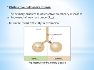
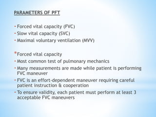
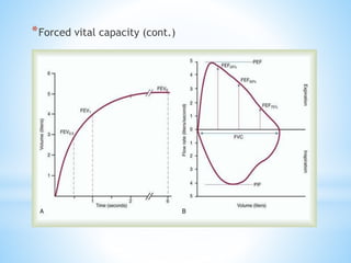
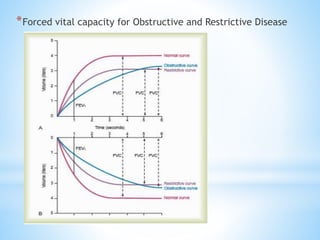
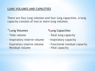
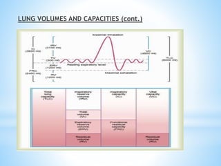
Pulmonary function tests (PFTs) measure lung function in three categories: dynamic air flow, lung volumes and capacities, and gas diffusion. PFTs identify and quantify changes from pulmonary disease, evaluate therapy effectiveness, and assess postoperative risk. Spirometry is the most common PFT and measures forced vital capacity (FVC) and forced expiratory volume in 1 second (FEV1) to detect airway obstruction. Other PFTs include slow vital capacity, maximal voluntary ventilation, flow-volume loops, and reversibility testing with bronchodilators. PFT interpretation considers ratios like FEV1/FVC and compares values to predicted normals to diagnose obstructive or restrictive lung disease.
Pulmonary function tests (PFTs) are noninvasive tests that show how well the lungs are working. The tests measure lung volume, capacity, rates of flow, and gas exchange. This information can help your healthcare provider diagnose and decide the treatment of certain lung disorders.
PULMONARY FUNCTION TESTS - LAB DATA INTERPRETATIONLincyAsha
PULMONARY FUNCTION TESTS
LAB DATA INTERPRETATION
CLINICAL PHARMACY PRACTICE
M.PHARMACY
PHARMACY PRACTICE
1ST YEAR
Pulmonary function tests are a series of tests performed to examine a patient’s respiratory system and identify the severity of pulmonary impairment.
These tests are performed to measure a patient’s lung volume, capacity, flow rate and gas exchange.
This allows medical professionals to obtain an accurate diagnosis and determine the best course of medical intervention for the patient.
In general there are two types of lung disorders that these tests can be used to assess
Obstructive lung diseases
Restrictive lung diseases
1.OBSTRUCTIVE LUNG DISEASES
It include conditions that make it difficult to exhale air out of the lungs
This results in shortness of breath that occurs from narrowing and constriction of the airways and causes the patient to have decreased flow rates. Eg. COPD, Asthma
2.RESTRICTIVE LUNG DISEASES
It include conditions that make it difficult to fully fill the lungs with air during inhalation.
When the lungs aren’t fully able to expand it causes the patient to have decreased lung volumes. Eg. Pulmonary fibrosis, interstitial lung disease
Pulmonary function tests would be indicated for the following:
On healthy patients as part of a routine physical exam
Evaluate signs and symptoms of lung disease
Diagnosis of certain medical conditions
Measure current stage of disease and evaluate its progress
Assess how a patient is responding to different treatments
Determine patient’s condition before surgery to assess the risk of respiratory complications
Screen people who are at risk of pulmonary disease
Determine how much a patient’s airways have narrowed due to disorders
In certain types of work environments to assess the health of employees.
Additionally PFTs may be indicated for the following
Chronic lung conditions
Restrictive airway problems
Asthma
COPD
Shortness of breath
Impairment or disability
Early morning wheezing
Chest muscle weakness
Lung cancer
Respiratory infections
STATIC LUNG VOLUMES
Lung volume is the amount of air breathed by an individual under a specific condition.
1.Tidal Volume (TV)
It is the volume of air inspired or expired during normal breathing at rest.
2.Inspiratory Reserve Volume (IRV)
It is the volume of air inspired with maximum effort over and above the normal tidal volume.
3.Expiratory Reserve Volume (ERV)
It is the volume of air expired forcefully after a normal respiration.
4.Residual Volume (RV)
It is the volume of air remaining in the lungs after a forceful expiration
STATIC LUNG CAPACITIES
1.Inspiratory capacity (IC)
It is the amount of air a person can inspire forcefully after a normal respiration.
IC = TV+IRV
2.Functional Residual Capacity (FRC)
It is the amount of air that remains in the lungs at the end of normal respiration.
FRC = ERV+RV
3.Vital Capacity (VC)
It is the maximum volume of air exhaled forcefully from the lungs after a maximum inspiration.
4.Total Lung Capacity
Pulmonary function tests (PFT) are series of tests that measure lung function and aid in the management of patients with respiratory disease.
They are performed using standardized equipment and can be used for diagnosis, prognostication, management and follow-up of patients with pulmonary pathology.
Although PFT may not identify the exact pathology, it broadly classifies respiratory disorders as either obstructive or restrictive. In this session , the role of PFT in the measurement of lung mechanics and diagnosis of various diseases will be discussed in detail.
Pulmonary function testing is the process of having the patient perform specific inspiratory and expiratory maneuvers while breathing in and out of tubing attached to the equipment that measure a variety of variables
MANAGEMENT OF ATRIOVENTRICULAR CONDUCTION BLOCK.pdfJim Jacob Roy
Cardiac conduction defects can occur due to various causes.
Atrioventricular conduction blocks ( AV blocks ) are classified into 3 types.
This document describes the acute management of AV block.
Knee anatomy and clinical tests 2024.pdfvimalpl1234
This includes all relevant anatomy and clinical tests compiled from standard textbooks, Campbell,netter etc..It is comprehensive and best suited for orthopaedicians and orthopaedic residents.
263778731218 Abortion Clinic /Pills In Harare ,sisternakatoto
263778731218 Abortion Clinic /Pills In Harare ,ABORTION WOMEN’S CLINIC +27730423979 IN women clinic we believe that every woman should be able to make choices in her pregnancy. Our job is to provide compassionate care, safety,affordable and confidential services. That’s why we have won the trust from all generations of women all over the world. we use non surgical method(Abortion pills) to terminate…Dr.LISA +27730423979women Clinic is committed to providing the highest quality of obstetrical and gynecological care to women of all ages. Our dedicated staff aim to treat each patient and her health concerns with compassion and respect.Our dedicated group ABORTION WOMEN’S CLINIC +27730423979 IN women clinic we believe that every woman should be able to make choices in her pregnancy. Our job is to provide compassionate care, safety,affordable and confidential services. That’s why we have won the trust from all generations of women all over the world. we use non surgical method(Abortion pills) to terminate…Dr.LISA +27730423979women Clinic is committed to providing the highest quality of obstetrical and gynecological care to women of all ages. Our dedicated staff aim to treat each patient and her health concerns with compassion and respect.Our dedicated group of receptionists, nurses, and physicians have worked together as a teamof receptionists, nurses, and physicians have worked together as a team wwww.lisywomensclinic.co.za/
Recomendações da OMS sobre cuidados maternos e neonatais para uma experiência pós-natal positiva.
Em consonância com os ODS – Objetivos do Desenvolvimento Sustentável e a Estratégia Global para a Saúde das Mulheres, Crianças e Adolescentes, e aplicando uma abordagem baseada nos direitos humanos, os esforços de cuidados pós-natais devem expandir-se para além da cobertura e da simples sobrevivência, de modo a incluir cuidados de qualidade.
Estas diretrizes visam melhorar a qualidade dos cuidados pós-natais essenciais e de rotina prestados às mulheres e aos recém-nascidos, com o objetivo final de melhorar a saúde e o bem-estar materno e neonatal.
Uma “experiência pós-natal positiva” é um resultado importante para todas as mulheres que dão à luz e para os seus recém-nascidos, estabelecendo as bases para a melhoria da saúde e do bem-estar a curto e longo prazo. Uma experiência pós-natal positiva é definida como aquela em que as mulheres, pessoas que gestam, os recém-nascidos, os casais, os pais, os cuidadores e as famílias recebem informação consistente, garantia e apoio de profissionais de saúde motivados; e onde um sistema de saúde flexível e com recursos reconheça as necessidades das mulheres e dos bebês e respeite o seu contexto cultural.
Estas diretrizes consolidadas apresentam algumas recomendações novas e já bem fundamentadas sobre cuidados pós-natais de rotina para mulheres e neonatos que recebem cuidados no pós-parto em unidades de saúde ou na comunidade, independentemente dos recursos disponíveis.
É fornecido um conjunto abrangente de recomendações para cuidados durante o período puerperal, com ênfase nos cuidados essenciais que todas as mulheres e recém-nascidos devem receber, e com a devida atenção à qualidade dos cuidados; isto é, a entrega e a experiência do cuidado recebido. Estas diretrizes atualizam e ampliam as recomendações da OMS de 2014 sobre cuidados pós-natais da mãe e do recém-nascido e complementam as atuais diretrizes da OMS sobre a gestão de complicações pós-natais.
O estabelecimento da amamentação e o manejo das principais intercorrências é contemplada.
Recomendamos muito.
Vamos discutir essas recomendações no nosso curso de pós-graduação em Aleitamento no Instituto Ciclos.
Esta publicação só está disponível em inglês até o momento.
Prof. Marcus Renato de Carvalho
www.agostodourado.com
Basavarajeeyam is an important text for ayurvedic physician belonging to andhra pradehs. It is a popular compendium in various parts of our country as well as in andhra pradesh. The content of the text was presented in sanskrit and telugu language (Bilingual). One of the most famous book in ayurvedic pharmaceutics and therapeutics. This book contains 25 chapters called as prakaranas. Many rasaoushadis were explained, pioneer of dhatu druti, nadi pareeksha, mutra pareeksha etc. Belongs to the period of 15-16 century. New diseases like upadamsha, phiranga rogas are explained.
Title: Sense of Smell
Presenter: Dr. Faiza, Assistant Professor of Physiology
Qualifications:
MBBS (Best Graduate, AIMC Lahore)
FCPS Physiology
ICMT, CHPE, DHPE (STMU)
MPH (GC University, Faisalabad)
MBA (Virtual University of Pakistan)
Learning Objectives:
Describe the primary categories of smells and the concept of odor blindness.
Explain the structure and location of the olfactory membrane and mucosa, including the types and roles of cells involved in olfaction.
Describe the pathway and mechanisms of olfactory signal transmission from the olfactory receptors to the brain.
Illustrate the biochemical cascade triggered by odorant binding to olfactory receptors, including the role of G-proteins and second messengers in generating an action potential.
Identify different types of olfactory disorders such as anosmia, hyposmia, hyperosmia, and dysosmia, including their potential causes.
Key Topics:
Olfactory Genes:
3% of the human genome accounts for olfactory genes.
400 genes for odorant receptors.
Olfactory Membrane:
Located in the superior part of the nasal cavity.
Medially: Folds downward along the superior septum.
Laterally: Folds over the superior turbinate and upper surface of the middle turbinate.
Total surface area: 5-10 square centimeters.
Olfactory Mucosa:
Olfactory Cells: Bipolar nerve cells derived from the CNS (100 million), with 4-25 olfactory cilia per cell.
Sustentacular Cells: Produce mucus and maintain ionic and molecular environment.
Basal Cells: Replace worn-out olfactory cells with an average lifespan of 1-2 months.
Bowman’s Gland: Secretes mucus.
Stimulation of Olfactory Cells:
Odorant dissolves in mucus and attaches to receptors on olfactory cilia.
Involves a cascade effect through G-proteins and second messengers, leading to depolarization and action potential generation in the olfactory nerve.
Quality of a Good Odorant:
Small (3-20 Carbon atoms), volatile, water-soluble, and lipid-soluble.
Facilitated by odorant-binding proteins in mucus.
Membrane Potential and Action Potential:
Resting membrane potential: -55mV.
Action potential frequency in the olfactory nerve increases with odorant strength.
Adaptation Towards the Sense of Smell:
Rapid adaptation within the first second, with further slow adaptation.
Psychological adaptation greater than receptor adaptation, involving feedback inhibition from the central nervous system.
Primary Sensations of Smell:
Camphoraceous, Musky, Floral, Pepperminty, Ethereal, Pungent, Putrid.
Odor Detection Threshold:
Examples: Hydrogen sulfide (0.0005 ppm), Methyl-mercaptan (0.002 ppm).
Some toxic substances are odorless at lethal concentrations.
Characteristics of Smell:
Odor blindness for single substances due to lack of appropriate receptor protein.
Behavioral and emotional influences of smell.
Transmission of Olfactory Signals:
From olfactory cells to glomeruli in the olfactory bulb, involving lateral inhibition.
Primitive, less old, and new olfactory systems with different path
Tom Selleck Health: A Comprehensive Look at the Iconic Actor’s Wellness Journeygreendigital
Tom Selleck, an enduring figure in Hollywood. has captivated audiences for decades with his rugged charm, iconic moustache. and memorable roles in television and film. From his breakout role as Thomas Magnum in Magnum P.I. to his current portrayal of Frank Reagan in Blue Bloods. Selleck's career has spanned over 50 years. But beyond his professional achievements. fans have often been curious about Tom Selleck Health. especially as he has aged in the public eye.
Follow us on: Pinterest
Introduction
Many have been interested in Tom Selleck health. not only because of his enduring presence on screen but also because of the challenges. and lifestyle choices he has faced and made over the years. This article delves into the various aspects of Tom Selleck health. exploring his fitness regimen, diet, mental health. and the challenges he has encountered as he ages. We'll look at how he maintains his well-being. the health issues he has faced, and his approach to ageing .
Early Life and Career
Childhood and Athletic Beginnings
Tom Selleck was born on January 29, 1945, in Detroit, Michigan, and grew up in Sherman Oaks, California. From an early age, he was involved in sports, particularly basketball. which played a significant role in his physical development. His athletic pursuits continued into college. where he attended the University of Southern California (USC) on a basketball scholarship. This early involvement in sports laid a strong foundation for his physical health and disciplined lifestyle.
Transition to Acting
Selleck's transition from an athlete to an actor came with its physical demands. His first significant role in "Magnum P.I." required him to perform various stunts and maintain a fit appearance. This role, which he played from 1980 to 1988. necessitated a rigorous fitness routine to meet the show's demands. setting the stage for his long-term commitment to health and wellness.
Fitness Regimen
Workout Routine
Tom Selleck health and fitness regimen has evolved. adapting to his changing roles and age. During his "Magnum, P.I." days. Selleck's workouts were intense and focused on building and maintaining muscle mass. His routine included weightlifting, cardiovascular exercises. and specific training for the stunts he performed on the show.
Selleck adjusted his fitness routine as he aged to suit his body's needs. Today, his workouts focus on maintaining flexibility, strength, and cardiovascular health. He incorporates low-impact exercises such as swimming, walking, and light weightlifting. This balanced approach helps him stay fit without putting undue strain on his joints and muscles.
Importance of Flexibility and Mobility
In recent years, Selleck has emphasized the importance of flexibility and mobility in his fitness regimen. Understanding the natural decline in muscle mass and joint flexibility with age. he includes stretching and yoga in his routine. These practices help prevent injuries, improve posture, and maintain mobilit
These simplified slides by Dr. Sidra Arshad present an overview of the non-respiratory functions of the respiratory tract.
Learning objectives:
1. Enlist the non-respiratory functions of the respiratory tract
2. Briefly explain how these functions are carried out
3. Discuss the significance of dead space
4. Differentiate between minute ventilation and alveolar ventilation
5. Describe the cough and sneeze reflexes
Study Resources:
1. Chapter 39, Guyton and Hall Textbook of Medical Physiology, 14th edition
2. Chapter 34, Ganong’s Review of Medical Physiology, 26th edition
3. Chapter 17, Human Physiology by Lauralee Sherwood, 9th edition
4. Non-respiratory functions of the lungs https://academic.oup.com/bjaed/article/13/3/98/278874
2. INTRODUCTION
-It is a group of procedures that measures the function of
the lungs.
-Pulmonary function tests can provide valuable information
about the important individual processes that support gas
exchange.
CATEGORIES OF PFT
There are 3 categories of PFT measuring
i. Dynamic flow rates of gases through airways.
ii. Lung volumes and capacities.
iii. The ability of the lungs to diffuse gases.
3. PURPOSE OF PFT
- To identify and quantify the changes in pulmonary function
due to any disease.
- Evaluate effectiveness of therapy.
- Perform epidemiological surveillance for pulmonary disease.
- Assess patients for risk of postoperative complications.
- Determine pulmonary disability.
4. PATHOPHYSIOLOGY
*Pulmonary disease are divided into two major categories:
-Obstructive pulmonary disease. Eg. COPD
-Restrictive pulmonary disease. Eg. Asthma
-Some pulmonary disease can cause both obstructive and
restrictive pulmonary disease.
*Comparison of obstructive & restrictive pulmonary disease
5. Fig. Obstructive Pulmonary Disease
* Obstructive pulmonary disease
- The primary problem in obstructive pulmonary disease is
an increased airway resistance (Raw).
- In simple terms difficulty in expiration.
6. Fig. Restrictive Pulmonary Disease
* Restrictive pulmonary disease
- The primary problem in restrictive pulmonary disease is
reduced lung compliance, lung volume or both.
- In simple terms difficulty in inspiration.
7. PULMONARY FUNCTION TEST EQUIPMENT
• Two general types of measuring devices exist, those that:
- Measure volume
- Measure flow
• Volume-measuring devices - spirometers
• Flow-measuring devices - pneumotachometers
• Every measuring device has capacity, accuracy, error,
resolution, precision, linearity, & output
8. SPIROMETRY
*It is the measurement of air entering and leaving lungs
which includes measurement of several values of forced
airflow and volume during inspiration and expiration.
• Tests of pulmonary mechanics:
-Forced vital capacity (FVC)
-Forced expiratory volume in 1 second (FEV1)
-Other forced expiratory flow measurements
-Maximum voluntary ventilation
• These measurements assess ability of lungs to move large
volumes of air quickly through airways
9. PURPOSE/IMPORTANCE OF SPIROMETRY
• The purpose of spirometry is to assess the ability of the
lungs to move large volumes of air quickly through the
airways to identify airway obstruction.
• Measuring flow rates is a surrogate for measuring airway
resistance.
• To a lesser extent spirometry can also identify and quantify
a restrictive type of pulmonary disease.
10. TYPES OF SPIROMETRY
-Computerized spirometer
-Incentive spirometer
-Tilt compensated spirometer
-Windmill type spirometer
-Tank type spirometer
12. INDICATIONS OF SPIROMETRY
It should be especially indicated for persons with complaint
of shortness of breath.
-Asthma evaluation
-COPD diagnosis
-Screening for occupational lung disease
-Pre-operative evaluation
-Chronic cough
-Unexplained dyspnoea
-Smoking cessation
-Nonspecific
13. CONTRAINDICATIONS OF SPIROMETRY
It should not be indicated for persons with,
-Haemoptysis of unknown origin
-Pneumothorax
-Unstable angina pectoris
-Recent myocardial infarction
-Thoracic aneurysms, abdominal aneurysms, cerebral
aneurysms.
-Recent eye surgery ( intraocular pressure during forced
expiration)
-History of syncope associated with forced expiration
-Patient with active Tuberculosis should not be tested
14. PRECAUTIONS TO BE TAKEN FOR SPIROMETRY
-Persons with high blood pressure
-Semi-comatic patients
-Patients with age of 80yrs or more
-The subject should be healthy (free from asthma)
-No air leaks in the apparatus (or else will give inaccurate
readings)
-The mouth piece should be sterilized
-The water chamber should not be overfilled (or it may
enter air tubes)
15. TERMINATION CRITERIA FOR SPIROMETRY
-Feeling of giddiness
-Gasping
-Dyspnoea
-Hesitation or false starts
-Coughs
-Variable efforts
-Glottis closure
-Air leaks
-Early termination before a plateau (6s) reached
-Laughing
16. TECHNIQUES OF PFT
*Spirometry
*Helium dilution method
- Based on fact that known amount of helium will be diluted by
size of patient’s RV
*Nitrogen washout method
- Based on fact that 79% of RV is nitrogen
- Volume of nitrogen exhaled ÷ 0.79 = RV
*Whole body plethysmography
- Boyle’s law
Unknown lung gas vol = Gas pressure of the box
Known box gas vol Gas pressure of the lungs
20. PARAMETERS OF PFT
-Forced vital capacity (FVC)
-Slow vital capacity (SVC)
-Maximal voluntary ventilation (MVV)
*Forced vital capacity
-Most common test of pulmonary mechanics
-Many measurements are made while patient is performing
FVC maneuver
-FVC is an effort-dependent maneuver requiring careful
patient instruction & cooperation
-To ensure validity, each patient must perform at least 3
acceptable FVC maneuvers
23. *Slow vital capacity
-Slow Vital Capacity is a spirometry test that displays the
volume of gas measured on a low complete expiration
after a maximal inspiration without forced or rapid effort.
-Ensure that at least four consecutive tidal breaths are
stable before beginning SVC.
-SVC’s should agree within 5% or 150ml.
-SVC’s can be performed as inspiratory, expiratory or mixed
IC/ERV efforts.
25. *Maximal voluntary ventilation
-Effort-dependent test: the patient should be instructed
to breathe as rapidly and as deeply as possible for at
least 12 seconds.
-The patient’s breathing is measured on a spirogram or
electronically for the specific number of seconds (t) and
the volume (V) breathed when the MVV is converted to
liters per minute.
• Results reflect:
-Patient effort
-Function of respiratory muscles
-Ability of chest wall to expand
-Patency of airways
26. *Maximal voluntary ventilation (cont.)
-Normal MVV for males is 160 to 180 L/min & slightly lower in
females
-MVV is reduced in patients with moderate to severe
obstructive lung disease
-MVV may be normal or slightly reduced in patients with
restrictive disease
-Undernourished patients may have reduced MVV
27. LUNG VOLUMES AND CAPACITIES
There are four lung volumes and four lung capacities. A lung
capacity consists of two or more lung volumes.
*Lung Volumes *Lung Capacities
- Tidal volume - Total lung capacity
- Inspiratory reserve volume - Inspiratory capacity
- Expiratory reserve volume - Functional residual capacity
- Residual volume - Vital capacity
29. FLOW VOLUME LOOP
-The flow-volume loop is a plot of inspiratory and
expiratory flow (on the Y-axis) against volume (on the X-
axis) during the performance of maximally forced
inspiratory and expiratory maneuvers
-The normal expiratory portion of the flow-volume curve is
characterized by a rapid rise to the peak flow rate
followed by a nearly linear fall in flow as the patient
exhales toward RV.
-The inspiratory curve, in contrast, is a relatively
symmetrical, saddle-shaped curve.
*NOTE – Answer is given above for the question how flow
volume loop is produced?
32. REVERSIBILITY TEST
-If obstruction is present, reversibility must be evaluated
-Done by performing spirometry before & after therapy
-Bronchodilator is administered by small-volume nebulizer
or MDI
-Reversibility indicates effective therapy
-Reversibility is defined as 15% or greater improvement in
FEV1 & at least 200-ml increase in FEV1
33. PFT REPORT INTERPRETATION
-FEV1/FVC ratio is good place to start with; reduced
(<70%) with obstructive lung disease
-If TLC less than 80% of predicted normal & FEV1/FVC is
normal - restrictive disease is present
*If DLCO is <80% of normal - diffusion defect is present
-Reduced surface area = emphysema
-Thickened AC membrane = pulmonary fibrosis
35. CONCLUSION
-Pulmonary function tests are an important tool in the
assessment of patients with suspected or known respiratory
disease.
-They are also important in the evaluation of patients prior to
major surgery.
-Interpretation of the tests, which requires knowledge of normal
values and appearance of flow volume curves, must be
combined with the patient’s clinical history and presentation.
REFERENCE:
-Egan’s Fundamentals of respiratory care
-PDF-Slow Vital Capacity - UTMB.edu
-Pubmed-www.ncbi.nlm.nih.gov/pmc/articles/PMC3229853/
-www.uptodate.com/contents/flow-volume-loops
-Article- Spirometry in the evaluation of pulmonary function