Distal Radius #
Dr. Ashish S
Asst. Professor
Department of Musculoskeletal Sciences
DVVPF’s College of Physiotherapy, Ahmednagar
EPIDEMIOLOGY
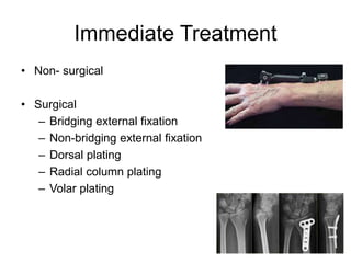
• Distal radius fractures are among the most common
fractures of the upper extremity.
• Fractures of the distal radius represent approximately
one-sixth of all fractures treated in emergency
departments.
• Risk factors for fractures of the distal radius in the elderly
include decreased bone mineral density, female sex,
white race, family history, and early menopause.
MECHANISM OF INJURY
MECHANISM OF INJURY
• Common mechanisms in younger individuals include
falls from a height, motor vehicle accident, or injuries
sustained during athletic participation. In elderly
individuals, distal radial fractures may arise from low-
energy mechanisms, such as a simple fall from a
standing height.
• The most common mechanism of injury is a fall onto an
outstretched hand with the wrist in dorsiflexion.
CLINICAL EVALUATION
• Patients typically present with variable wrist deformity and
displacement of the hand in relation to the wrist The wrist is
typically swollen with ecchymosis, tenderness, and painful
range of motion.
• The ipsilateral elbow and shoulder should be examined for
associated injuries.
• A careful neurovascular assessment should be performed,
with particular attention to median nerve function. Carpal
tunnel compression symptoms are common (13% to 23%)
owing to traction during forced hyperextension of the wrist,
direct trauma from fracture fragments, hematoma formation,
or increased compartment pressure.
RADIOGRAPHIC EVALUATION
• Posteroanterior and lateral views of the wrist should be
obtained, with oblique views for further fracture
definition, if necessary. Shoulder or elbow symptoms
should be evaluated radiographically.
Classification by fernandez
• Type 1- bending type , extra articular , metaphyseal #.
Displacement dorsal- colle’s, volar – smith’s.
• Type 2- articular # with volar or dorsal with radial styloid
#(chauffeur’s #) and lunet facet.
• Type 3- compression # with displacement of radial
articular surface, separation of scaphoid and lunet facet.
• Type 4 – radiocarpal # dislocation with avulsion #.
• Type 5- combined features of all other types and may
also involve forearm compartmental syndrome, open
wound or associated injury forearm or elbow.
Eponyms
• Colle’s fracture
• Smith fracture (reverse Colle’s fracture)
• Barton fracture
• Radial styloid fracture
COMPLICATIONS
• Stiffness of joints
• Mal-union
• Subluxation of inferior radio-ulnar joint
• Carpel- tunnel syndrome
• Sudeck’s osteodystrophy
• Rupture of the extensor pollicis longus tendon
Immediate Treatment
• Non- surgical
• Surgical
– Bridging external fixation
– Non-bridging external fixation
– Dorsal plating
– Radial column plating
– Volar plating
Goals of Rehabilitation
• Short term goals
– Control pain
– Reduce contractures
– Reduce inflammation
• Long term goals
– Improve and maintain ROM
– Improve and mainatin strength
– Allow patient to be psychologically ready to return
Thank You

Distal radius

  • 1.
    Distal Radius # Dr.Ashish S Asst. Professor Department of Musculoskeletal Sciences DVVPF’s College of Physiotherapy, Ahmednagar
  • 2.
    EPIDEMIOLOGY • Distal radiusfractures are among the most common fractures of the upper extremity. • Fractures of the distal radius represent approximately one-sixth of all fractures treated in emergency departments. • Risk factors for fractures of the distal radius in the elderly include decreased bone mineral density, female sex, white race, family history, and early menopause.
  • 3.
  • 4.
    MECHANISM OF INJURY •Common mechanisms in younger individuals include falls from a height, motor vehicle accident, or injuries sustained during athletic participation. In elderly individuals, distal radial fractures may arise from low- energy mechanisms, such as a simple fall from a standing height. • The most common mechanism of injury is a fall onto an outstretched hand with the wrist in dorsiflexion.
  • 5.
    CLINICAL EVALUATION • Patientstypically present with variable wrist deformity and displacement of the hand in relation to the wrist The wrist is typically swollen with ecchymosis, tenderness, and painful range of motion. • The ipsilateral elbow and shoulder should be examined for associated injuries. • A careful neurovascular assessment should be performed, with particular attention to median nerve function. Carpal tunnel compression symptoms are common (13% to 23%) owing to traction during forced hyperextension of the wrist, direct trauma from fracture fragments, hematoma formation, or increased compartment pressure.
  • 6.
    RADIOGRAPHIC EVALUATION • Posteroanteriorand lateral views of the wrist should be obtained, with oblique views for further fracture definition, if necessary. Shoulder or elbow symptoms should be evaluated radiographically.
  • 8.
    Classification by fernandez •Type 1- bending type , extra articular , metaphyseal #. Displacement dorsal- colle’s, volar – smith’s. • Type 2- articular # with volar or dorsal with radial styloid #(chauffeur’s #) and lunet facet. • Type 3- compression # with displacement of radial articular surface, separation of scaphoid and lunet facet.
  • 9.
    • Type 4– radiocarpal # dislocation with avulsion #. • Type 5- combined features of all other types and may also involve forearm compartmental syndrome, open wound or associated injury forearm or elbow.
  • 11.
    Eponyms • Colle’s fracture •Smith fracture (reverse Colle’s fracture) • Barton fracture • Radial styloid fracture
  • 12.
    COMPLICATIONS • Stiffness ofjoints • Mal-union • Subluxation of inferior radio-ulnar joint • Carpel- tunnel syndrome • Sudeck’s osteodystrophy • Rupture of the extensor pollicis longus tendon
  • 13.
    Immediate Treatment • Non-surgical • Surgical – Bridging external fixation – Non-bridging external fixation – Dorsal plating – Radial column plating – Volar plating
  • 14.
    Goals of Rehabilitation •Short term goals – Control pain – Reduce contractures – Reduce inflammation • Long term goals – Improve and maintain ROM – Improve and mainatin strength – Allow patient to be psychologically ready to return
  • 15.