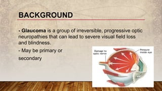
PRIMARY OPEN-
ANGLE GLAUCOMA
By
Dr. Amr Mounir
Lecturer of Ophthalmology
Sohag University
BACKGROUND
- Glaucoma is a group of irreversible, progressive optic
neuropathies that can lead to severe visual field loss
and blindness.
- May be primary or
secondary
BACKGROUND
In primary glaucoma, the elevation of IOP is not
associated with any other ocular disorder
In secondary glaucoma a recognizable ocular or
non-ocular disorder alters aqueous outflow which, in
turn, results in elevation of IOP.
DEFINITION
Also referred to as chronic simple glaucoma, is a generally bilateral, but not
always symmetrical disease,
characterized by:
1- Adult onset.
2- An IOP > 20 mmHg.
3- An open angle of normal appearance.
4- Glaucomatous optic nerve head damage.
5- Visual Field loss.
INCIDENCE
- The commonest type of
glaucoma.
- Population: Affecting 1% of
the general population > 40
years.
- Sex: It affects both sexes
equally.
- Age: most cases > 65 years. It
is unusual < 40 years.
- Race: more common, develops
earlier, and is more severe, in
black people.
- Laterality: Bilateral (usually
one eye before the other).
RISK FACTORS AND ASSOCIATIONS:
1) Family history and
inheritance: POAG is
frequently inherited.
2) Myopia: is associated
with an increased incidence
of POAG.
3) Retinal disease:
- Central retinal vein
occlusion.
- Retinitis pigmentosa.
SYMPTOMS
1) Early: Asymptomatic
2) Late: Significant loss
of visual field occurs
gradually.
3) Bilaterality:
Patients therefore
frequently present
with significant visual
field loss in one eye
and less advanced
disease in the other.
SIGNS
A) IOP:
1) Raised IOP:
- IOPs > 20 mmHg.
2) Diurnal fluctuations:
Diurnal fluctuations up to 5
mmHg
- In POAG: this fluctuation is
exaggerated .
3) Asymmetry of the IOP
between the two eyes
If 5 mmHg or more, should
arouse suspicion
SIGNS
B) Optic disc changes:
1) Baring of the curve linear blood
vessels
2) Optic disc hemorrhage (Splinter or
flame shaped).
3) Nasalization of central blood vessels.
4) Enlargement of the physiological cup.
5) Asymmetry cup / disc ratio between
both eyes.
SIGNS
6) Specific glaucomatous
cupping:
- Narrow opening.
- Size: large.
- Depth: very deep.
- Edges: overhanging.
- Increase cup/disc ratio (normal = 0.3).
- In advanced cases lamina cribrosa can
be seen.
SIGNS
C) Visual fields
(Perimetry): Central changes
1) Paracentral scotomata: Are
isolated scotomata which later
becomes connected with the blind
spot to form the arcuate scotoma.
2) Baring of the blind spot: Is
exclusion of the blind spot from
the central field (due to localized
contraction of the temporal edge
of the central field)
SIGNS
C) Visual fields
(Perimetry):Central changes
3) Siedel scotoma: Is a tapering
wing like elongation of the upper
and/or lower pole of the blind spot.
4) Bjerrum (arcuate) scotoma:
Upper and/or lower bundle defect
around and joining the blind spot and
is concentric with the fixation point.
5) Annular scotoma: By fusion of
the 2 (upper and lower) Bjerrum
scotomata in the nasal field.
SIGNS
C) Visual fields
(Perimetry):Peripheral
changes
1) Nasal field contraction: Occurs
earlier than the temporal field because
the temporal retinal fibres are more
dense and so suffer more.
2) Roenne's nasal step: Is a
sectorial nasal field defect (upper or
lower) with a sharply horizontal edge.
3) Concentric contraction.
4) Tubular vision.
HUMPHREY VISUAL FIELD MACHINE
SIGNS
D) Gonioscopy:
Shows a normal open
angle
- Target pressure:
- It is an IOP level below which further damage is
unlikely. Therapy should maintain the IOP at or
below the target level.
Monitoring
is performed of the optic nerve and visual fields. In the
event of further damage the target IOP is reset at a
lower level.
- The primary aim of
treatment is to prevent
functional impairment
of vision within the
patient's lifetime by
slowing the rate of
ganglion cell.
- Method of achieving this
goal is by lowering of
IOP.
AIM
1) Medical.
2) Laser.
3) Surgical.
LINES OF TREATMENT:
Basic principles
1) Any chosen drug
should be used in
its lowest
concentration.
2) Initial treatment is
usually with one
drug.
3) Follow-up
1) MEDICAL THERAPY
1) B-blockers
2) Alpha-2 Agonist
3) Prostaglandin analogues
4) Topical Carbonic anhydrase
inhibitors
5) Systemic Carbonic
anhydrase inhibitors
6) Hyperosmotic agents
ANTIGLAUCOMA DRUGS
Laser trabeculoplasty:
In this procedure discrete
argon or diode laser burns
are applied to the
trabeculum to enhance
aqueous outflow and lower
IOP.
- Often transient effect , lasting
a few years, so that laser
therapy may delay the need
for filtration surgery.
2) LASER TREATMENT
Trabeculectomy
This involves the surgical creation
of a fistula between the angle of
the anterior chamber and the
sub-Tenon space, which allows
passage of aqueous from the
anterior chamber into a
'drainage bleb' under the upper
eyelid
Indications:
1) Failed medical therapy and/or
laser trabeculoplasty.
2) Advanced disease requiring a
very low target pressure.
3) SURGICAL TREATMENT:
The Simoom 1878
· · · Ludwig Hans Fischer
(1848-1915)
Austrian
Thank you

Primary open angle glaucoma

  • 1.
    PRIMARY OPEN- ANGLE GLAUCOMA By Dr.Amr Mounir Lecturer of Ophthalmology Sohag University
  • 2.
    BACKGROUND - Glaucoma isa group of irreversible, progressive optic neuropathies that can lead to severe visual field loss and blindness. - May be primary or secondary
  • 3.
    BACKGROUND In primary glaucoma,the elevation of IOP is not associated with any other ocular disorder In secondary glaucoma a recognizable ocular or non-ocular disorder alters aqueous outflow which, in turn, results in elevation of IOP.
  • 5.
    DEFINITION Also referred toas chronic simple glaucoma, is a generally bilateral, but not always symmetrical disease, characterized by: 1- Adult onset. 2- An IOP > 20 mmHg. 3- An open angle of normal appearance. 4- Glaucomatous optic nerve head damage. 5- Visual Field loss.
  • 6.
    INCIDENCE - The commonesttype of glaucoma. - Population: Affecting 1% of the general population > 40 years. - Sex: It affects both sexes equally. - Age: most cases > 65 years. It is unusual < 40 years. - Race: more common, develops earlier, and is more severe, in black people. - Laterality: Bilateral (usually one eye before the other).
  • 7.
    RISK FACTORS ANDASSOCIATIONS: 1) Family history and inheritance: POAG is frequently inherited. 2) Myopia: is associated with an increased incidence of POAG. 3) Retinal disease: - Central retinal vein occlusion. - Retinitis pigmentosa.
  • 8.
    SYMPTOMS 1) Early: Asymptomatic 2)Late: Significant loss of visual field occurs gradually. 3) Bilaterality: Patients therefore frequently present with significant visual field loss in one eye and less advanced disease in the other.
  • 9.
    SIGNS A) IOP: 1) RaisedIOP: - IOPs > 20 mmHg. 2) Diurnal fluctuations: Diurnal fluctuations up to 5 mmHg - In POAG: this fluctuation is exaggerated . 3) Asymmetry of the IOP between the two eyes If 5 mmHg or more, should arouse suspicion
  • 10.
    SIGNS B) Optic discchanges: 1) Baring of the curve linear blood vessels 2) Optic disc hemorrhage (Splinter or flame shaped). 3) Nasalization of central blood vessels. 4) Enlargement of the physiological cup. 5) Asymmetry cup / disc ratio between both eyes.
  • 11.
    SIGNS 6) Specific glaucomatous cupping: -Narrow opening. - Size: large. - Depth: very deep. - Edges: overhanging. - Increase cup/disc ratio (normal = 0.3). - In advanced cases lamina cribrosa can be seen.
  • 12.
    SIGNS C) Visual fields (Perimetry):Central changes 1) Paracentral scotomata: Are isolated scotomata which later becomes connected with the blind spot to form the arcuate scotoma. 2) Baring of the blind spot: Is exclusion of the blind spot from the central field (due to localized contraction of the temporal edge of the central field)
  • 13.
    SIGNS C) Visual fields (Perimetry):Centralchanges 3) Siedel scotoma: Is a tapering wing like elongation of the upper and/or lower pole of the blind spot. 4) Bjerrum (arcuate) scotoma: Upper and/or lower bundle defect around and joining the blind spot and is concentric with the fixation point. 5) Annular scotoma: By fusion of the 2 (upper and lower) Bjerrum scotomata in the nasal field.
  • 14.
    SIGNS C) Visual fields (Perimetry):Peripheral changes 1)Nasal field contraction: Occurs earlier than the temporal field because the temporal retinal fibres are more dense and so suffer more. 2) Roenne's nasal step: Is a sectorial nasal field defect (upper or lower) with a sharply horizontal edge. 3) Concentric contraction. 4) Tubular vision.
  • 15.
  • 16.
  • 18.
    - Target pressure: -It is an IOP level below which further damage is unlikely. Therapy should maintain the IOP at or below the target level. Monitoring is performed of the optic nerve and visual fields. In the event of further damage the target IOP is reset at a lower level. - The primary aim of treatment is to prevent functional impairment of vision within the patient's lifetime by slowing the rate of ganglion cell. - Method of achieving this goal is by lowering of IOP. AIM
  • 19.
    1) Medical. 2) Laser. 3)Surgical. LINES OF TREATMENT:
  • 20.
    Basic principles 1) Anychosen drug should be used in its lowest concentration. 2) Initial treatment is usually with one drug. 3) Follow-up 1) MEDICAL THERAPY
  • 21.
    1) B-blockers 2) Alpha-2Agonist 3) Prostaglandin analogues 4) Topical Carbonic anhydrase inhibitors 5) Systemic Carbonic anhydrase inhibitors 6) Hyperosmotic agents ANTIGLAUCOMA DRUGS
  • 22.
    Laser trabeculoplasty: In thisprocedure discrete argon or diode laser burns are applied to the trabeculum to enhance aqueous outflow and lower IOP. - Often transient effect , lasting a few years, so that laser therapy may delay the need for filtration surgery. 2) LASER TREATMENT
  • 23.
    Trabeculectomy This involves thesurgical creation of a fistula between the angle of the anterior chamber and the sub-Tenon space, which allows passage of aqueous from the anterior chamber into a 'drainage bleb' under the upper eyelid Indications: 1) Failed medical therapy and/or laser trabeculoplasty. 2) Advanced disease requiring a very low target pressure. 3) SURGICAL TREATMENT:
  • 24.
    The Simoom 1878 ·· · Ludwig Hans Fischer (1848-1915) Austrian Thank you