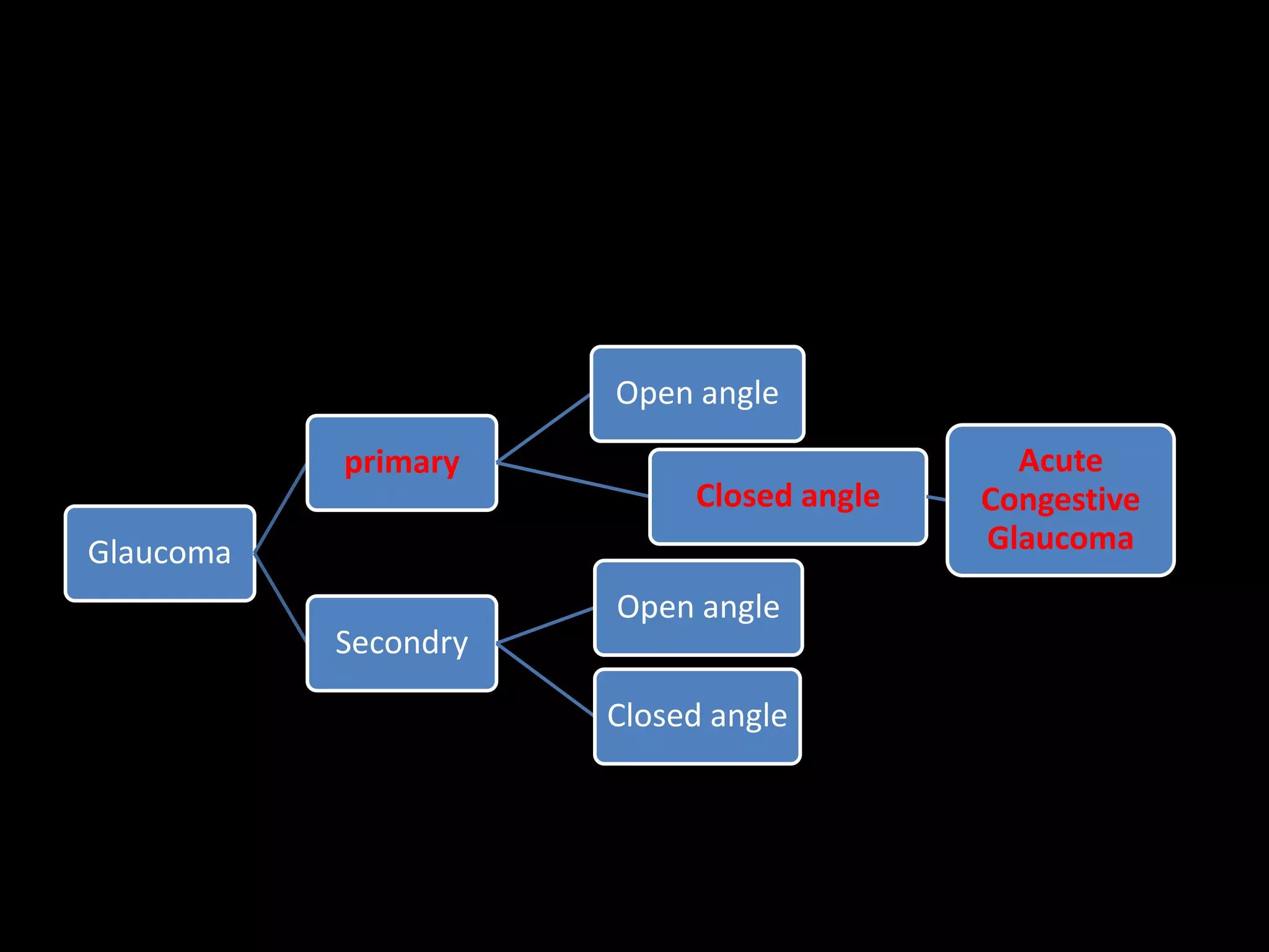
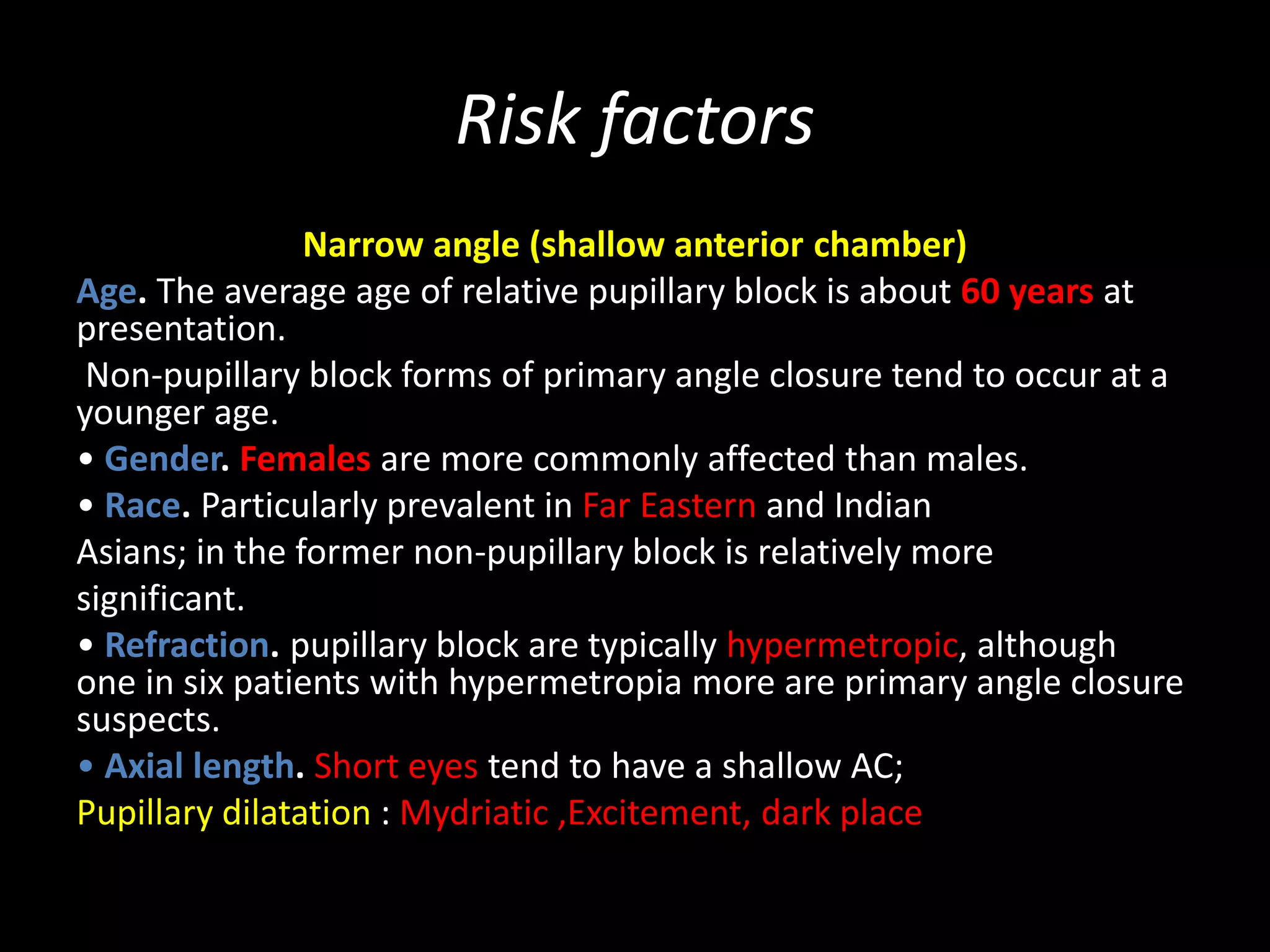
Acute Congestive Glaucoma
By . M. N. O.
4th year medical Student(MBBCh)
Glaucoma
primary
Open angle
Closed angle
Acute
Congestive
Glaucoma
Secondry
Open angle
Closed angle
Objective
• Overview
• mechanism
• Risk factors
• classification
• Symptoms and signs
• Management
overview
• PACG may be responsible for up to half of all
cases of glaucoma globally, with a particularly
highof Far Eastern descent.
• The term ‘angle closure’ refers to occlusion of
the trabecular meshwork
• by the peripheral iris (iridotrabecular contact –
ITC), obstructing aqueous outflow
Mechanism
• Relative Pupillary block
• Non-pupillary block
Relative Pupillary block Mechanism
• Relative pupillary block :
• Normally, aqueous humor is produced in the ciliary body, flows through the pupil
into the anterior chamber, and drains into the trabecular meshwork to exit the
eye.
• When the pupil is mid-dilated, the distance between the iris and the lens is the
shortest, and the two structures can come into contact with each other in
individuals at risk for angle closure.
• When this occurs, aqueous humor cannot flow through the pupil into the
anterior chamber (pupillary block), pushing the iris forward. When the iris is
pushed against the trabecular meshwork, aqueous humor cannot flow out of the
eye (angle closure), increasing IOP.
Non-pupillary block Mechanism
Non-pupillary block :
Thought to be important in many Far Eastern patients.
plateau iris younger than those with pure pupillary block.
plateau; iris (anteriorly positioned/rotated ciliary processes , and a thicker or more
anteriorly positioned iris a ‘thick peripheral iris roll’
iris-induced mechanisms
Risk factors
Narrow angle (shallow anterior chamber)
Age. The average age of relative pupillary block is about 60 years at
presentation.
Non-pupillary block forms of primary angle closure tend to occur at a
younger age.
• Gender. Females are more commonly affected than males.
• Race. Particularly prevalent in Far Eastern and Indian
Asians; in the former non-pupillary block is relatively more
significant.
• Refraction. pupillary block are typically hypermetropic, although
one in six patients with hypermetropia more are primary angle closure
suspects.
• Axial length. Short eyes tend to have a shallow AC;
Pupillary dilatation : Mydriatic ,Excitement, dark place
Classification
• Intermittent(prodromal) stage
• Acute stage (Acute Congestive Glaucoma)
• Chronic angle closure
Intermittent(prodromal) stage
• Transient mild attack of Headache.
• blurred vision & halos around light
• The attack Is relived by
• sleep & exposure to light
• (due to miosis → lead to open angle)
Diagnosis
• History (Recuttent attacks of headache & blurred Vission)
• Gonioscopy : Narrow angle
• Tonometry Normal btw attacks ↑durring attack
• Provocative tests :
• darkroom test
• Dark room/prone provocative test (DRPPT)
• Mydriatic test
• ; positive if IOP rises >8mmHg
Tono-Pen Tonometry
Indentation Tonometry
Applanation Tonometry
Acute stage
(Acute Congestive Glaucoma)
Ophthalmic Emergency
symptoms
• ocular/periocular pain and headache;
• blurring (‘smoke-filled room’) &
• Colored haloes ☼(‘rainbow around lights’)
due to corneal epithelial edema
• Markedly↓Decreased vision early ,late
• Redness
• nausea and vomiting abdominal pain and
other gastrointestinal symptoms may occur.
Medical Emergency
`
Signs
• Conjunctiva→: Red eye (ciliary injection )circumcorneal
injection.
• Cornea→: cloudy/edema
• Anterior chamber→ : shallow
• Iris→ : Iris bombe
• Pupil→ : semi-dilated, non-reactive ,oval vertical
• IOP→: is usually very high (50–100 mmHg). STONY HARD
• VA→: ↓
• Sequelae of ischemia→:
segmental iris atrophy (focal iris stroma necrosis)
dilated irregular pupil (sphincter and dilator necrosis)
glaukomflecken (focal anterior lens opacities due to epithelial
necrosis)
Treatment
• Hospitalization :patient should assume a supine position to
encourage the lens to shift posteriorly under the influence of
gravity.
Systemic Treatment :
• IV Acetazolamide((Diamox)) [C/I sulfonamide allergy]
• IV mannitol [Circulatory overload C/I in heart failure]
Topical Treatment
• M3 agonist( Pilocarpine) → Miosis→ (↓IOP)
• β-blocker (Timolol ) → ↓AH syn. → (↓IOP)
• α2-agonist( Apraclonidine) → ↓A.H. syn. → (↓IOP)
• Topical steroids for inflammation
• Analgesia and an Antiemetic may be required
• peripheral Iridotomy or iridectomy
• Prophylactic iridotomy or iridoectomy to other eye
References :
Kanski’s Clinical Ophthalmology A SYSTEMATIC APPROACH 8th ed.
Review of Ophthalmology, 2nd Ed 2012
https://emedicine.medscape.com
https://www.webmd.com/
primary closed angle glaucoma (Acute congestive glaucoma)

primary closed angle glaucoma (Acute congestive glaucoma)

  • 1.
    Acute Congestive Glaucoma By. M. N. O. 4th year medical Student(MBBCh)
  • 2.
  • 3.
    Objective • Overview • mechanism •Risk factors • classification • Symptoms and signs • Management
  • 4.
    overview • PACG maybe responsible for up to half of all cases of glaucoma globally, with a particularly highof Far Eastern descent. • The term ‘angle closure’ refers to occlusion of the trabecular meshwork • by the peripheral iris (iridotrabecular contact – ITC), obstructing aqueous outflow
  • 6.
    Mechanism • Relative Pupillaryblock • Non-pupillary block
  • 7.
    Relative Pupillary blockMechanism • Relative pupillary block : • Normally, aqueous humor is produced in the ciliary body, flows through the pupil into the anterior chamber, and drains into the trabecular meshwork to exit the eye. • When the pupil is mid-dilated, the distance between the iris and the lens is the shortest, and the two structures can come into contact with each other in individuals at risk for angle closure. • When this occurs, aqueous humor cannot flow through the pupil into the anterior chamber (pupillary block), pushing the iris forward. When the iris is pushed against the trabecular meshwork, aqueous humor cannot flow out of the eye (angle closure), increasing IOP.
  • 10.
    Non-pupillary block Mechanism Non-pupillaryblock : Thought to be important in many Far Eastern patients. plateau iris younger than those with pure pupillary block. plateau; iris (anteriorly positioned/rotated ciliary processes , and a thicker or more anteriorly positioned iris a ‘thick peripheral iris roll’ iris-induced mechanisms
  • 11.
    Risk factors Narrow angle(shallow anterior chamber) Age. The average age of relative pupillary block is about 60 years at presentation. Non-pupillary block forms of primary angle closure tend to occur at a younger age. • Gender. Females are more commonly affected than males. • Race. Particularly prevalent in Far Eastern and Indian Asians; in the former non-pupillary block is relatively more significant. • Refraction. pupillary block are typically hypermetropic, although one in six patients with hypermetropia more are primary angle closure suspects. • Axial length. Short eyes tend to have a shallow AC; Pupillary dilatation : Mydriatic ,Excitement, dark place
  • 13.
    Classification • Intermittent(prodromal) stage •Acute stage (Acute Congestive Glaucoma) • Chronic angle closure
  • 14.
    Intermittent(prodromal) stage • Transientmild attack of Headache. • blurred vision & halos around light • The attack Is relived by • sleep & exposure to light • (due to miosis → lead to open angle)
  • 15.
    Diagnosis • History (Recuttentattacks of headache & blurred Vission) • Gonioscopy : Narrow angle • Tonometry Normal btw attacks ↑durring attack • Provocative tests : • darkroom test • Dark room/prone provocative test (DRPPT) • Mydriatic test • ; positive if IOP rises >8mmHg
  • 17.
  • 18.
    Acute stage (Acute CongestiveGlaucoma) Ophthalmic Emergency
  • 19.
    symptoms • ocular/periocular painand headache; • blurring (‘smoke-filled room’) & • Colored haloes ☼(‘rainbow around lights’) due to corneal epithelial edema • Markedly↓Decreased vision early ,late • Redness • nausea and vomiting abdominal pain and other gastrointestinal symptoms may occur.
  • 20.
  • 21.
  • 22.
    Signs • Conjunctiva→: Redeye (ciliary injection )circumcorneal injection. • Cornea→: cloudy/edema • Anterior chamber→ : shallow • Iris→ : Iris bombe • Pupil→ : semi-dilated, non-reactive ,oval vertical • IOP→: is usually very high (50–100 mmHg). STONY HARD • VA→: ↓ • Sequelae of ischemia→: segmental iris atrophy (focal iris stroma necrosis) dilated irregular pupil (sphincter and dilator necrosis) glaukomflecken (focal anterior lens opacities due to epithelial necrosis)
  • 29.
    Treatment • Hospitalization :patientshould assume a supine position to encourage the lens to shift posteriorly under the influence of gravity. Systemic Treatment : • IV Acetazolamide((Diamox)) [C/I sulfonamide allergy] • IV mannitol [Circulatory overload C/I in heart failure] Topical Treatment • M3 agonist( Pilocarpine) → Miosis→ (↓IOP) • β-blocker (Timolol ) → ↓AH syn. → (↓IOP) • α2-agonist( Apraclonidine) → ↓A.H. syn. → (↓IOP) • Topical steroids for inflammation • Analgesia and an Antiemetic may be required • peripheral Iridotomy or iridectomy • Prophylactic iridotomy or iridoectomy to other eye
  • 31.
    References : Kanski’s ClinicalOphthalmology A SYSTEMATIC APPROACH 8th ed. Review of Ophthalmology, 2nd Ed 2012 https://emedicine.medscape.com https://www.webmd.com/