3. GLAUCOMA
• Chronic, progressive optic neuropathy
characterised by morphological changes at the
optic disc and retinal nerve fibre layer leading to
characteristic visual field changes, in the absence
of other ocular diseases or congenital anomalies
(with or without a raised IOP).
3
4. • k/a chronic simple glaucoma
• Most prevalent of all glaucoma (1/3rd
cases of all glaucomas)
• Affects both sexes equally
• Affects about 1 in 100 of the general
population above the age of 40 years
4
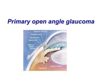
Primary open angle glaucoma
(POAG)
5. • Slowly progressive raied IOP > 21 mm hg
• Glaucomatous optic nerve damage
• An open normal appearing anterior
chamber angle
• Characteristic visual field loss
• Absence of signs of secondary glaucoma
or a non-glaucomatous cause for the optic
neuropathy
5
6. Etiopathogenesis
• Multifactorial aetiology
• Risk factors include:
Elevated Intra Ocular Pressure(IOP)
(More than 21 mm Hg)
Optic disc cupping
Increasing Age : More common in 5th to 7th decades
Race: More common and severe in Black population
6
7. Etiopathogenesis
Heredity/Family History: Risk of about 10% in
siblings;4% in off springs(MYOC,OPTN ,WDR36)
Diabetes
Systemic Hypertension
Myopia
Thin central corneas
Steroid usage
??Migraine, Cigarette smoking,Graves disease
7
8. Pathophysiology of POAG
• Decrease in aqueous outflow facility due to
increased resistance to outflow leads to rise in IOP
• Two theories of axonal loss in optic disc
• 1. Mechanical: Distortion of lamina cribrosa
leading to impaired axoplasmic flow
2. Vascular: Optic disc ischaemia with
defective autoregulation of blood vessels 8
9. Clinical features of POAG
Symptoms
• Usually asymptomatic in early cases
• Mild headache and eye ache
• Frequent changes in presbyopic glasses
• Delayed dark adaptation
• Loss of peripheral vision
• Loss of central vision(late cases)
9
10. Signs of POAG
• Normal anterior segment
• Pupil reaction to light may be sluggish(in
advanced cases only)
• Elevated IOP(More than 21 mm Hg) with
diurnal variation more than 5-8 mmHg
• Optic disc changes (Progressive,
asymmetric)
• Visual field defects
10
13. Optic disc changes in glaucoma
• Early changes
o Vertically oval cup
o Asymmetry of the cups(More than 0.2
difference)
o Large cup(CD more than 0.6)
o Splinter haemorrhages
o Pallor areas on the disc
o Retinal nerve fibre layer atrophy (red free
filter ) 13
14. Advanced glaucomatous disc changes
• Marked cupping (More than 0.7)
• Thinning of NRR (Neuroretinal rim)
• Lamellar dot sign
• Vascular alterations
o Nasal shifting of retinal vessels
o Bayonetting sign(convoluted path due to NRR
loss)
o Baring of circumlinear vessels and overpass
vessels
• Glaucomatous optic atrophy
14
19. Visual field defects in glaucoma
• Arcuate nerve fibres in the
superior and inferior
temporal portions of the
optic disc:
Most sensitive to damage
• Macular fibres : Most
resistant to damage
CENTRAL VISION IS
PRESERVED TILL THE
LAST IN GLAUCOMA
19
20. Progression of field defects
• Isopter contraction: Generalised field
constriction
• Baring of blind spot : Non specific
(Exclusion of blind spot from central field)
• Paracentral scotoma: Wing shaped and
occurs above or below the blind spot in the
Bjerrum’s area(10-25 degrees from
fixation)
Is the earliest clinically significant defect
20
21. Progression of field defects
• Seidel’s scotoma: sickle shaped
Due to joining of blind spot and
paracentral scotoma
• Bjerrum’s/Arcuate scotoma:
Extension of Seidel’s scotoma to reach the
horizontal line.
• Double arcuate/ring scotoma
21
22. Progression of field defects
• Roenne’s central nasal step:
Sharp right angled defect at the horizontal
meridian when arcuate scotomas run in
different arcs
• Peripheral field defects
• Advanced defects
Residual Tubular vision
Temporal island of vision
22
24. Quantification of visual field defects
• Visual field analyzer
Kinetic perimeter
Static perimeter (automated)
Testing more than once is required before
final interpretation
24
31. Diagnostic work up/Investigations
• Tonometry
• Goniscopy: Open angles
• Perimetry: To detect visual field defects
• Slit lamp examination: To rule out causes
of secondary open angle glaucoma
• Fundus examination to document optic
disc changes
• Diurnal variation testing
• Provocative testing: Water drinking test
31
32. Diagnosis
• POAG: Raised IOP(More than 21 mm Hg),
glaucomatous optic disc cupping, visual
field changes.
• Ocular hypertension/glaucoma suspect:
Raised IOP
• NTG(Normal tension glaucoma):
Glaucomatous optic disc cupping with or
without visual field changes with normal IOP
32
33. Management of POAG
• Therapeutic choices
Medical therapy
Argon/Diode Laser Trabeculoplasty
Filtration surgery
33
34. Basic principles of therapy
• Make a correct diagnosis
• Set a target IOP
• Start with a single drug to lower IOP
• Switch to another group of drugs if needed
• Control IOP on minimal medications
• Monitor therapy and reset target IOP
whenever needed
34
36. Systemic drugs used for POAG therapy
• Used rarely, for short term control of IOP
• Oral carbonic anhydrase inhibitors
Acetazolamide, Methazolamide
36
37. Laser treatment
• Indications
Target IOP not achieved with medical
therapy
Non compliance of medical therapy
Argon/ Diode Laser Trabeculoplasty (ALT)
Selective Laser Trabeculoplasty (SLT)
37
38. Surgical therapy
• Indications
Target IOP not achieved with maximal
tolerated medical therapy and laser
trabeculoplasty
Non compliance of medical therapy
Non availability of laser therapy
Advanced glaucoma
38
39. Surgical therapy
• Filtration surgery : Trabeculectomy
• Modified trabeculectomy :
Use of antifibrotic agents
Mitomycin/5FU
• Aqueous drainage devices:
Ahmed glaucoma valve
In cases with no/poor visual potential:
Cycloablative therapy with laser/cryotherapy
39