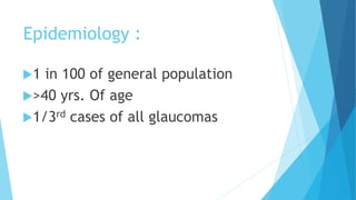
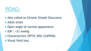
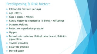
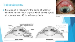
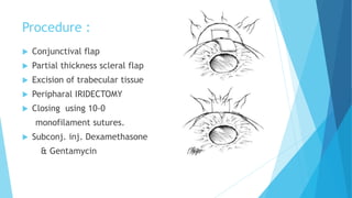
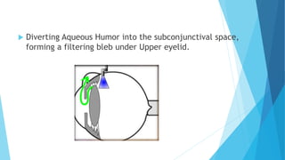
PRIMARY OPEN ANGLE
GLAUCOMA
-Dr. Saurabh Kumar
(Intern, PMCH Dhanbad)
Department of EYE
Tue,(17/12/2019)
Definition-
Chronic, progressive optic neuropathy
caused by a group of ocular
conditions,which lead to damage of
optic nerve with loss of visual
function,commonest risk factor being
raised IOP.
Epidemiology :
1 in 100 of general population
>40 yrs. Of age
1/3rd cases of all glaucomas
POAG:
 Also called as Chronic Simple Glaucoma
 Adult onset
 Open angle of normal appearance
 IOP : >21 mmHg
 Characteristic OPTIC DISC CUPPING
 Visual field loss.
Predisposing & Risk factor:
 Intraocular Pressure (m/imp)
 Age >40 yrs.
 Race : Blacks > Whites
 Family history & Inheritance : Siblings > Offsprings
 Diabetes Mellitus
 Reduction in perfusion pressure
 Myopia
 Retinal vein occlusion, Retinal detachment, Retinitis
pigmentosa
 Thyroid disorders
 Cigarrete smoking
 Steroid usage
Pathogenesis : rise in IOP
 Thickening & sclerosis of Trabecular Meshwork
 Narrowing of Intertrabecular spaces
 Amorphous material deposition in
Juxtacanalicular Space
 Collapse of Schlemm’s Canal
Pathogenesis : Retinal Ganglion Cell death
 Blockage of transport of GF(Neurotrophins) from
Brain to RGC.
Symptoms :
 Insidious & asymptomatic onset
 Gradual painless loss of vision
 Headache, Eyeache
 Visual field defect (SCOTOMA)
 Frequent change in presbyopic glasses
 Delayed dark adaptation
 Significant loss of vision & blindness
Signs :
 Anterior segment signs:
-Corneal haze
-Sluggish pupil
IOP changes:
-Morning rise (20%)
-Afternoon rise (25%)
-Biphasic rise (55%)
Variation in IOP over 5mmHg is suspicious,
& over 8mmHg is diagnostic Of Glaucoma.
Optic nerve head evaluation :
 Vertically oval Cup
(loss of neural rim tissue in Inf & Sup. Poles)
 Assymmetry of cups (>0.2)
{N’mlly : Cup/Disc diam .=0.3}
 Large cup (>0.6)
 Splinter Hemorrhage
 Pallor areas
 Atrophy (Retinal nerve fiber layers)
{seen with RED FREE light}
Early changes :
Advanced changes:
 Marked Cupping (0.7-0.9)
 Neuroretinal Rim thinning
N’lly ; I S N T Rule
 Nasal shifting of Retinal vessels
(Bayonetting sign)
 Retinal arteriols Pulsations
 Lamellor Dot Sign
(slit shaped pores in lamina cribrosa)
Localised RNFL loss :
Localised Wedge shaped dark area
Five Rules for assement of
the Optic Disc in Glaucoma:
 Observe the scleral Ring to identify the
limits of the optic disc and its size.
 Identify the size of the Rim.
 Examine the RNFL.
 Examine the Region of parapapillary atrophy.
 Look for Retinal &optic disc haemorrhage.
Visual Field Defects :
 Isopter contraction
 Barring of Blind Spot
 Small wing shaped Paracentral Scotoma
 Seidel Scotoma
 Arcuate/Bjerrum’s Scotoma
 Ring/Double Arcuate Scotoma
 Roenne’s central nasal step
 Peripheral field defects
Visual Field Defects :
Investigations:
 Tonometry (Applanation > Schiotz)
 Central Corneal Thickness
 Diurnal variation test
 Gonioscopy
 Optic disc changes documentation
 Slit lamp
 Perimetry
 Nerve Fibre Layer Analyser
 Provocative test (water drinking test)
 Slit Lamp +78D /90D Indirect Opthalmoscopy is useful in diagnosing early
Glaucoma since Optic disc changes occur before loss of Visual field is visible.
Tonometry
Treatment :
 Aim : To lower IOP to a level where further visual loss does not occur.
 1) Prostaglandn analogue (Inc. Uveoscleral outflow) :
LATANOPROST (causes pigmentation of eyelids)
TRAVOPROST
BIMATOPROST
 2) B - blocker (topical) (Dec. Aq. Humour secretion) :
TIMOLOL MALEATE
LEVOBUNOLOL (longest action)
BETAXOLOL
3) a Agonists (Inc. Aq. Outflow)
BRIMONIDINE (2nd Drug of choice)
4) CA Inhibitors (Dec. Aq. Production)
DORZOLAMIDE {bitter taste}
BRIZOLAMIDE
5) Cholinergic drug (Inc. Aq outflow)
PILOCARPINE
Argon/Diode Laser Trabeculoplasty :
 Indications:
1) avoidance of polypharmacy
2)avoidance of surgery
3) primary therapy in patients
with non compliance
 Mech of action : Inc. outflow by causing
shrinkage of trabecular meshwork.
40-50 spots on the Ant. half of trabecular
meshwork over 180* using a Goniolens.
Surgical management :
Indications-
 Uncontrolled Glaucoma despite maximal
medical (3 drugs) & laser trabeculoplasty.
 Non-compliance of medical therapy
 Advanced disease requiring a very low target
IOP
Trabeculectomy
 Creation of a fistula b/w the angle of anterior
chamber & sub-tenon’s space which allows egress
of aqueous from AC to a drainage bleb.
Procedure :
 Conjunctival flap
 Partial thickness scleral flap
 Excision of trabecular tissue
 Peripharal IRIDECTOMY
 Closing using 10-0
monofilament sutures.
 Subconj. inj. Dexamethasone
& Gentamycin
 Diverting Aqueous Humor into the subconjunctival space,
forming a filtering bleb under Upper eyelid.
THANK YOU
Have A Nice Day
Regards - saurabh

POAG

  • 1.
    PRIMARY OPEN ANGLE GLAUCOMA -Dr.Saurabh Kumar (Intern, PMCH Dhanbad) Department of EYE Tue,(17/12/2019)
  • 2.
    Definition- Chronic, progressive opticneuropathy caused by a group of ocular conditions,which lead to damage of optic nerve with loss of visual function,commonest risk factor being raised IOP.
  • 3.
    Epidemiology : 1 in100 of general population >40 yrs. Of age 1/3rd cases of all glaucomas
  • 4.
    POAG:  Also calledas Chronic Simple Glaucoma  Adult onset  Open angle of normal appearance  IOP : >21 mmHg  Characteristic OPTIC DISC CUPPING  Visual field loss.
  • 5.
    Predisposing & Riskfactor:  Intraocular Pressure (m/imp)  Age >40 yrs.  Race : Blacks > Whites  Family history & Inheritance : Siblings > Offsprings  Diabetes Mellitus  Reduction in perfusion pressure  Myopia  Retinal vein occlusion, Retinal detachment, Retinitis pigmentosa  Thyroid disorders  Cigarrete smoking  Steroid usage
  • 6.
    Pathogenesis : risein IOP  Thickening & sclerosis of Trabecular Meshwork  Narrowing of Intertrabecular spaces  Amorphous material deposition in Juxtacanalicular Space  Collapse of Schlemm’s Canal Pathogenesis : Retinal Ganglion Cell death  Blockage of transport of GF(Neurotrophins) from Brain to RGC.
  • 7.
    Symptoms :  Insidious& asymptomatic onset  Gradual painless loss of vision  Headache, Eyeache  Visual field defect (SCOTOMA)  Frequent change in presbyopic glasses  Delayed dark adaptation  Significant loss of vision & blindness
  • 8.
    Signs :  Anteriorsegment signs: -Corneal haze -Sluggish pupil IOP changes: -Morning rise (20%) -Afternoon rise (25%) -Biphasic rise (55%) Variation in IOP over 5mmHg is suspicious, & over 8mmHg is diagnostic Of Glaucoma.
  • 9.
    Optic nerve headevaluation :  Vertically oval Cup (loss of neural rim tissue in Inf & Sup. Poles)  Assymmetry of cups (>0.2) {N’mlly : Cup/Disc diam .=0.3}  Large cup (>0.6)  Splinter Hemorrhage  Pallor areas  Atrophy (Retinal nerve fiber layers) {seen with RED FREE light} Early changes :
  • 10.
    Advanced changes:  MarkedCupping (0.7-0.9)  Neuroretinal Rim thinning N’lly ; I S N T Rule  Nasal shifting of Retinal vessels (Bayonetting sign)  Retinal arteriols Pulsations  Lamellor Dot Sign (slit shaped pores in lamina cribrosa)
  • 11.
    Localised RNFL loss: Localised Wedge shaped dark area
  • 12.
    Five Rules forassement of the Optic Disc in Glaucoma:  Observe the scleral Ring to identify the limits of the optic disc and its size.  Identify the size of the Rim.  Examine the RNFL.  Examine the Region of parapapillary atrophy.  Look for Retinal &optic disc haemorrhage.
  • 13.
    Visual Field Defects:  Isopter contraction  Barring of Blind Spot  Small wing shaped Paracentral Scotoma  Seidel Scotoma  Arcuate/Bjerrum’s Scotoma  Ring/Double Arcuate Scotoma  Roenne’s central nasal step  Peripheral field defects
  • 14.
  • 15.
    Investigations:  Tonometry (Applanation> Schiotz)  Central Corneal Thickness  Diurnal variation test  Gonioscopy  Optic disc changes documentation  Slit lamp  Perimetry  Nerve Fibre Layer Analyser  Provocative test (water drinking test)  Slit Lamp +78D /90D Indirect Opthalmoscopy is useful in diagnosing early Glaucoma since Optic disc changes occur before loss of Visual field is visible.
  • 16.
  • 17.
    Treatment :  Aim: To lower IOP to a level where further visual loss does not occur.  1) Prostaglandn analogue (Inc. Uveoscleral outflow) : LATANOPROST (causes pigmentation of eyelids) TRAVOPROST BIMATOPROST  2) B - blocker (topical) (Dec. Aq. Humour secretion) : TIMOLOL MALEATE LEVOBUNOLOL (longest action) BETAXOLOL
  • 18.
    3) a Agonists(Inc. Aq. Outflow) BRIMONIDINE (2nd Drug of choice) 4) CA Inhibitors (Dec. Aq. Production) DORZOLAMIDE {bitter taste} BRIZOLAMIDE 5) Cholinergic drug (Inc. Aq outflow) PILOCARPINE
  • 19.
    Argon/Diode Laser Trabeculoplasty:  Indications: 1) avoidance of polypharmacy 2)avoidance of surgery 3) primary therapy in patients with non compliance  Mech of action : Inc. outflow by causing shrinkage of trabecular meshwork. 40-50 spots on the Ant. half of trabecular meshwork over 180* using a Goniolens.
  • 20.
    Surgical management : Indications- Uncontrolled Glaucoma despite maximal medical (3 drugs) & laser trabeculoplasty.  Non-compliance of medical therapy  Advanced disease requiring a very low target IOP
  • 21.
    Trabeculectomy  Creation ofa fistula b/w the angle of anterior chamber & sub-tenon’s space which allows egress of aqueous from AC to a drainage bleb.
  • 22.
    Procedure :  Conjunctivalflap  Partial thickness scleral flap  Excision of trabecular tissue  Peripharal IRIDECTOMY  Closing using 10-0 monofilament sutures.  Subconj. inj. Dexamethasone & Gentamycin
  • 23.
     Diverting AqueousHumor into the subconjunctival space, forming a filtering bleb under Upper eyelid.
  • 24.
    THANK YOU Have ANice Day Regards - saurabh