Polymerase Chain Reaction
History of PCR
Instrumentation of PCR
Principle of PCR
Components of PCR
Steps of PCR
Optimal PCR Factors
Applications of PCR
A real-time polymerase chain reaction is a laboratory technique of molecular biology based on the polymerase chain reaction (PCR). It monitors the amplification of a targeted DNA molecule during the PCR, i.e. in real-time, and not at its end, as in conventional PCR.
https://www.patreon.com/biotechlive
SUPPORT EDUCATION... SUPPORT US
It is called “polymerase” because the only enzyme used in this reaction is DNA polymerase.
It is called “chain” because the products of the first reaction become substrates of the following one, and so on.
published a DNA sequencing method in 1977 based on chemical modification of DNA and subsequent cleavage at specific bases. Also known as chemical sequencing, this method allowed purified samples of double-stranded DNA to be used without further cloning.
Maxam-Gilbert sequencing requires radioactive labeling at one 5' end of the DNA and purification of the DNA fragment to be sequenced. Chemical treatment then generates breaks at a small proportion of one or two of the four nucleotide bases in each of four reactions (G, A+G, C, C+T). The concentration of the modifying chemicals is controlled to introduce on average one modification per DNA molecule. Thus a series of labeled fragments is generated, from the radiolabeled end to the first "cut" site in each molecule. The fragments in the four reactions are electrophoresed side by side in denaturing acrylamide gels for size separation. To visualize the fragments, the gel is exposed to X-ray film for autoradiography, yielding a series of dark bands each corresponding to a radiolabeled DNA fragment, from which the sequence may be inferred.
PCR is a technique which is used to amplify the number of copies of a specific region of DNA, in order to produce enough DNA to be adequately tested.
Cell-free amplification for synthesizing multiple identical copies (billions) of any DNA of interest.
Basic tool for the molecular biologist.
The purpose of a PCR is to make a huge number of copies of a gene. As a result, it now becomes possible to analyze and characterize DNA fragments found in minute quantities in places like a drop of blood at a crime scene or a cell from an extinct dinosaur.
Like Xerox machine for gene copying.
Polymerase chain reaction (abbreviated PCR) is a laboratory technique for rapidly producing (amplifying) millions to billions of copies of a specific segment of DNA, which can then be studied in greater detail.
PCR presentation
Consists of the main parts of PCR , type of PCR including the most advanced and most efficient.
It also includes history of PCR.
PCR is a machine used to make multiple copies of gene, while gene is a part of DNA. it has 3 steps , initiaition, elongation, termination. which required different temperature for different step. these slides includes most information about PCR.
Polymerase chain reaction (PCR)
Polymerase chain reaction (PCR) is a common laboratory technique used to make many copies (millions or billions) of a particular region of DNA.
RECOMBINATION MOLECULAR BIOLOGY PPT UPDATED new.pptxSabahat Ali
This ppt is about recombination and where it occurs. Types of recombination and models of recombination along with many factors in prokaryotic and eukaryotic recombination
Folding depends upon sequence of Amino Acids not the Composition. Folding starts with the secondary structure and ends at quaternary structure.
Denaturation occur at secondary, tertiary & quaternary level but not at primary level.
Tertiary Structure basically of Hydrophobic interactions, (interactions in side chains), hydrogen bonding, salt bridges, Vander Waals interactions.
e.g. Globular proteins & Fibrous Proteins
This presentation explores a brief idea about the structural and functional attributes of nucleotides, the structure and function of genetic materials along with the impact of UV rays and pH upon them.
Richard's entangled aventures in wonderlandRichard Gill
Since the loophole-free Bell experiments of 2020 and the Nobel prizes in physics of 2022, critics of Bell's work have retreated to the fortress of super-determinism. Now, super-determinism is a derogatory word - it just means "determinism". Palmer, Hance and Hossenfelder argue that quantum mechanics and determinism are not incompatible, using a sophisticated mathematical construction based on a subtle thinning of allowed states and measurements in quantum mechanics, such that what is left appears to make Bell's argument fail, without altering the empirical predictions of quantum mechanics. I think however that it is a smoke screen, and the slogan "lost in math" comes to my mind. I will discuss some other recent disproofs of Bell's theorem using the language of causality based on causal graphs. Causal thinking is also central to law and justice. I will mention surprising connections to my work on serial killer nurse cases, in particular the Dutch case of Lucia de Berk and the current UK case of Lucy Letby.
THE IMPORTANCE OF MARTIAN ATMOSPHERE SAMPLE RETURN.Sérgio Sacani
The return of a sample of near-surface atmosphere from Mars would facilitate answers to several first-order science questions surrounding the formation and evolution of the planet. One of the important aspects of terrestrial planet formation in general is the role that primary atmospheres played in influencing the chemistry and structure of the planets and their antecedents. Studies of the martian atmosphere can be used to investigate the role of a primary atmosphere in its history. Atmosphere samples would also inform our understanding of the near-surface chemistry of the planet, and ultimately the prospects for life. High-precision isotopic analyses of constituent gases are needed to address these questions, requiring that the analyses are made on returned samples rather than in situ.
This pdf is about the Schizophrenia.
For more details visit on YouTube; @SELF-EXPLANATORY;
https://www.youtube.com/channel/UCAiarMZDNhe1A3Rnpr_WkzA/videos
Thanks...!
Professional air quality monitoring systems provide immediate, on-site data for analysis, compliance, and decision-making.
Monitor common gases, weather parameters, particulates.
Earliest Galaxies in the JADES Origins Field: Luminosity Function and Cosmic ...Sérgio Sacani
We characterize the earliest galaxy population in the JADES Origins Field (JOF), the deepest
imaging field observed with JWST. We make use of the ancillary Hubble optical images (5 filters
spanning 0.4−0.9µm) and novel JWST images with 14 filters spanning 0.8−5µm, including 7 mediumband filters, and reaching total exposure times of up to 46 hours per filter. We combine all our data
at > 2.3µm to construct an ultradeep image, reaching as deep as ≈ 31.4 AB mag in the stack and
30.3-31.0 AB mag (5σ, r = 0.1” circular aperture) in individual filters. We measure photometric
redshifts and use robust selection criteria to identify a sample of eight galaxy candidates at redshifts
z = 11.5 − 15. These objects show compact half-light radii of R1/2 ∼ 50 − 200pc, stellar masses of
M⋆ ∼ 107−108M⊙, and star-formation rates of SFR ∼ 0.1−1 M⊙ yr−1
. Our search finds no candidates
at 15 < z < 20, placing upper limits at these redshifts. We develop a forward modeling approach to
infer the properties of the evolving luminosity function without binning in redshift or luminosity that
marginalizes over the photometric redshift uncertainty of our candidate galaxies and incorporates the
impact of non-detections. We find a z = 12 luminosity function in good agreement with prior results,
and that the luminosity function normalization and UV luminosity density decline by a factor of ∼ 2.5
from z = 12 to z = 14. We discuss the possible implications of our results in the context of theoretical
models for evolution of the dark matter halo mass function.
Deep Behavioral Phenotyping in Systems Neuroscience for Functional Atlasing a...Ana Luísa Pinho
Functional Magnetic Resonance Imaging (fMRI) provides means to characterize brain activations in response to behavior. However, cognitive neuroscience has been limited to group-level effects referring to the performance of specific tasks. To obtain the functional profile of elementary cognitive mechanisms, the combination of brain responses to many tasks is required. Yet, to date, both structural atlases and parcellation-based activations do not fully account for cognitive function and still present several limitations. Further, they do not adapt overall to individual characteristics. In this talk, I will give an account of deep-behavioral phenotyping strategies, namely data-driven methods in large task-fMRI datasets, to optimize functional brain-data collection and improve inference of effects-of-interest related to mental processes. Key to this approach is the employment of fast multi-functional paradigms rich on features that can be well parametrized and, consequently, facilitate the creation of psycho-physiological constructs to be modelled with imaging data. Particular emphasis will be given to music stimuli when studying high-order cognitive mechanisms, due to their ecological nature and quality to enable complex behavior compounded by discrete entities. I will also discuss how deep-behavioral phenotyping and individualized models applied to neuroimaging data can better account for the subject-specific organization of domain-general cognitive systems in the human brain. Finally, the accumulation of functional brain signatures brings the possibility to clarify relationships among tasks and create a univocal link between brain systems and mental functions through: (1) the development of ontologies proposing an organization of cognitive processes; and (2) brain-network taxonomies describing functional specialization. To this end, tools to improve commensurability in cognitive science are necessary, such as public repositories, ontology-based platforms and automated meta-analysis tools. I will thus discuss some brain-atlasing resources currently under development, and their applicability in cognitive as well as clinical neuroscience.
Introduction:
RNA interference (RNAi) or Post-Transcriptional Gene Silencing (PTGS) is an important biological process for modulating eukaryotic gene expression.
It is highly conserved process of posttranscriptional gene silencing by which double stranded RNA (dsRNA) causes sequence-specific degradation of mRNA sequences.
dsRNA-induced gene silencing (RNAi) is reported in a wide range of eukaryotes ranging from worms, insects, mammals and plants.
This process mediates resistance to both endogenous parasitic and exogenous pathogenic nucleic acids, and regulates the expression of protein-coding genes.
What are small ncRNAs?
micro RNA (miRNA)
short interfering RNA (siRNA)
Properties of small non-coding RNA:
Involved in silencing mRNA transcripts.
Called “small” because they are usually only about 21-24 nucleotides long.
Synthesized by first cutting up longer precursor sequences (like the 61nt one that Lee discovered).
Silence an mRNA by base pairing with some sequence on the mRNA.
Discovery of siRNA?
The first small RNA:
In 1993 Rosalind Lee (Victor Ambros lab) was studying a non- coding gene in C. elegans, lin-4, that was involved in silencing of another gene, lin-14, at the appropriate time in the
development of the worm C. elegans.
Two small transcripts of lin-4 (22nt and 61nt) were found to be complementary to a sequence in the 3' UTR of lin-14.
Because lin-4 encoded no protein, she deduced that it must be these transcripts that are causing the silencing by RNA-RNA interactions.
Types of RNAi ( non coding RNA)
MiRNA
Length (23-25 nt)
Trans acting
Binds with target MRNA in mismatch
Translation inhibition
Si RNA
Length 21 nt.
Cis acting
Bind with target Mrna in perfect complementary sequence
Piwi-RNA
Length ; 25 to 36 nt.
Expressed in Germ Cells
Regulates trnasposomes activity
MECHANISM OF RNAI:
First the double-stranded RNA teams up with a protein complex named Dicer, which cuts the long RNA into short pieces.
Then another protein complex called RISC (RNA-induced silencing complex) discards one of the two RNA strands.
The RISC-docked, single-stranded RNA then pairs with the homologous mRNA and destroys it.
THE RISC COMPLEX:
RISC is large(>500kD) RNA multi- protein Binding complex which triggers MRNA degradation in response to MRNA
Unwinding of double stranded Si RNA by ATP independent Helicase
Active component of RISC is Ago proteins( ENDONUCLEASE) which cleave target MRNA.
DICER: endonuclease (RNase Family III)
Argonaute: Central Component of the RNA-Induced Silencing Complex (RISC)
One strand of the dsRNA produced by Dicer is retained in the RISC complex in association with Argonaute
ARGONAUTE PROTEIN :
1.PAZ(PIWI/Argonaute/ Zwille)- Recognition of target MRNA
2.PIWI (p-element induced wimpy Testis)- breaks Phosphodiester bond of mRNA.)RNAse H activity.
MiRNA:
The Double-stranded RNAs are naturally produced in eukaryotic cells during development, and they have a key role in regulating gene expression .
Comparative structure of adrenal gland in vertebrates
Polymerase Chain Reaction(PCR)
1. Polymerase Chain Reaction
(PCR)
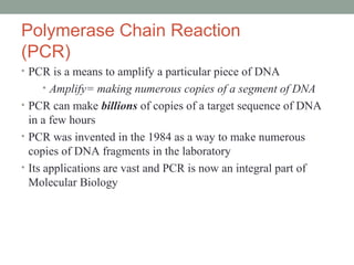
• PCR is a means to amplify a particular piece of DNA
• Amplify= making numerous copies of a segment of DNA
• PCR can make billions of copies of a target sequence of DNA
in a few hours
• PCR was invented in the 1984 as a way to make numerous
copies of DNA fragments in the laboratory
• Its applications are vast and PCR is now an integral part of
Molecular Biology
2. DNA Replication vs. PCR
• PCR is a laboratory version of DNA Replication in cells
• The laboratory version is commonly called “in vitro” since
it occurs in a test tube while “in vivo” signifies occurring in
a living cell.
3. DNA Replication in Cells (in vivo)
• DNA replication is the copying of
DNA
• It typically takes a cell just a few
hours to copy all of its DNA
• DNA replication is semi-
conservative (i.e. one strand of the
DNA is used as the template for the
growth of a new DNA strand)
• This process occurs with very few
errors (on average there is one error
per 1 billion nucleotides copied)
• More than a dozen enzymes and
proteins participate in DNA
replication
4. Key enzymes involved in DNA
Replication
• DNA Polymerase
• DNA Ligase
• Primase
• Helicase
• Topoisomerase
• Single strand binding protein
5. DNA Replication enzymes:
DNA Polymerase
• catalyzes the elongation of DNA by adding nucleoside
triphosphates to the 3’ end of the growing strand
• A nucleotide triphosphate is a 1 sugar + 1 base + 3
phosphates
• When a nucleoside triphosphate joins the DNA strand, two
phosphates are removed.
• DNA polymerase can only add nucleotides to 3’ end of growing
strand
6. Complementary Base-Pairing in DNA
• DNA is a double helix, made up of nucleotides, with a sugar-
phosphate backbone on the outside of the helix.
• Note: a nucleotide is a sugar + phosphate + nitrogenous
base
• The two strands of DNA are held together by pairs of
nitrogenous bases that are attached to each other via hydrogen
bonds.
• The nitrogenous base adenine will only pair with thymine
• The nitrogenous base guanine will only pair with cytosine
• During replication, once the DNA strands are separated, DNA
polymerase uses each strand as a template to synthesize new
strands of DNA with the precise, complementary order of
nucleotides.
7. DNA Replication enzymes:
DNA Ligase
• The two strands of DNA in a double helix are antiparallel (i.e.
they are oriented in opposite directions with one strand
oriented from 5’ to 3’ and the other strand oriented from 3’ to 5’
• 5’ and 3’ refer to the numbers assigned to the carbons in
the 5 carbon sugar
• Given the antiparallel nature of DNA and the fact that DNA
ploymerases can only add nucleotides to the 3’ end, one strand
(referred to as the leading strand) of DNA is synthesized
continuously and the other strand (referred to as the lagging
strand) in synthesized in fragments (called Okazaki
fragments) that are joined together by DNA ligase.
8. DNA Replication enzymes: Primase
• DNA Polymerase cannot initiate the synthesis of DNA
• Remember that DNA polymerase can only add nucleotides
to 3’ end of an already existing strand of DNA
• In humans, primase is the enzyme that can start an RNA chain
from scratch and it creates a primer (a short stretch RNA
with an available 3’ end) that DNA polymerase can add
nucleotides to during replication.
Note that the RNA primer is subsequently replaced with
DNA
9. DNA Replication enzymes:
Helicase, Topoisomerase and Single-strand binding protein
• Helicase untwists the two parallel DNA strands
• Topoisomerase relieves the stress of this twisting
• Single-strand binding protein binds to and stabilizes the
unpaired DNA strands
10. PCR: the in vitro version of DNA Replication
The following components are needed to perform
PCR in the laboratory:
1) DNA (your DNA of interest that contains the target
sequence you wish to copy)
2) A heat-stable DNA Polymerase (like Taq Polymerase)
3) All four nucleotide triphosphates
4) Buffers
5) Two short, single-stranded DNA molecules that serve
as primers
6) Thin walled tubes
7) Thermal cycler (a device that can change temperatures
dramatically in a very short period of time)
11. Common PCR additives
BSA (usually at 0.1 to 0.8 µg/µL final concentration)
Stabilize Taq polymerase & overcome PCR inhibitors
DMSO (usually at 2-5% v/v, inhibitory at ≤ 10% v/v)
Denaturant - good at keeping GC rich template/primer strands from
forming secondary structures.
Glycerol (usually at 5-10% v/v)
Increases apparent concentration of primer/template mix, and often
increases PCR efficiency at high temperatures.
Non-ionic detergents (Triton X, Tween 20 or Nonidet P-40) (0.1–1%)
NOT SDS (0.01% SDS cuts Taq activity to ~10% of normal)
Stabilize Taq polymerase & suppress formation of 2º structure
12. PCR
The DNA, DNA
polymerase, buffer,
nucleoside triphosphates,
and primers are placed in
a thin-walled tube and
then these tubes are
placed in the PCR
thermal cycler
PCR Thermocycler
13. The three main steps of PCR
• The basis of PCR is temperature changes and the effect that these
temperature changes have on the DNA.
• In a PCR reaction, the following series of steps is repeated 20-40 times
(note: 25 cycles usually takes about 2 hours and amplifies the DNA
fragment of interest 100,000 fold)
Step 1: Denature DNA
At 95°C, the DNA is denatured (i.e. the two strands are separated)
Step 2: Primers Anneal
At 40°C- 65°C, the primers anneal (or bind to) their complementary
sequences on the single strands of DNA
Step 3: DNA polymerase Extends the DNA chain
At 72°C, DNA Polymerase extends the DNA chain by adding nucleotides
to the 3’ ends of the primers.
14. Heat-stable DNA Polymerase
• Given that PCR involves very high temperatures, it is
imperative that a heat-stable DNA polymerase be used in the
reaction.
• Most DNA polymerases would denature (and thus not
function properly) at the high temperatures of PCR.
• Taq DNA polymerase was purified from the hot springs
bacterium Thermus aquaticus in 1976
• Taq has maximal enzymatic activity at 75 °C to 80 °C, and
substantially reduced activities at lower temperatures.
16. Step 2 Annealing or Primers Binding
Primers bind to the complimentary sequence on the
target DNA. Primers are chosen such that one is
complimentary to the one strand at one end of the
target sequence and that the other is complimentary
to the other strand at the other end of the target
sequence.
Forward Primer
Reverse Primer
17. Step 3 Extension or Primer Extension
DNA polymerase catalyzes the extension of the
strand in the 5-3 direction, starting at the
primers, attaching the appropriate nucleotide
(A-T, C-G)
extension
extension
18. • The next cycle will begin by denaturing the new DNA
strands formed in the previous cycle
19. The Size of the DNA Fragment Produced in
PCR is Dependent on the Primers
• The PCR reaction will amplify the DNA section between the two
primers.
• If the DNA sequence is known, primers can be developed to amplify
any piece of an organism’s DNA.
Forward primer
Reverse primer
Size of fragment that is amplified
20. PCR has become a very powerful tool
in molecular biology
• One can start with a single sperm cell or stand of hair and
amplify the DNA sufficiently to allow for DNA analysis and a
distinctive band on an agarose gel.
• One can amplify fragments of interest in an organism’s DNA by
choosing the right primers.
• One can use the selectivity of the primers to identify the
likelihood of an individual carrying a particular allele of a gene.
21. More about Primers
• PCR primers are short, single stranded DNA molecules (15-40
bp)
• They are manufactured commercially and can be ordered to
match any DNA sequence
• Primers are sequence specific, they will bind to a particular
sequence in a genome
• As you design primers with a longer length (15 → 40 bp), the
primers become more selective.
• DNA polymerase requires primers to initiate replication
23. Selectivity of Primers
• Primers bind to their complementary sequence
on the target DNA
• A primer composed of only 3 letter, ACC, for example,
would be very likely to encounter its complement in a
genome.
• As the size of the primer is increased, the likelihood of,
for example, a primer sequence of 35 base letters
repeatedly encountering a perfect complementary
section on the target DNA become remote.