Downloaded 19 times


![Circular layer
1. Superiorpharyngeal
Constrictors Muscle
2. Middlepharyngeal
Constrictors Muscle
3. Inferior Pharyngeal
Constrictors Muscle
Innervation =Vagus
nerve [X]
Muscle of Pharynx](https://image.slidesharecdn.com/chapter-3-pharynxesophagus-200309055326/75/Chapter-3-pharynx-esophagus-6-2048.jpg)


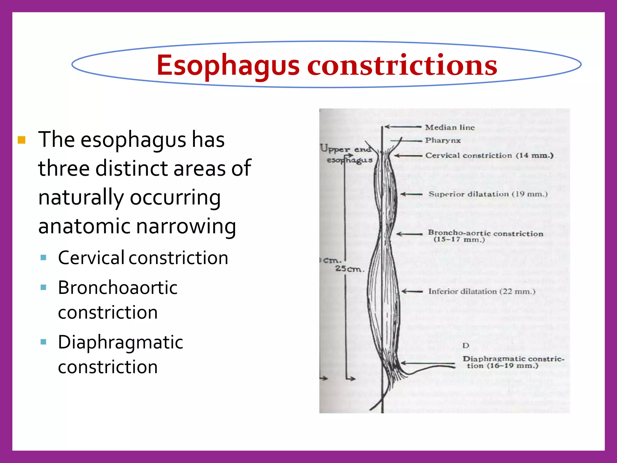


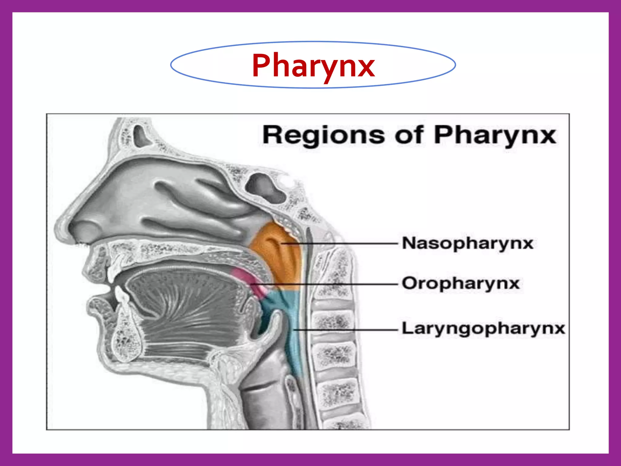
The document summarizes the anatomy and physiology of the pharynx and esophagus. It describes the pharynx as a funnel-shaped passageway divided into three regions (nasopharynx, oropharynx, laryngopharynx) with two layers of skeletal muscle. It then discusses the esophagus, describing it as a muscular tube that connects the pharynx to the stomach and passes through the diaphragm. Key details about the parts, constrictions, histology, blood supply, innervation and physiology of swallowing for both the pharynx and esophagus are provided.