Downloaded 14 times
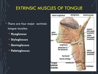
![EXTRINSIC MUSCLES
Muscle Origin Insertion Innervation Function
Genioglossus
Superiormental
tubercles
Body of hyoid;
entire lengthof
tongue
Hypoglossalnerve
[XII]
Protrudes tongue;
depresses center of
tongue
Hyoglossus Greater horn and
adjacent part of
body of hyoid bone
Lateral surfaceof
tongue
Hypoglossalnerve
[XII]
Depresses tongue
Styloglossus Styloid process
(anterolateral
surface)
Lateral surfaceof
tongue
Hypoglossalnerve
[XII]
Elevates and
retracts tongue
Palatoglossus
Inferior surfaceof
palatine
aponeurosis
Lateral margin of
tongue
Vagus nerve [X]
(via pharyngeal
branch to
pharyngealplexus)
Depressespalate;
moves
palatoglossal fold
toward midline;
elevates back of
the tongue](https://image.slidesharecdn.com/chapter-2-salivaryglands-200309055016/85/Chapter-2-salivary-glands-15-320.jpg)
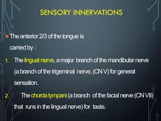
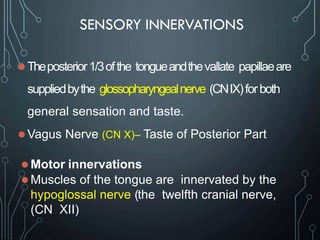
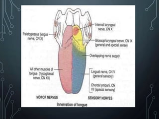
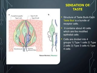
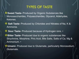
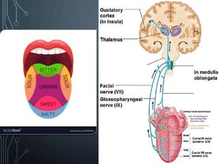
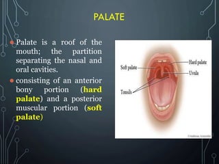
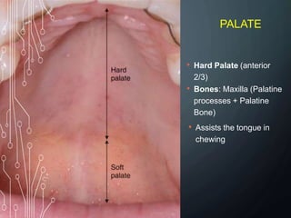
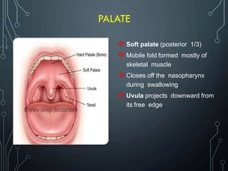
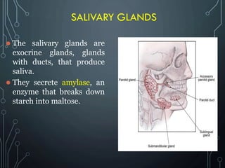
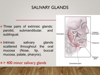
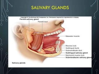
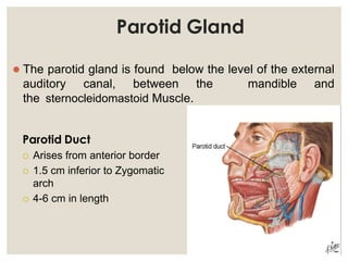
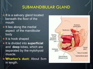
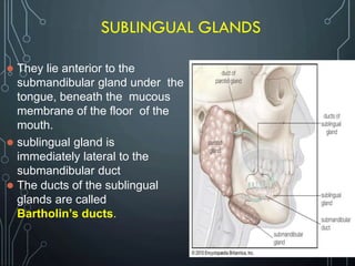

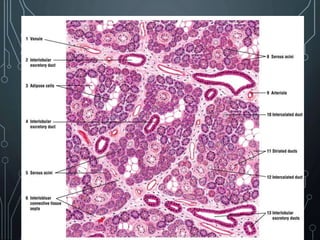
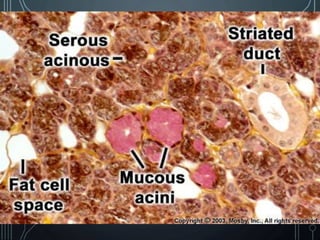


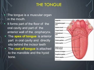
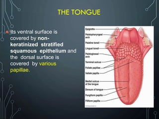
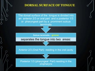
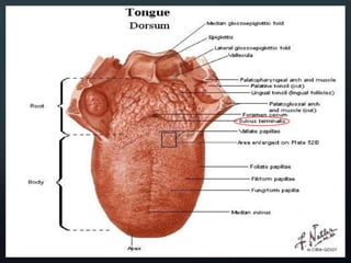
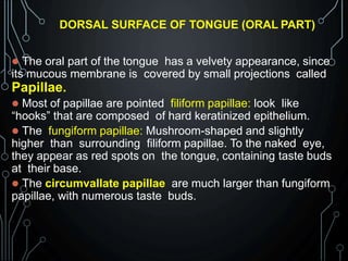
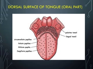
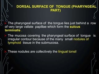
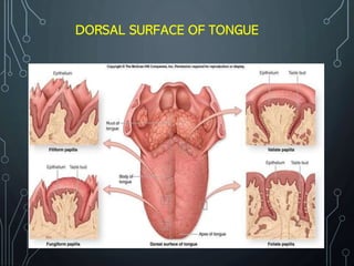
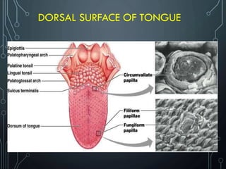
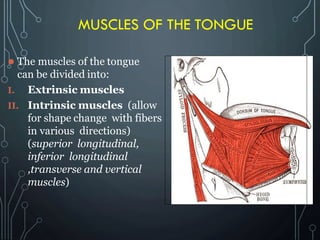
The document provides detailed information about the anatomy of the mouth, including the tongue, palate, salivary glands, and taste sensation. It describes the tongue's structure, muscles, blood and lymph supply. The dorsal surface of the tongue contains various papillae that provide texture and contain taste buds. The document outlines the three pairs of major salivary glands - parotid, submandibular, and sublingual glands - and their locations, ducts and blood supply. Taste buds are located on papillae on the tongue and contain receptor cells that detect the five basic tastes: sweet, salty, sour, bitter and umami.


