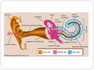
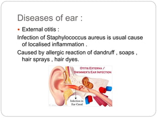
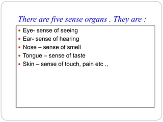
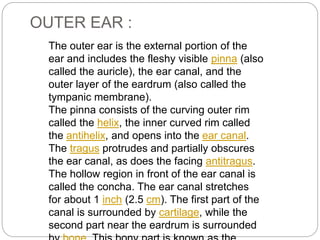
SENSE ORGANS
EAR
PRESENTED BY :
KEERTHI.NEELU FOR
1ST PHARM.D
Y17PHD0411
HINDU COLLEGE OF PHARMACY
UNDER GUIDANCE OF :
Mrs. A. PRATYUSHA M.PHARM
(PH.D)
DEPARTMENT OF
PHARMACOLOGY HINDU
COLLEGE OF PHARMACY
CONTENTS :
 INTRODUCTION OF SENSE ORGANS
 ANATOMY OF EAR
 HEARING AND BALANCE OF EAR
 DISORDERS OF EARS
 REFERENCES
There are five sense organs . They are :
 Eye- sense of seeing
 Ear- sense of hearing
 Nose – sense of smell
 Tongue – sense of taste
 Skin – sense of touch, pain etc .,
Now we will discuss about ear
 The ear is organ of hearing
 Supplied by 8th cranial nerve, i.e. the cochlear
part of vestibulocochlear nerve which is
stimulated by vibrations caused by sound waves
 Exception of auricle [pinna] structures that form
ear are encased within petrous portion of
temporal bone
STRUCTURE OF EAR : They are 3 parts
 OUTER EAR
 MIDDLE EAR [tympanic cavity]
 INNER EAR
The outer ear is the external portion of the
ear and includes the fleshy visible pinna (also
called the auricle), the ear canal, and the
outer layer of the eardrum (also called the
tympanic membrane).
The pinna consists of the curving outer rim
called the helix, the inner curved rim called
the antihelix, and opens into the ear canal.
The tragus protrudes and partially obscures
the ear canal, as does the facing antitragus.
The hollow region in front of the ear canal is
called the concha. The ear canal stretches
for about 1 inch (2.5 cm). The first part of the
canal is surrounded by cartilage, while the
second part near the eardrum is surrounded
OUTER EAR :
MIDDLE EAR :
 Middle ear
 Main article: Middle ear
 The middle ear
 The middle ear lies between the outer ear and the inner ear. It consists of an air-
filled cavity called the tympanic cavity and includes the three ossicles and their
attaching ligaments; the auditory tube; and the round and oval windows. The
ossicles are three small bones that function together to receive, amplify, and
transmit the sound from the eardrum to the inner ear. The ossicles are the
malleus (hammer), incus (anvil), and the stapes (stirrup). The stapes is the
smallest named bone in the body. The middle ear also connects to the upper
throat at the nasopharynx via the pharyngeal opening of the Eustachian tube.[3][7]
 The three ossicles transmit sound from the outer ear to the inner ear. The
malleus receives vibrations from sound pressure on the eardrum, where it is
connected at its longest part (the manubrium or handle) by a ligament. It
transmits vibrations to the incus, which in turn transmits the vibrations to the
small stapes bone. The wide base of the stapes rests on the oval window. As the
stapes vibrates, vibrations are transmitted through the oval window, causing
movement of fluid within the cochlea.[3]
 The round window allows for the fluid within the inner ear to move. As the stapes
pushes the secondary tympanic membrane, fluid in the inner ear moves and
pushes the membrane of the round window out by a corresponding amount into
the middle ear. The ossicles help amplify sound waves by nearly 15–20 times.[2]
The inner ear sits within the temporal bone in a complex cavity
called the bony labyrinth. A central area known as the vestibule
contains two small fluid-filled recesses, the utricle and saccule.
These connect to the semicircular canals and the cochlea.
There are three semicircular canals angled at right angles to
each other which are responsible for dynamic balance. The
cochlea is a spiral shell-shaped organ responsible for the
sense of hearing. These structures together create the
membranous labyrinth.[8]
The bony labyrinth refers to the bony compartment which
contains the membranous labyrinth, contained within the
temporal bone. The inner ear structurally begins at the oval
window, which receives vibrations from the incus of the middle
ear. Vibrations are transmitted into the inner ear into a fluid
called endolymph, which fills the membranous labyrinth. The
endolymph is situated in two vestibules, the utricle and
saccule, and eventually transmits to the cochlea, a spiral-
shaped structure. The cochlea consists of three fluid-filled
spaces: the vestibular duct, the cochlear duct, and the
tympanic duct.[3] Hair cells responsible for transduction—
INNER EAR :
Hearing is the process by which the ear transforms
sound vibrations in the external environment into nerve
impulses that are conveyed to the brain, where they
are interpreted as sounds. Sounds are produced when
vibrating objects, such as the plucked string of a guitar,
produce pressure pulses of vibrating air molecules,
better known as sound waves. The ear can distinguish
different subjective aspects of a sound, such as its
loudness and pitch, by detecting and analyzing
different physical characteristics of the waves. Pitch is
the perception of the frequency of sound waves—i.e.,
the number of wavelengths that pass a fixed point in a
unit of time. Frequency is usually measured in cycles
per second, or hertz. The human ear is most sensitive
to and most easily detects frequencies of 1,000 to
4,000 hertz, but at least for normal young ears the
Physiology of hearing :
Physiology of balance :
 The vestibular system is the sensory apparatus of
the inner ear that helps the body maintain its
postural equilibrium. The information furnished by
the vestibular system is also essential for
coordinating the position of the head and the
movement of the eyes. There are two sets of end
organs in the inner ear, or labyrinth: the
semicircular canals, which respond to rotational
movements (angular acceleration); and the utricle
and saccule within the vestibule, which respond
to changes in the position of the head with
respect to gravity (linear acceleration).
Although the vestibular organs
and the cochlea are derived
embryologically from the same
formation, the otic vesicle, their
association in the inner ear
seems to be a matter more of
convenience than of necessity.
From both the developmental
and the structural point of view,
the kinship of the vestibular
organs with the lateral line
system of the fish is readily
Diseases of ear :
 External otitis :
Infection of Staphylococcus aureus is usual cause
of localised inflammation .
Caused by allergic reaction of dandruff , soaps ,
hair sprays , hair dyes.
Acute otitis media :
•Inflammation of middle ear
•Common in children
•Caused by upward spread of microbes from an upper respiratory
tract infection via the auditory tube
Deafness :
Total or significant loss
of hearing
 Causes :
 Wax or foreign body
 Barotrauma
 Otosclerosis
 Presbycusis etc .,
References :
 https://www.britannica.com/science/
ear/The-physiology-of-balance-
vestibular-function
 intranet.tdmu.edu.ua/...%20physiolo
gy%20of%20work/02%20physiolog
y%20of%20.
 https://en.wikipedia.org/wiki/Ear
 ANATOMY AND PHYSIOLOGY –
ROSS AND WILSON
Sense organs ear

Sense organs ear

  • 1.
    SENSE ORGANS EAR PRESENTED BY: KEERTHI.NEELU FOR 1ST PHARM.D Y17PHD0411 HINDU COLLEGE OF PHARMACY UNDER GUIDANCE OF : Mrs. A. PRATYUSHA M.PHARM (PH.D) DEPARTMENT OF PHARMACOLOGY HINDU COLLEGE OF PHARMACY
  • 2.
    CONTENTS :  INTRODUCTIONOF SENSE ORGANS  ANATOMY OF EAR  HEARING AND BALANCE OF EAR  DISORDERS OF EARS  REFERENCES
  • 3.
    There are fivesense organs . They are :  Eye- sense of seeing  Ear- sense of hearing  Nose – sense of smell  Tongue – sense of taste  Skin – sense of touch, pain etc .,
  • 4.
    Now we willdiscuss about ear  The ear is organ of hearing  Supplied by 8th cranial nerve, i.e. the cochlear part of vestibulocochlear nerve which is stimulated by vibrations caused by sound waves  Exception of auricle [pinna] structures that form ear are encased within petrous portion of temporal bone
  • 5.
    STRUCTURE OF EAR: They are 3 parts  OUTER EAR  MIDDLE EAR [tympanic cavity]  INNER EAR
  • 7.
    The outer earis the external portion of the ear and includes the fleshy visible pinna (also called the auricle), the ear canal, and the outer layer of the eardrum (also called the tympanic membrane). The pinna consists of the curving outer rim called the helix, the inner curved rim called the antihelix, and opens into the ear canal. The tragus protrudes and partially obscures the ear canal, as does the facing antitragus. The hollow region in front of the ear canal is called the concha. The ear canal stretches for about 1 inch (2.5 cm). The first part of the canal is surrounded by cartilage, while the second part near the eardrum is surrounded OUTER EAR :
  • 8.
    MIDDLE EAR : Middle ear  Main article: Middle ear  The middle ear  The middle ear lies between the outer ear and the inner ear. It consists of an air- filled cavity called the tympanic cavity and includes the three ossicles and their attaching ligaments; the auditory tube; and the round and oval windows. The ossicles are three small bones that function together to receive, amplify, and transmit the sound from the eardrum to the inner ear. The ossicles are the malleus (hammer), incus (anvil), and the stapes (stirrup). The stapes is the smallest named bone in the body. The middle ear also connects to the upper throat at the nasopharynx via the pharyngeal opening of the Eustachian tube.[3][7]  The three ossicles transmit sound from the outer ear to the inner ear. The malleus receives vibrations from sound pressure on the eardrum, where it is connected at its longest part (the manubrium or handle) by a ligament. It transmits vibrations to the incus, which in turn transmits the vibrations to the small stapes bone. The wide base of the stapes rests on the oval window. As the stapes vibrates, vibrations are transmitted through the oval window, causing movement of fluid within the cochlea.[3]  The round window allows for the fluid within the inner ear to move. As the stapes pushes the secondary tympanic membrane, fluid in the inner ear moves and pushes the membrane of the round window out by a corresponding amount into the middle ear. The ossicles help amplify sound waves by nearly 15–20 times.[2]
  • 10.
    The inner earsits within the temporal bone in a complex cavity called the bony labyrinth. A central area known as the vestibule contains two small fluid-filled recesses, the utricle and saccule. These connect to the semicircular canals and the cochlea. There are three semicircular canals angled at right angles to each other which are responsible for dynamic balance. The cochlea is a spiral shell-shaped organ responsible for the sense of hearing. These structures together create the membranous labyrinth.[8] The bony labyrinth refers to the bony compartment which contains the membranous labyrinth, contained within the temporal bone. The inner ear structurally begins at the oval window, which receives vibrations from the incus of the middle ear. Vibrations are transmitted into the inner ear into a fluid called endolymph, which fills the membranous labyrinth. The endolymph is situated in two vestibules, the utricle and saccule, and eventually transmits to the cochlea, a spiral- shaped structure. The cochlea consists of three fluid-filled spaces: the vestibular duct, the cochlear duct, and the tympanic duct.[3] Hair cells responsible for transduction— INNER EAR :
  • 12.
    Hearing is theprocess by which the ear transforms sound vibrations in the external environment into nerve impulses that are conveyed to the brain, where they are interpreted as sounds. Sounds are produced when vibrating objects, such as the plucked string of a guitar, produce pressure pulses of vibrating air molecules, better known as sound waves. The ear can distinguish different subjective aspects of a sound, such as its loudness and pitch, by detecting and analyzing different physical characteristics of the waves. Pitch is the perception of the frequency of sound waves—i.e., the number of wavelengths that pass a fixed point in a unit of time. Frequency is usually measured in cycles per second, or hertz. The human ear is most sensitive to and most easily detects frequencies of 1,000 to 4,000 hertz, but at least for normal young ears the Physiology of hearing :
  • 14.
    Physiology of balance:  The vestibular system is the sensory apparatus of the inner ear that helps the body maintain its postural equilibrium. The information furnished by the vestibular system is also essential for coordinating the position of the head and the movement of the eyes. There are two sets of end organs in the inner ear, or labyrinth: the semicircular canals, which respond to rotational movements (angular acceleration); and the utricle and saccule within the vestibule, which respond to changes in the position of the head with respect to gravity (linear acceleration).
  • 15.
    Although the vestibularorgans and the cochlea are derived embryologically from the same formation, the otic vesicle, their association in the inner ear seems to be a matter more of convenience than of necessity. From both the developmental and the structural point of view, the kinship of the vestibular organs with the lateral line system of the fish is readily
  • 16.
    Diseases of ear:  External otitis : Infection of Staphylococcus aureus is usual cause of localised inflammation . Caused by allergic reaction of dandruff , soaps , hair sprays , hair dyes.
  • 17.
    Acute otitis media: •Inflammation of middle ear •Common in children •Caused by upward spread of microbes from an upper respiratory tract infection via the auditory tube
  • 18.
    Deafness : Total orsignificant loss of hearing  Causes :  Wax or foreign body  Barotrauma  Otosclerosis  Presbycusis etc .,
  • 19.
    References :  https://www.britannica.com/science/ ear/The-physiology-of-balance- vestibular-function intranet.tdmu.edu.ua/...%20physiolo gy%20of%20work/02%20physiolog y%20of%20.  https://en.wikipedia.org/wiki/Ear  ANATOMY AND PHYSIOLOGY – ROSS AND WILSON