Downloaded 25 times


![Innervation of The Teeth And Gingivae
• All nerves that innervate the teeth and
gingivae are branches of the trigeminal nerve
[V]](https://image.slidesharecdn.com/chapter-1-oralcavity-200309054803/85/Chapter-1-oral-cavity-51-320.jpg)


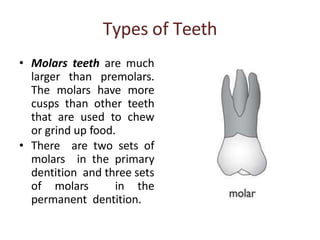
This document provides information about the anatomy of the mouth. It describes the oral cavity and its components including the lips and teeth. The oral cavity is lined with mucous membrane and consists of the vestibule and oral cavity proper. The lips are soft, pliable structures that form the mouth's margin. Teeth are made of enamel, dentin, cementum, and pulp and come in different types including incisors, canines, premolars, and molars. The document also discusses the blood supply, innervation, and lymphatic drainage of the oral structures.