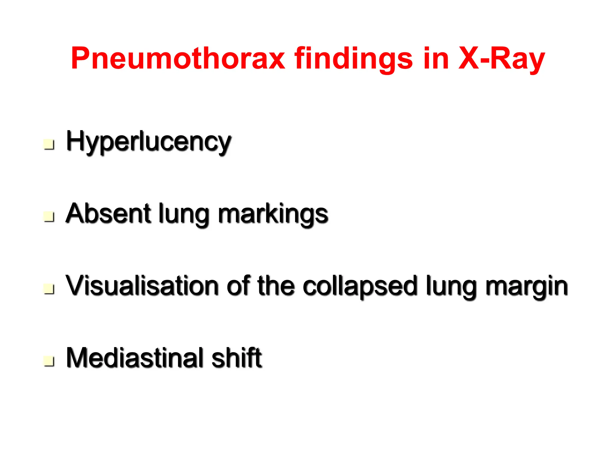
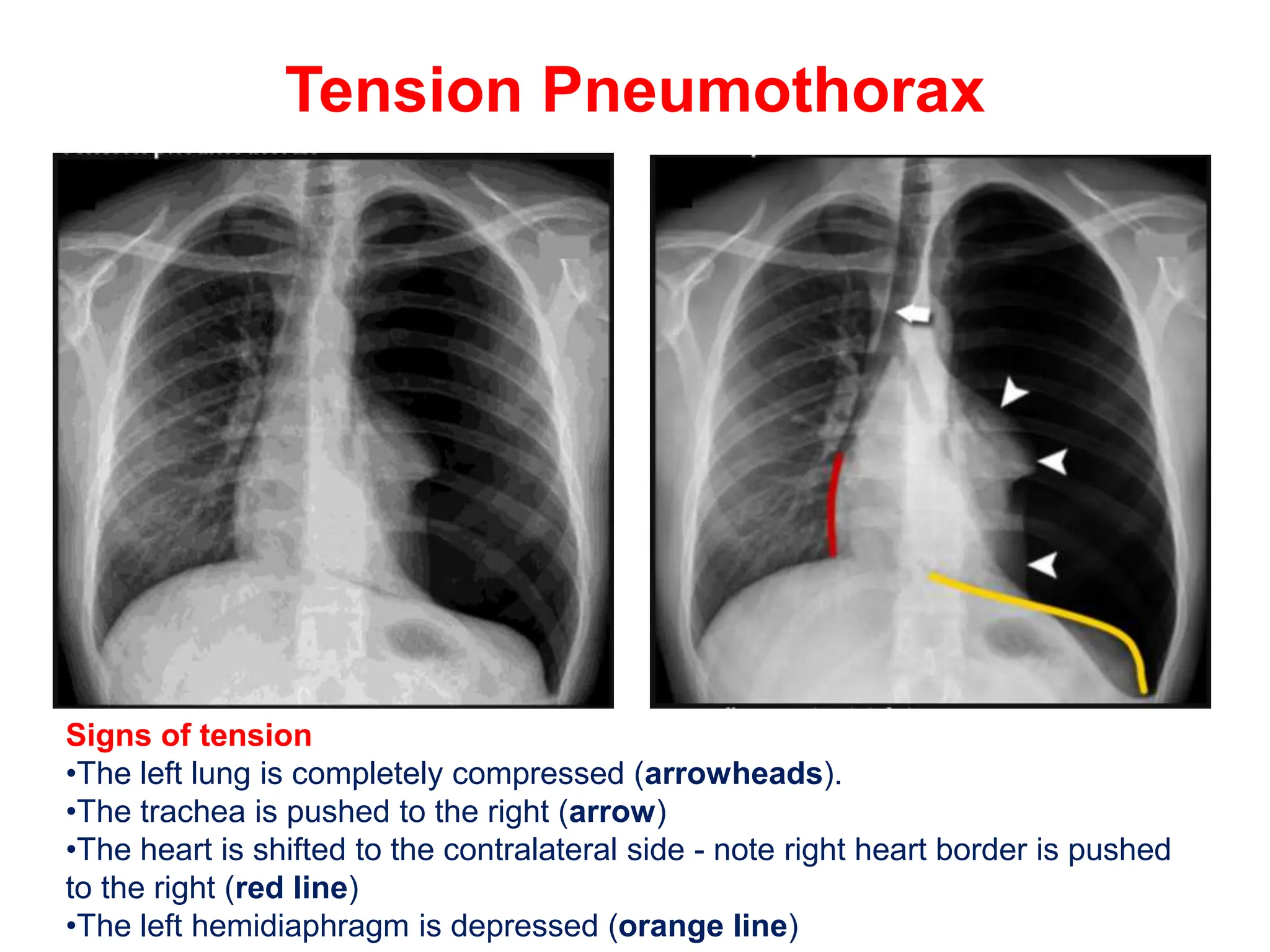
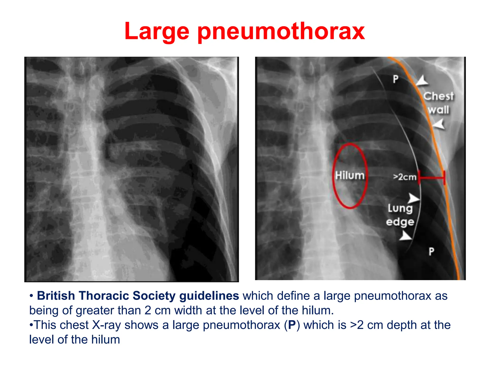
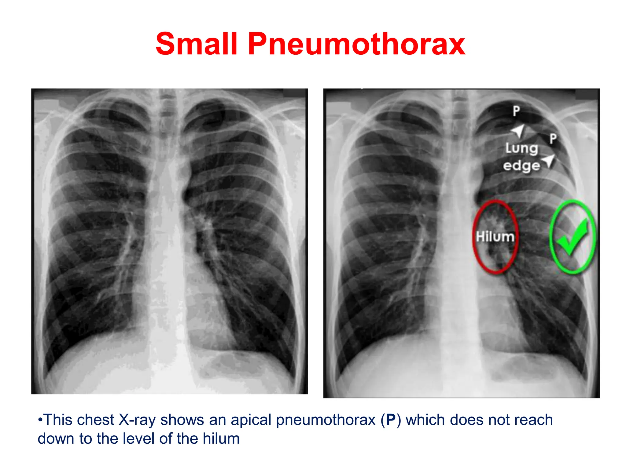
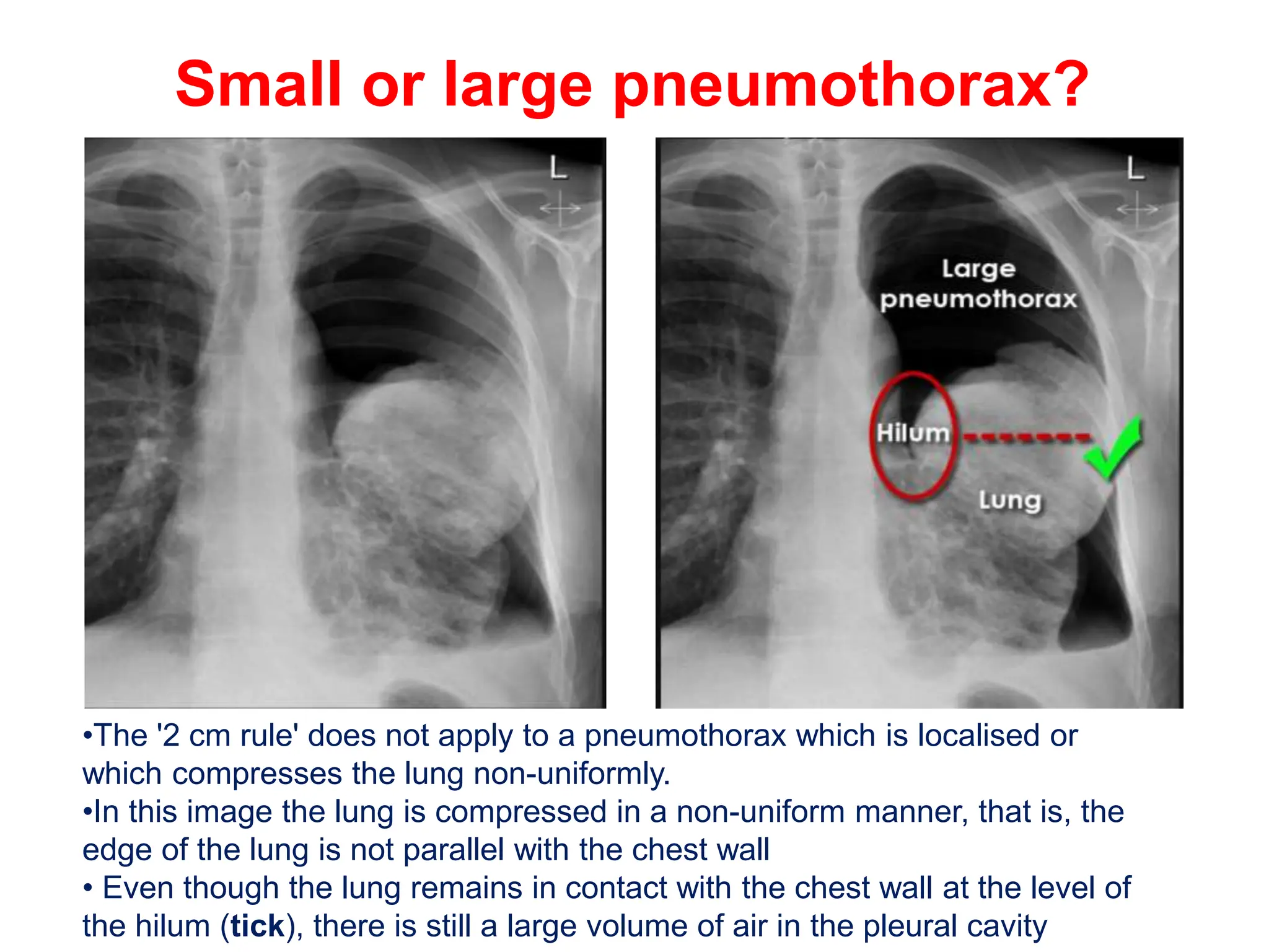
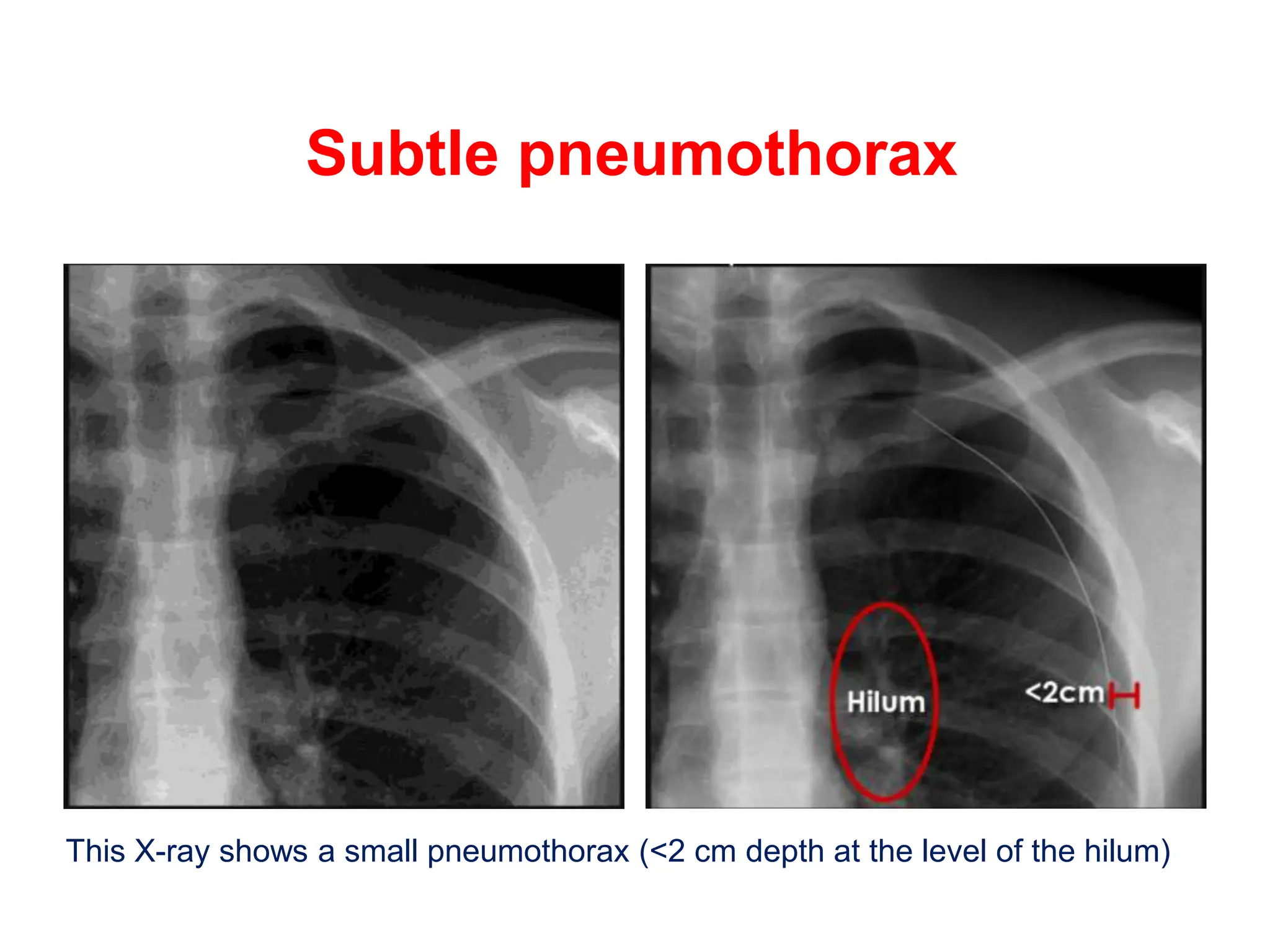
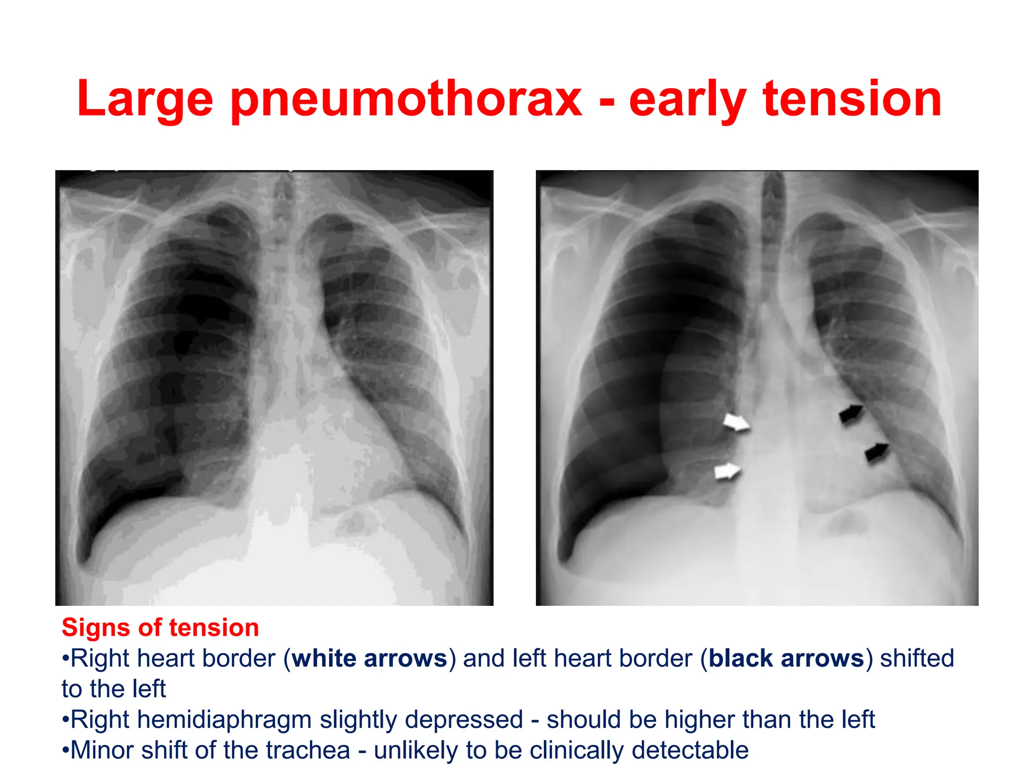
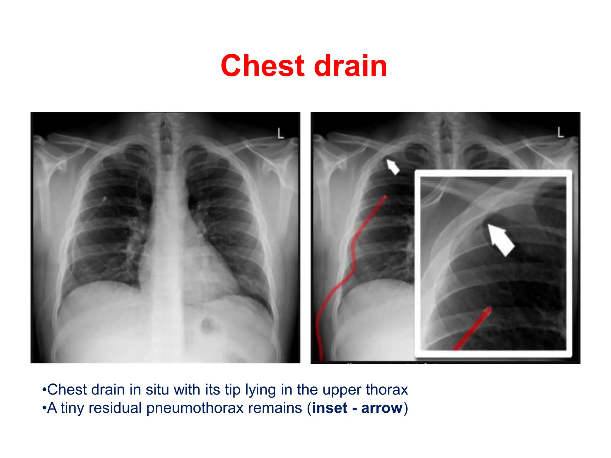
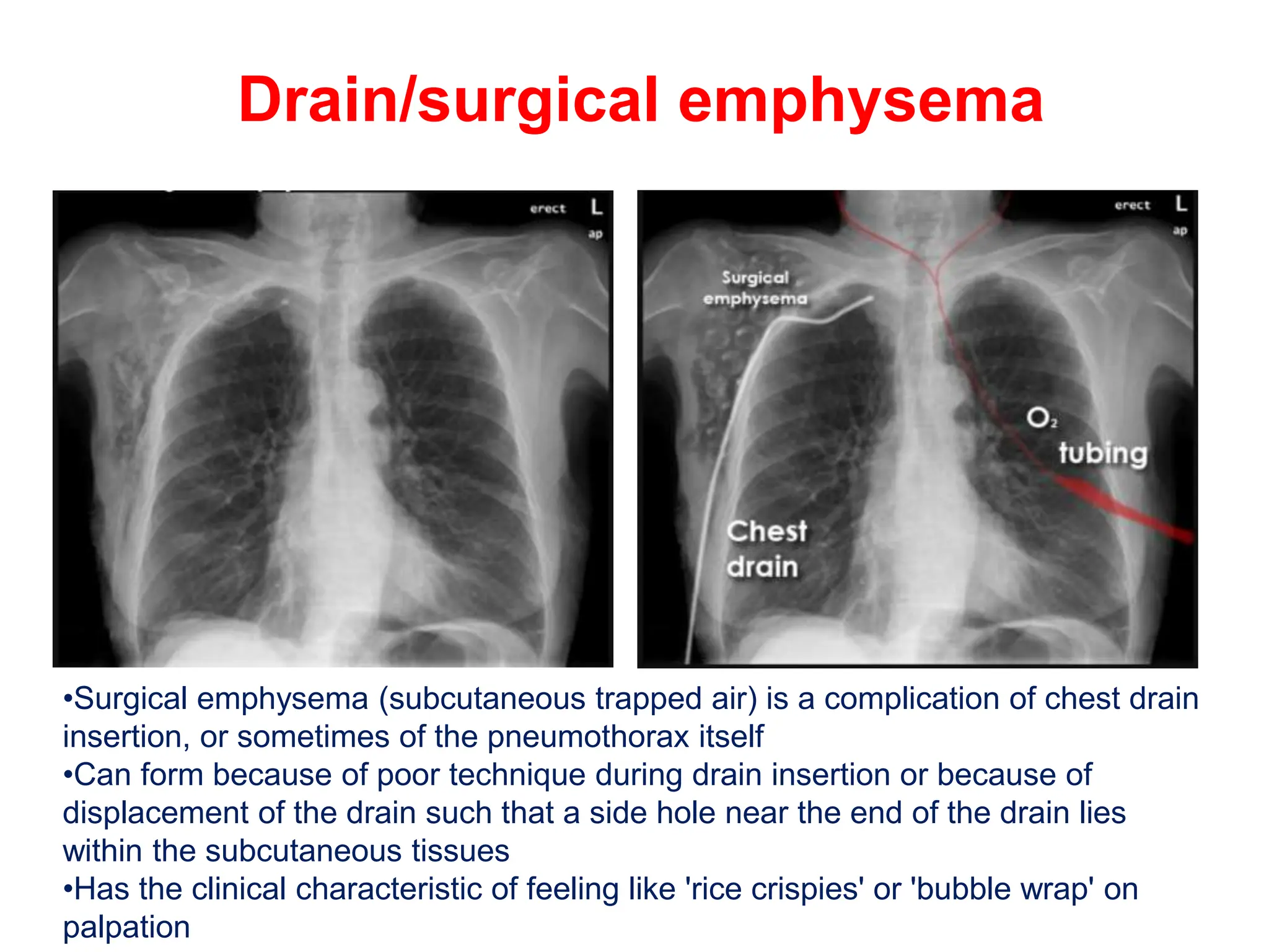
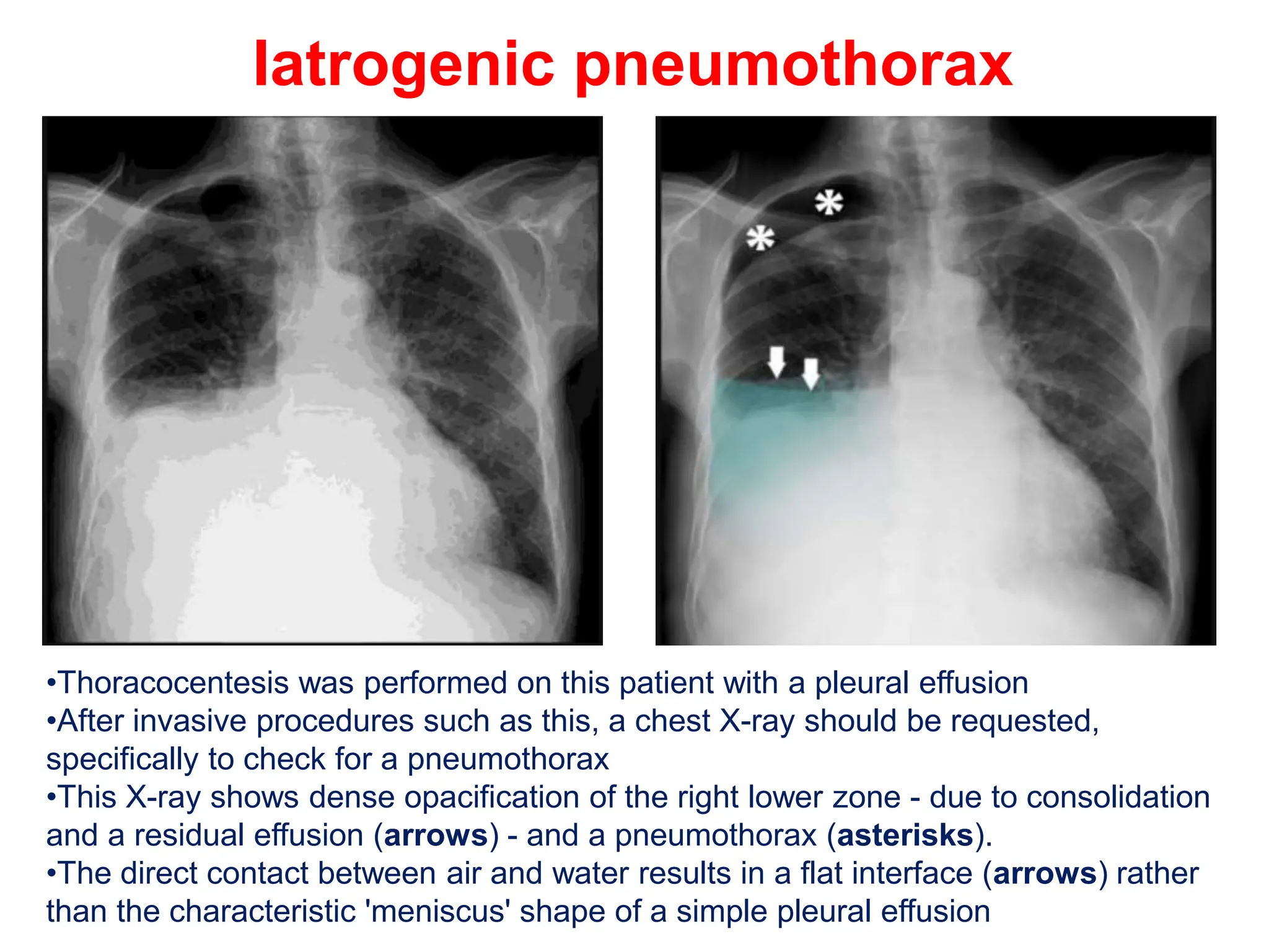
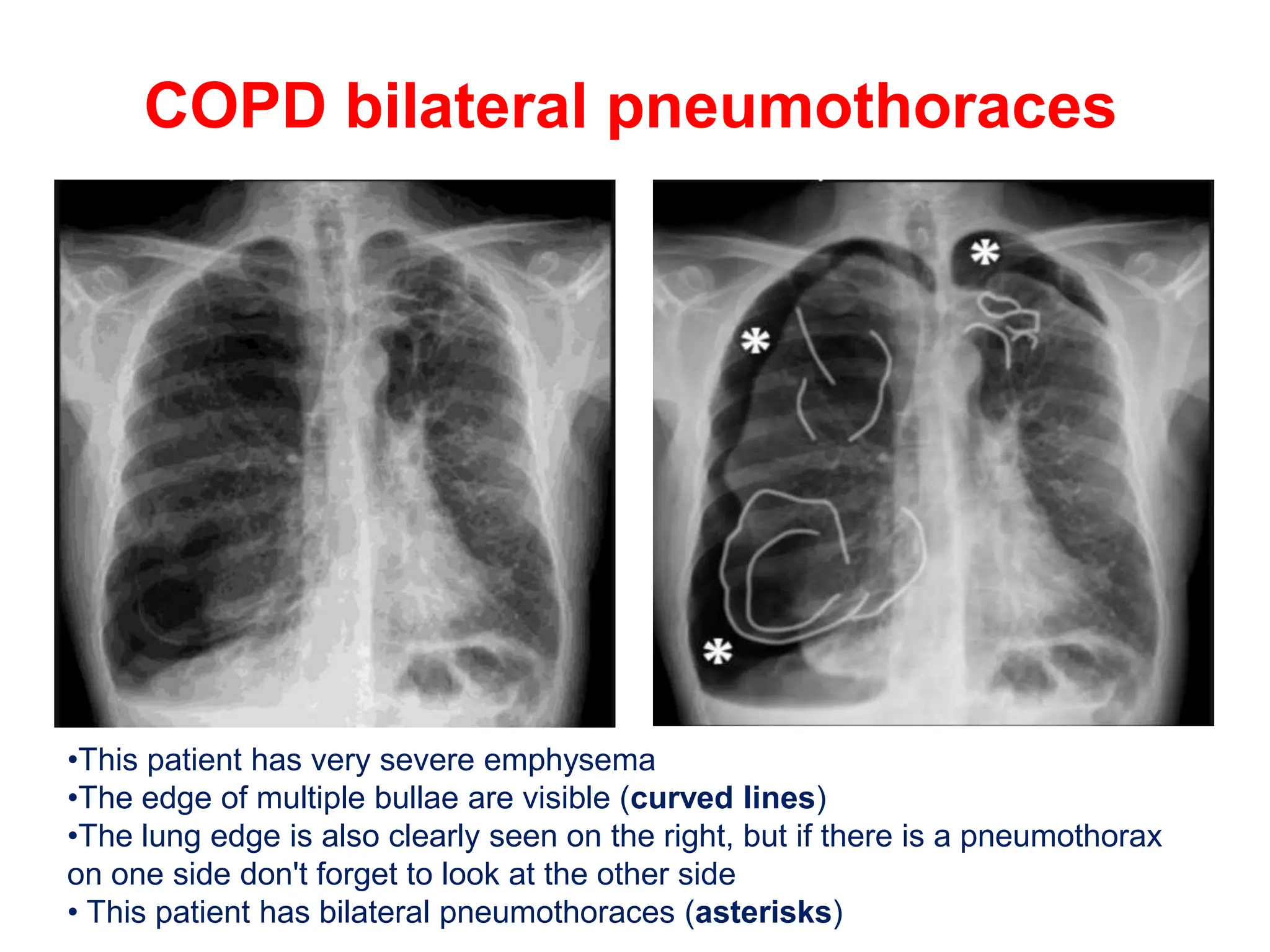
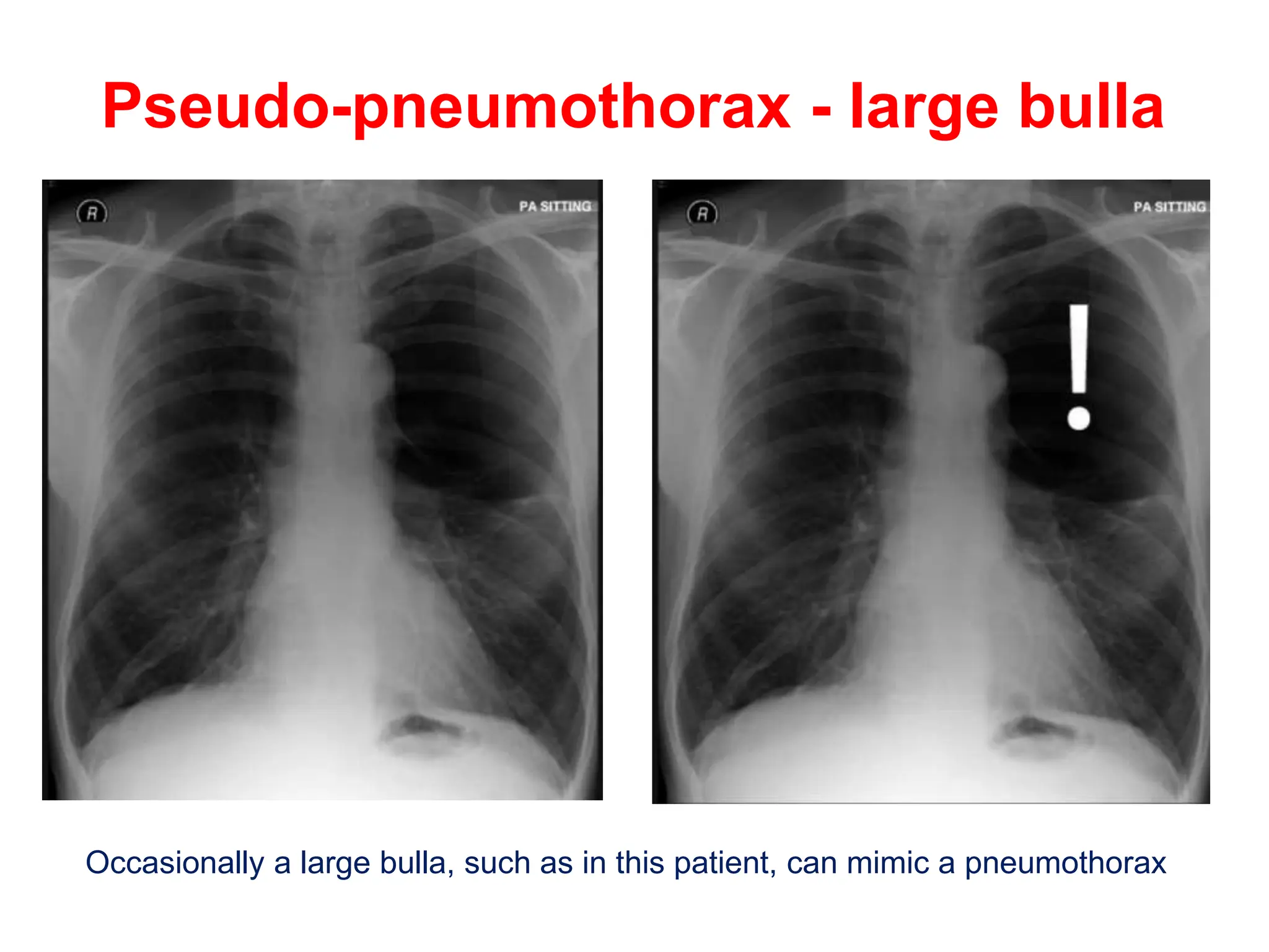
2. Pneumothorax findings in X-Ray
Hyperlucency
Absent lung markings
Visualisation of the collapsed lung margin
Mediastinal shift
3. Tension Pneumothorax
Signs of tension
•The left lung is completely compressed (arrowheads).
•The trachea is pushed to the right (arrow)
•The heart is shifted to the contralateral side - note right heart border is pushed
to the right (red line)
•The left hemidiaphragm is depressed (orange line)
4. Large pneumothorax
• British Thoracic Society guidelines which define a large pneumothorax as
being of greater than 2 cm width at the level of the hilum.
•This chest X-ray shows a large pneumothorax (P) which is >2 cm depth at the
level of the hilum
5. Small Pneumothorax
•This chest X-ray shows an apical pneumothorax (P) which does not reach
down to the level of the hilum
6. Small or large pneumothorax?
•The '2 cm rule' does not apply to a pneumothorax which is localised or
which compresses the lung non-uniformly.
•In this image the lung is compressed in a non-uniform manner, that is, the
edge of the lung is not parallel with the chest wall
• Even though the lung remains in contact with the chest wall at the level of
the hilum (tick), there is still a large volume of air in the pleural cavity
8. Large pneumothorax - early tension
Signs of tension
•Right heart border (white arrows) and left heart border (black arrows) shifted
to the left
•Right hemidiaphragm slightly depressed - should be higher than the left
•Minor shift of the trachea - unlikely to be clinically detectable
9. Chest drain
•Chest drain in situ with its tip lying in the upper thorax
•A tiny residual pneumothorax remains (inset - arrow)
10. Drain/surgical emphysema
•Surgical emphysema (subcutaneous trapped air) is a complication of chest drain
insertion, or sometimes of the pneumothorax itself
•Can form because of poor technique during drain insertion or because of
displacement of the drain such that a side hole near the end of the drain lies
within the subcutaneous tissues
•Has the clinical characteristic of feeling like 'rice crispies' or 'bubble wrap' on
palpation
11. Iatrogenic pneumothorax
nic pneumothorax
•Thoracocentesis was performed on this patient with a pleural effusion
•After invasive procedures such as this, a chest X-ray should be requested,
specifically to check for a pneumothorax
•This X-ray shows dense opacification of the right lower zone - due to consolidation
and a residual effusion (arrows) - and a pneumothorax (asterisks).
•The direct contact between air and water results in a flat interface (arrows) rather
than the characteristic 'meniscus' shape of a simple pleural effusion
12. COPD bilateral pneumothoraces
•This patient has very severe emphysema
•The edge of multiple bullae are visible (curved lines)
•The lung edge is also clearly seen on the right, but if there is a pneumothorax
on one side don't forget to look at the other side
• This patient has bilateral pneumothoraces (asterisks)
13. Pseudo-pneumothorax - large bulla
Occasionally a large bulla, such as in this patient, can mimic a pneumothorax