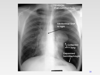
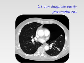
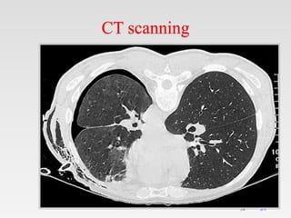
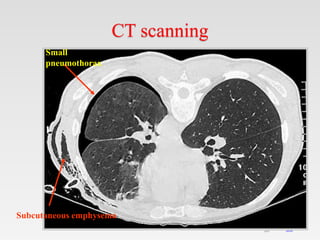
Tips on using my ppt.
1. You can freely download, edit, modify and put your
name etc.
2. Don’t be concerned about number of slides. Half the
slides are blanks except for the title.
3. First show the blank slides (eg. Aetiology ) > Ask
students what they already know about ethology of
today's topic. > Then show next slide which enumerates
aetiologies.
4. At the end rerun the show – show blank> ask questions >
show next slide.
5. This will be an ACTIVE LEARNING SESSION x
three revisions.
6. Good for self study also.
7. See notes for bibliography.
Introduction & history
Pathophysiology
3
Pathophysiology
• The probable cause of pneumothorax is rupture
of an apical bleb or bulla
• Smoking causes a 9-fold increase in the
relative risk of a pneumothorax in females
• A 22-fold increase in male smokers
4
Mechanism
5
Mechanism
• In normal people, the pressure in pleural space is
negative during the entire respiratory cycle.
• When a communication develops between
pleural space, and intrapulmonary air space or
exterior and air will flow into the pleural space
until there is no longer a pressure difference or the
communication is sealed
– Compress lungs,blood vessels and heart
– Decreased cardiac output
– Impaired venous return
– Hypotension
6
Etiology
Etiology
Pneumothorax
Spontaneous
Primary Secondary
Traumatic
Iatrogenic
Interventional
procedures.
Positive pressure
ventilation
Non iatrogenic
Penetrating
trauma
Blunt trauma.
Primary spontaneous pneumothorax
• It occurs in young healthy individuals
without underlying lung disease
• It is due to rupture of apical
sub-pleural bleb or bullae
Predisposing factors:
 Smoking.
 Tall, thin male.
 Airway inflammation (distal)
 Structural abnormalities of bronchial tree
 Genetic contribution 9
Secondary spontaneous pneumothorax
Common causes.
• TB
• Asthma
• COPD
• Suppurative pneumonia
• Cystic fibrosis
• Rare cause
• ILD
• Eosinophilic granuloma
• Sarcoidosis
• Lymphangioleiomyomatosis
• AIDS.
10
• Primary lung carcinoma
• Complication of
chemotherapy
• Connective tissue disease
• Scleroderma
• Marfans syndrome
• Histiocytosis- x
• Rheumatoid disease
• Pulmonary infarct
• Wegener’s granulomatosis…
Traumatic pneumothorax
Accidental trauma:(non-iatrogenic)
Blunt trauma: with fracture ribs.
Penetrating trauma: stab wound or gun shot injury.
Iatrogenic :
Positive pressure ventilation:
Alveolar rupture  interstitial emphysema
pneumothorax.(B/L PNX)
Interventional procedures:
Biopsy, thoraco-centesis, CVP line,trachestomy etc..
11
Clinical Features
12
Clinical Features
Constant
• Dyspnea
• Chest pain ( pleuritic)
Uncommon manifestation
• Cough
• Hemoptysis
• Orthopnea
• Cyanosis
• Tachycardia
• Anxiety,
• vague presenting symptoms (eg, general malaise,
fatigue)
13
14
Clinical types
Pneumothorax
Closed
pneumothorax
Open
pneumothorax
Tension
pneumothorax
15
Closed
pneumothorax
Open
pneumothorax
Tension
pneumothorax
The pleural tear
Is sealed
The pleural tear
is open
The pleural tear
act as a ball &
valve mechanism
The pleural
cavity pressure
is < the
atmospheric
pressure
The pleural
cavity pressure
is = the
atmospheric
pressure
The pleural cavity
pressure is > the
atmospheric
pressure
Tension pneumothorax
• Tension pneumothorax is classically characterized by
hypotension and hypoxia.
Tension pneumothorax
• It is life threatening condition.
• Rapidly progressive breathlessness and circulatory
collapse (tachycardia, hypotension & sweating).
• Jugular venous distention
• The pleural pressure is more than the atmospheric pressure.
Radiological manifestations of large pneumothorax
• Mediastinal shift,
• Flattening of the hemidiaphragm &
• Lung collapse.
It is more common with
• Positive pressure ventilation &
• Traumatic pneumothorax.
Clinical features: IN ICU
– Patients on Mechanical ventilation or
cardiopulmonary resuscitation who
suddenly deteriorate clinically,with
– RAPIDLY PROGRESSIVE
DYSPNOEA.
– Cyanosis
– Marked tachycardia
– Hypotension
– The airway pressure alarms are triggered.
Physical examination
• Depend on size of pneumothorax
• The vital signs usually normal
• Unilateral Chest movements
• The trachea may be shifted toward the
contralateral side if the pneumothorax is
large
• Tactile fremitus is absent
• The percussion note is hyperresonant
• The breath sounds are reduced or absent on
the affected side
• The lower edge of the liver may be shifted
inferiorly with a right-side pneumothorax
Pneumothorax
in erect position
Pneumothorax
in supine position
Air in apicolateral pleural
space
Air in anteromedial pleural
space.
Small pneumothorax
22
23
Small pneumothorax
LARGE PNEUMOTHORAX
24
25
Visceral pleural line
26
DD of visceral pleural line
Skin fold:
• Positive mash band (optical edge enhancement).
• Extend beyond the chest wall.
• Lung markings extend beyond it.
DD of visceral pleural line
Scapular edge
Tension pneumothorax
29
30
CT scanning
It is recommended in difficult cases such
as patients in whom the lungs are
obscured by overlying surgical
emphysema
To differentiate a pneumothorax from
suspected bulla in complex cystic lung
disease
31
CT can diagnose easily
pneumothroax
CT can diagnose easily
pneumothroax
34
34
CT scanning
35
35
CT scanning
Small
pneumothorax
Subcutaneous emphysema
U/S in pneumothorax
• Ultrasound found to be more sensitive than
CXR in diagnosis of pneumothorax.
Treatment
Treatment
Traumatic pneumothorax
• Intercostal tube drainage for trauma.
• Aspiration is the technique of choice for
iatrogenic pneumothoraces, because
recurrence is usually not a factor. Tube
thoracostomy is reserved for very
symptomatic patients.
43
Simple aspiration
 Simple aspiration is recommended as first line
treatment for all PSP requiring intervention
 Simple aspiration is less likely to succeed in
secondary pneumothoraces and in this situation, is
only recommended as an initial treatment in small
(<2 cm) pneumothoraces in minimally breathless
patients under the age of 50 years
 Patients should be admitted to hospital and observed
for at least 24 hours before discharge.
44
 Repeated aspiration is reasonable for primary
pneumothorax when the first aspiration has
been successful
 A volume of < 2.5 L has been aspirated on the
first attempt
 The aspiration can be done by needle or
catheter
Catheter aspiration
Chemical pleurodesis
45
 Goals
• To prevent pneumothorax recurrence
• To produce inflammation of pleura and adhesions
• Indications:
• Persistent air leak and repeated pneumothorax
• Bilateral pneumothorax
• Complicated with bullae
• Lung dysfunction, cannot tolerate operation.
Surgical treatment
46
 Indications
◦ No response to medical treatment
◦ Persistant air leak
◦ Hemopneumothorax
◦ Bilateral pneumothoraces
◦ Recurrent pneumothorax
◦ Tension pneumothorax failed to drainage
◦ Thickened pleura making lung unable to reexpand
◦ Multiple blebs or bullae
Complications of pneumothorax
Recurrence of spontaneous pneumothorax
Tension pneumothorax
Hydropneumothorax
Encysted pneumothorax
Failure of expansion of the collapsed lung
Re-expansion pulmonary edema
Broncho-pleural fistula
Pneumomediastinum
Recurrence of spontaneous
pneumothorax
• 50% on the same side.
• 15% on the contralateral side.
More common in
• secondary spontaneous pneumothorax.
Tension pneumothorax
Tension pneumothorax:
Management
• Aspiration followed by ICD
Catamenial pneumothorax
• Catamenial pneumothorax is a condition
of pneumothorax occurring in conjunction
with menstrual periods(catamenial -
menstruation), believed to be caused
primarily by endometriosis of the pleura
Get this ppt in mobile
1. Download Microsoft
PowerPoint from play
store.
2. Open Google assistant
3. Open Google lens.
4. Scan qr code from
next slide.
Get this ppt in mobile
Get my ppt collection
• https://www.slideshare.net/drpradeeppande/
edit_my_uploads
• https://www.dropbox.com/sh/x600md3cvj8
5woy/AACVMHuQtvHvl_K8ehc3ltkEa?dl
=0
• https://www.facebook.com/doctorpradeeppa
nde/?ref=pages_you_manage

Pneumothorax.pptx

  • 1.
    Tips on usingmy ppt. 1. You can freely download, edit, modify and put your name etc. 2. Don’t be concerned about number of slides. Half the slides are blanks except for the title. 3. First show the blank slides (eg. Aetiology ) > Ask students what they already know about ethology of today's topic. > Then show next slide which enumerates aetiologies. 4. At the end rerun the show – show blank> ask questions > show next slide. 5. This will be an ACTIVE LEARNING SESSION x three revisions. 6. Good for self study also. 7. See notes for bibliography.
  • 2.
  • 3.
  • 4.
    Pathophysiology • The probablecause of pneumothorax is rupture of an apical bleb or bulla • Smoking causes a 9-fold increase in the relative risk of a pneumothorax in females • A 22-fold increase in male smokers 4
  • 5.
  • 6.
    Mechanism • In normalpeople, the pressure in pleural space is negative during the entire respiratory cycle. • When a communication develops between pleural space, and intrapulmonary air space or exterior and air will flow into the pleural space until there is no longer a pressure difference or the communication is sealed – Compress lungs,blood vessels and heart – Decreased cardiac output – Impaired venous return – Hypotension 6
  • 7.
  • 8.
  • 9.
    Primary spontaneous pneumothorax •It occurs in young healthy individuals without underlying lung disease • It is due to rupture of apical sub-pleural bleb or bullae Predisposing factors:  Smoking.  Tall, thin male.  Airway inflammation (distal)  Structural abnormalities of bronchial tree  Genetic contribution 9
  • 10.
    Secondary spontaneous pneumothorax Commoncauses. • TB • Asthma • COPD • Suppurative pneumonia • Cystic fibrosis • Rare cause • ILD • Eosinophilic granuloma • Sarcoidosis • Lymphangioleiomyomatosis • AIDS. 10 • Primary lung carcinoma • Complication of chemotherapy • Connective tissue disease • Scleroderma • Marfans syndrome • Histiocytosis- x • Rheumatoid disease • Pulmonary infarct • Wegener’s granulomatosis…
  • 11.
    Traumatic pneumothorax Accidental trauma:(non-iatrogenic) Blunttrauma: with fracture ribs. Penetrating trauma: stab wound or gun shot injury. Iatrogenic : Positive pressure ventilation: Alveolar rupture  interstitial emphysema pneumothorax.(B/L PNX) Interventional procedures: Biopsy, thoraco-centesis, CVP line,trachestomy etc.. 11
  • 12.
  • 13.
    Clinical Features Constant • Dyspnea •Chest pain ( pleuritic) Uncommon manifestation • Cough • Hemoptysis • Orthopnea • Cyanosis • Tachycardia • Anxiety, • vague presenting symptoms (eg, general malaise, fatigue) 13
  • 14.
  • 15.
    15 Closed pneumothorax Open pneumothorax Tension pneumothorax The pleural tear Issealed The pleural tear is open The pleural tear act as a ball & valve mechanism The pleural cavity pressure is < the atmospheric pressure The pleural cavity pressure is = the atmospheric pressure The pleural cavity pressure is > the atmospheric pressure
  • 16.
    Tension pneumothorax • Tensionpneumothorax is classically characterized by hypotension and hypoxia.
  • 17.
    Tension pneumothorax • Itis life threatening condition. • Rapidly progressive breathlessness and circulatory collapse (tachycardia, hypotension & sweating). • Jugular venous distention • The pleural pressure is more than the atmospheric pressure. Radiological manifestations of large pneumothorax • Mediastinal shift, • Flattening of the hemidiaphragm & • Lung collapse. It is more common with • Positive pressure ventilation & • Traumatic pneumothorax.
  • 18.
  • 19.
    Clinical features: INICU – Patients on Mechanical ventilation or cardiopulmonary resuscitation who suddenly deteriorate clinically,with – RAPIDLY PROGRESSIVE DYSPNOEA. – Cyanosis – Marked tachycardia – Hypotension – The airway pressure alarms are triggered.
  • 20.
    Physical examination • Dependon size of pneumothorax • The vital signs usually normal • Unilateral Chest movements • The trachea may be shifted toward the contralateral side if the pneumothorax is large • Tactile fremitus is absent • The percussion note is hyperresonant • The breath sounds are reduced or absent on the affected side • The lower edge of the liver may be shifted inferiorly with a right-side pneumothorax
  • 21.
    Pneumothorax in erect position Pneumothorax insupine position Air in apicolateral pleural space Air in anteromedial pleural space.
  • 22.
  • 23.
  • 24.
  • 25.
  • 26.
  • 27.
    DD of visceralpleural line Skin fold: • Positive mash band (optical edge enhancement). • Extend beyond the chest wall. • Lung markings extend beyond it.
  • 28.
    DD of visceralpleural line Scapular edge
  • 29.
  • 30.
  • 31.
    CT scanning It isrecommended in difficult cases such as patients in whom the lungs are obscured by overlying surgical emphysema To differentiate a pneumothorax from suspected bulla in complex cystic lung disease 31
  • 32.
    CT can diagnoseeasily pneumothroax
  • 33.
    CT can diagnoseeasily pneumothroax
  • 34.
  • 35.
  • 36.
    U/S in pneumothorax •Ultrasound found to be more sensitive than CXR in diagnosis of pneumothorax.
  • 37.
  • 38.
    Treatment Traumatic pneumothorax • Intercostaltube drainage for trauma. • Aspiration is the technique of choice for iatrogenic pneumothoraces, because recurrence is usually not a factor. Tube thoracostomy is reserved for very symptomatic patients.
  • 39.
    43 Simple aspiration  Simpleaspiration is recommended as first line treatment for all PSP requiring intervention  Simple aspiration is less likely to succeed in secondary pneumothoraces and in this situation, is only recommended as an initial treatment in small (<2 cm) pneumothoraces in minimally breathless patients under the age of 50 years  Patients should be admitted to hospital and observed for at least 24 hours before discharge.
  • 40.
    44  Repeated aspirationis reasonable for primary pneumothorax when the first aspiration has been successful  A volume of < 2.5 L has been aspirated on the first attempt  The aspiration can be done by needle or catheter Catheter aspiration
  • 41.
    Chemical pleurodesis 45  Goals •To prevent pneumothorax recurrence • To produce inflammation of pleura and adhesions • Indications: • Persistent air leak and repeated pneumothorax • Bilateral pneumothorax • Complicated with bullae • Lung dysfunction, cannot tolerate operation.
  • 42.
    Surgical treatment 46  Indications ◦No response to medical treatment ◦ Persistant air leak ◦ Hemopneumothorax ◦ Bilateral pneumothoraces ◦ Recurrent pneumothorax ◦ Tension pneumothorax failed to drainage ◦ Thickened pleura making lung unable to reexpand ◦ Multiple blebs or bullae
  • 43.
    Complications of pneumothorax Recurrenceof spontaneous pneumothorax Tension pneumothorax Hydropneumothorax Encysted pneumothorax Failure of expansion of the collapsed lung Re-expansion pulmonary edema Broncho-pleural fistula Pneumomediastinum
  • 44.
    Recurrence of spontaneous pneumothorax •50% on the same side. • 15% on the contralateral side. More common in • secondary spontaneous pneumothorax.
  • 45.
  • 46.
  • 47.
    Catamenial pneumothorax • Catamenialpneumothorax is a condition of pneumothorax occurring in conjunction with menstrual periods(catamenial - menstruation), believed to be caused primarily by endometriosis of the pleura
  • 48.
    Get this pptin mobile 1. Download Microsoft PowerPoint from play store. 2. Open Google assistant 3. Open Google lens. 4. Scan qr code from next slide.
  • 49.
    Get this pptin mobile
  • 50.
    Get my pptcollection • https://www.slideshare.net/drpradeeppande/ edit_my_uploads • https://www.dropbox.com/sh/x600md3cvj8 5woy/AACVMHuQtvHvl_K8ehc3ltkEa?dl =0 • https://www.facebook.com/doctorpradeeppa nde/?ref=pages_you_manage

Editor's Notes

  • #2 drpradeeppande@gmail.com 7697305442