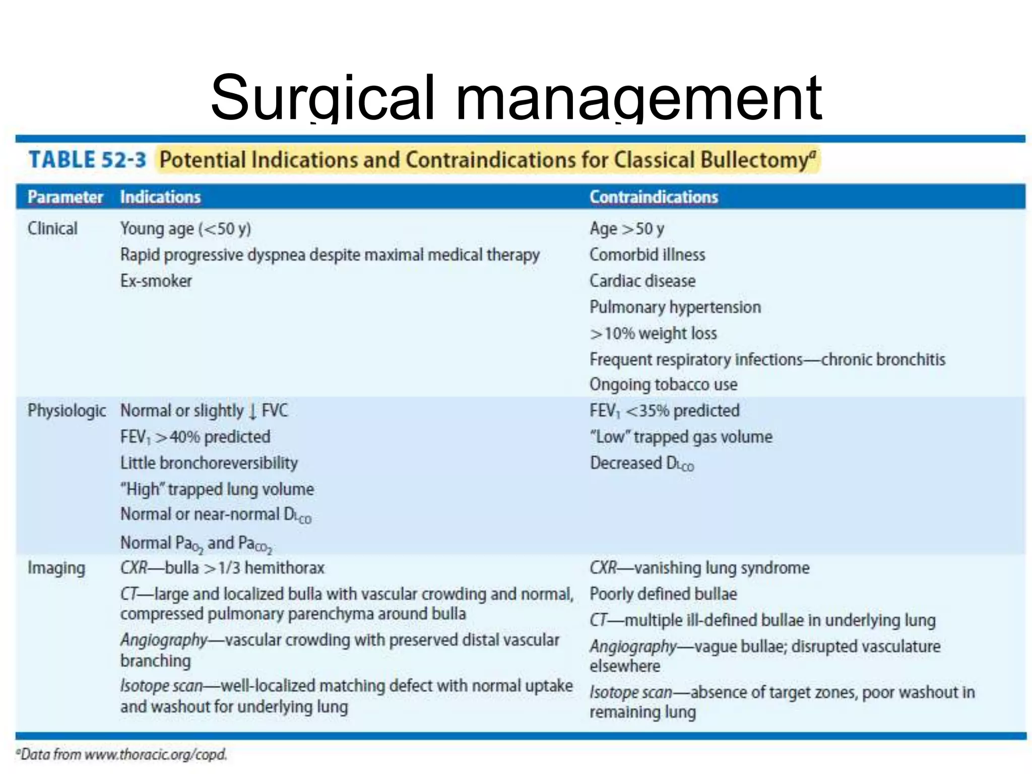
Approach to bullous lung disease
Dr. Abhishek Tandon
MD (Respiratory Medicine) Resident
Pt. B.D. Sharma PGIMS Rohtak
Introduction
• Bullous lung disease is characterized by the development of bullae within
the lung parenchyma.
• A bulla is an air-containing space within the lung parenchyma that arises
from destruction, dilatation, and confluence of airspaces distal to terminal
bronchioles and is larger than 1 cm in diameter and its walls are composed
of attenuated and compressed parenchyma.
• Bullae occur in various clinical contexts: (1) with emphysema (“bullous
emphysema”); (2) with pulmonary fibrosis, as in the late stages of
sarcoidosis or complicated pneumoconiosis; (3) in so-called “vanishing
lung,” in which the parenchyma is rapidly replaced by multiple bullae; and
(4) in lungs that are otherwise normal (“bullous lung disease”)
• Distinctions are drawn between bullae, blebs, and cysts
• A bleb is an accumulation of air between the two layers
of the visceral pleura that arises when the thin covering
of the bleb ruptures and permits entry of air
• Cysts are epithelial-lined cavities that may resemble
bullae on radiographs.
Etiology
• Bullae may originate in a variety of clinical and
pathogenetic set tings: (1) with emphysema of distal
acini; (2) in the setting of cigarette smoking; (3) in
conjunction with scar tissue formation, which “traps”
areas of normal lung, enlarges airspaces by traction on
surrounding intact alveoli, or produces retraction or
shrinkage of intact walls of adherent alveoli; (4) in the
setting of intravenous drug abuse; (5) as a result of
chronic inflammation and destructive changes in terminal
and first-order respiratory bronchioles, resulting in
airspace distention from delayed emptying; and (6) with
a1-antitrypsin deficiency.
Pathogenesis
• Several hypotheses have been proposed over the years for how
bullae develop, although none have been proved.
• These include (1) weakness of the alveolar walls predisposes to the
formation of bullae, particularly at the apices of the lungs, where
pleural pressures are most negative. This theory underscores the
proclivity of bullae for the upper lobes and stresses the influence of
mechanical forces acting upon flawed tissue. (2) Inflammatory
disease of a bronchiole leads to progressive air trapping and
“tension airspaces.” (3) Disordered collateral ventilation produces
the findings. (4) The same mechanisms responsible for generalized
emphysema are operative in the formation of bullae. (5) Underlying
paraseptal emphysema produces bullous disease.
• Of all the hypotheses, that of underlying paraseptal emphysema is
the most popular.
Classification
Evaluation and diagnosis
symptoms
Bullae cause symptoms via two distinct mechanisms:
•Bullae may interfere with ventilation of adjacent areas of
preserved lung, preventing them from expanding fully with
inspiration or, in severe cases, causing frank atelectasis.
•Bullae are space-occupying lesions that take up relatively
large volumes of the chest cavity without contributing to
functional gas exchange. Because they may enlarge with
exercise as a result of air-trapping, bullae may contribute to
dynamic hyperinflation, altering chest wall mechanics (e.g.,
diaphragm flattening and barrel chest) and increasing the
work of breathing.
• Sometimes asymptomatic and found only incidentally on
imaging.
• Typically, the symptoms are those of COPD:
 Shortness of breath or chest tightness, particularly with
exertion
 Cough
 Sputum production
 Occasionally, a sense of abdominal fullness or bloating,
usually associated with severe obstruction and
prominent air-trapping on pulmonary function testing
 Rarely chest pain due to air-trapping within a bulla,
causing distention of visceral pleura
• Symptoms are typically insidious,
• But if sudden and severe dyspnea or chest pain in a
patient with bullous lung disease suspect -
pneumothorax due to a ruptured bulla or bleb.
• Rarely, fever and malaise +/- increased sputum
production, may signal an infected bulla.
C-Xray
HRCT
CT provides valuable anatomic information
about the size, number, and relationships
of bullae, as well as crowding of adjacent
lung and disposition of the pulmonary
vasculature
• High resolution computed
tomography (HRCT) shows that
large bullae are frequently
associated not only with distal
acinar (paraseptal) emphysema,
but also with centriacinar
emphysema—the type of
emphysema usually associated
with cigarette smoking.
• Pulmonary function testing may be normal, particularly
in patients whose bullae are surrounded by normal lung
parenchyma.
• However, they usually demonstrate obstructive lung
disease. Common abnormalities include airflow
obstruction, hyperinflation (i.e., elevated total lung
capacity), air trapping (i.e., elevated residual volume),
and reduced diffusion capacity.
• Other signs of bullous lung disease may include
hypoxemia, particularly with exertion; hypercapnea; and
reduced exercise capacity.
On chest CT, giant bullae are predominantly located in the upper lobes
and are generally subpleural. However, in patients with a1-antitrypsin
deficiency, bullae are most commonly located at the lung base.
Approximately half of the patients have bilateral bullae and,
occasionally, deviation of the mediastinal structures to the contralateral
side may be noted.
CT has been used to create three-dimensional reconstructions of
bullae, which can then be used to calculate bullae volumes
Nuclear imaging
PFT
• Pulmonary function testing is not
diagnostic of bullous lung disease, but it is
critical in evaluating its functional
significance, guiding medical therapy, and
evaluating the likelihood of benefit from
surgical intervention
• Spirometry establishes the presence or absence of
airflow obstruction and reflects its severity. The forced
expiratory volume in 1 second (FEV1) is used to grade
severity, as well as selects appropriate candidates for
surgical bullectomy.
• Measurement of total lung capacity (TLC) and residual
volume (RV), is used to evaluate for hyperinflation and
air-trapping respectively. Because of the frequency of
severe air trapping in patients with bullous lung disease,
gas dilution methods may underestimate true lung
volumes. Consequently, plethysmography when it is
available is the preferred method for lung volume
measurement.
• A decreased diffusing capacity for carbon
monoxide (DLCO) can support the diagnosis of
emphysema or bullous disease, but is
nonspecific. The DLCO is also used to select
candidates for surgical treatment.
• Diagnostic procedures like bronchoscopy and lung
biopsies have essentially no role in the evaluation of
bullous lung disease.
• In surgical candidates, cardiac testing like
echocardiography, stress testing, and cardiac
catheterization may be considered as appropriate, in
evaluating for pulmonary arterial hypertension. In
addition, testing for coexistent IHD may be warranted.
These conditions may be contraindications for
bullectomy and lung volume reduction surgery.
Complication
• The major complications of bullous lung
disease are fluid accumulation(including
infection) in the bulla, spontaneous
pneumothorax, bronchogenic cancer,
chest pain, and hemoptysis.
Surgical management
Thankyou

Approach to Bullous lung disease

  • 1.
    Approach to bullouslung disease Dr. Abhishek Tandon MD (Respiratory Medicine) Resident Pt. B.D. Sharma PGIMS Rohtak
  • 2.
    Introduction • Bullous lungdisease is characterized by the development of bullae within the lung parenchyma. • A bulla is an air-containing space within the lung parenchyma that arises from destruction, dilatation, and confluence of airspaces distal to terminal bronchioles and is larger than 1 cm in diameter and its walls are composed of attenuated and compressed parenchyma. • Bullae occur in various clinical contexts: (1) with emphysema (“bullous emphysema”); (2) with pulmonary fibrosis, as in the late stages of sarcoidosis or complicated pneumoconiosis; (3) in so-called “vanishing lung,” in which the parenchyma is rapidly replaced by multiple bullae; and (4) in lungs that are otherwise normal (“bullous lung disease”)
  • 4.
    • Distinctions aredrawn between bullae, blebs, and cysts • A bleb is an accumulation of air between the two layers of the visceral pleura that arises when the thin covering of the bleb ruptures and permits entry of air • Cysts are epithelial-lined cavities that may resemble bullae on radiographs.
  • 7.
    Etiology • Bullae mayoriginate in a variety of clinical and pathogenetic set tings: (1) with emphysema of distal acini; (2) in the setting of cigarette smoking; (3) in conjunction with scar tissue formation, which “traps” areas of normal lung, enlarges airspaces by traction on surrounding intact alveoli, or produces retraction or shrinkage of intact walls of adherent alveoli; (4) in the setting of intravenous drug abuse; (5) as a result of chronic inflammation and destructive changes in terminal and first-order respiratory bronchioles, resulting in airspace distention from delayed emptying; and (6) with a1-antitrypsin deficiency.
  • 8.
    Pathogenesis • Several hypotheseshave been proposed over the years for how bullae develop, although none have been proved. • These include (1) weakness of the alveolar walls predisposes to the formation of bullae, particularly at the apices of the lungs, where pleural pressures are most negative. This theory underscores the proclivity of bullae for the upper lobes and stresses the influence of mechanical forces acting upon flawed tissue. (2) Inflammatory disease of a bronchiole leads to progressive air trapping and “tension airspaces.” (3) Disordered collateral ventilation produces the findings. (4) The same mechanisms responsible for generalized emphysema are operative in the formation of bullae. (5) Underlying paraseptal emphysema produces bullous disease. • Of all the hypotheses, that of underlying paraseptal emphysema is the most popular.
  • 9.
  • 10.
  • 11.
    symptoms Bullae cause symptomsvia two distinct mechanisms: •Bullae may interfere with ventilation of adjacent areas of preserved lung, preventing them from expanding fully with inspiration or, in severe cases, causing frank atelectasis. •Bullae are space-occupying lesions that take up relatively large volumes of the chest cavity without contributing to functional gas exchange. Because they may enlarge with exercise as a result of air-trapping, bullae may contribute to dynamic hyperinflation, altering chest wall mechanics (e.g., diaphragm flattening and barrel chest) and increasing the work of breathing.
  • 12.
    • Sometimes asymptomaticand found only incidentally on imaging. • Typically, the symptoms are those of COPD:  Shortness of breath or chest tightness, particularly with exertion  Cough  Sputum production  Occasionally, a sense of abdominal fullness or bloating, usually associated with severe obstruction and prominent air-trapping on pulmonary function testing  Rarely chest pain due to air-trapping within a bulla, causing distention of visceral pleura
  • 13.
    • Symptoms aretypically insidious, • But if sudden and severe dyspnea or chest pain in a patient with bullous lung disease suspect - pneumothorax due to a ruptured bulla or bleb. • Rarely, fever and malaise +/- increased sputum production, may signal an infected bulla.
  • 14.
  • 15.
    HRCT CT provides valuableanatomic information about the size, number, and relationships of bullae, as well as crowding of adjacent lung and disposition of the pulmonary vasculature • High resolution computed tomography (HRCT) shows that large bullae are frequently associated not only with distal acinar (paraseptal) emphysema, but also with centriacinar emphysema—the type of emphysema usually associated with cigarette smoking.
  • 16.
    • Pulmonary functiontesting may be normal, particularly in patients whose bullae are surrounded by normal lung parenchyma. • However, they usually demonstrate obstructive lung disease. Common abnormalities include airflow obstruction, hyperinflation (i.e., elevated total lung capacity), air trapping (i.e., elevated residual volume), and reduced diffusion capacity. • Other signs of bullous lung disease may include hypoxemia, particularly with exertion; hypercapnea; and reduced exercise capacity.
  • 17.
    On chest CT,giant bullae are predominantly located in the upper lobes and are generally subpleural. However, in patients with a1-antitrypsin deficiency, bullae are most commonly located at the lung base. Approximately half of the patients have bilateral bullae and, occasionally, deviation of the mediastinal structures to the contralateral side may be noted. CT has been used to create three-dimensional reconstructions of bullae, which can then be used to calculate bullae volumes
  • 20.
  • 21.
    PFT • Pulmonary functiontesting is not diagnostic of bullous lung disease, but it is critical in evaluating its functional significance, guiding medical therapy, and evaluating the likelihood of benefit from surgical intervention
  • 22.
    • Spirometry establishesthe presence or absence of airflow obstruction and reflects its severity. The forced expiratory volume in 1 second (FEV1) is used to grade severity, as well as selects appropriate candidates for surgical bullectomy. • Measurement of total lung capacity (TLC) and residual volume (RV), is used to evaluate for hyperinflation and air-trapping respectively. Because of the frequency of severe air trapping in patients with bullous lung disease, gas dilution methods may underestimate true lung volumes. Consequently, plethysmography when it is available is the preferred method for lung volume measurement.
  • 23.
    • A decreaseddiffusing capacity for carbon monoxide (DLCO) can support the diagnosis of emphysema or bullous disease, but is nonspecific. The DLCO is also used to select candidates for surgical treatment.
  • 24.
    • Diagnostic procedureslike bronchoscopy and lung biopsies have essentially no role in the evaluation of bullous lung disease. • In surgical candidates, cardiac testing like echocardiography, stress testing, and cardiac catheterization may be considered as appropriate, in evaluating for pulmonary arterial hypertension. In addition, testing for coexistent IHD may be warranted. These conditions may be contraindications for bullectomy and lung volume reduction surgery.
  • 25.
    Complication • The majorcomplications of bullous lung disease are fluid accumulation(including infection) in the bulla, spontaneous pneumothorax, bronchogenic cancer, chest pain, and hemoptysis.
  • 31.
  • 34.