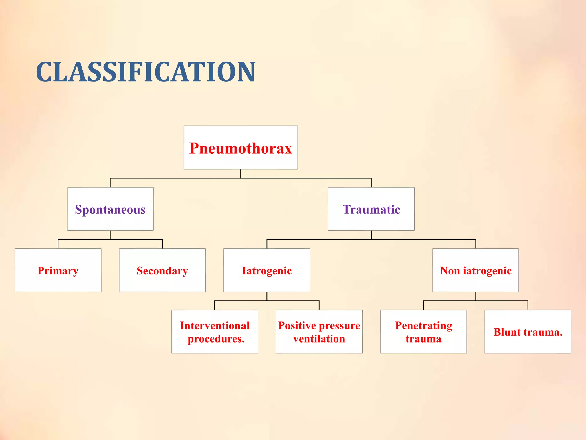
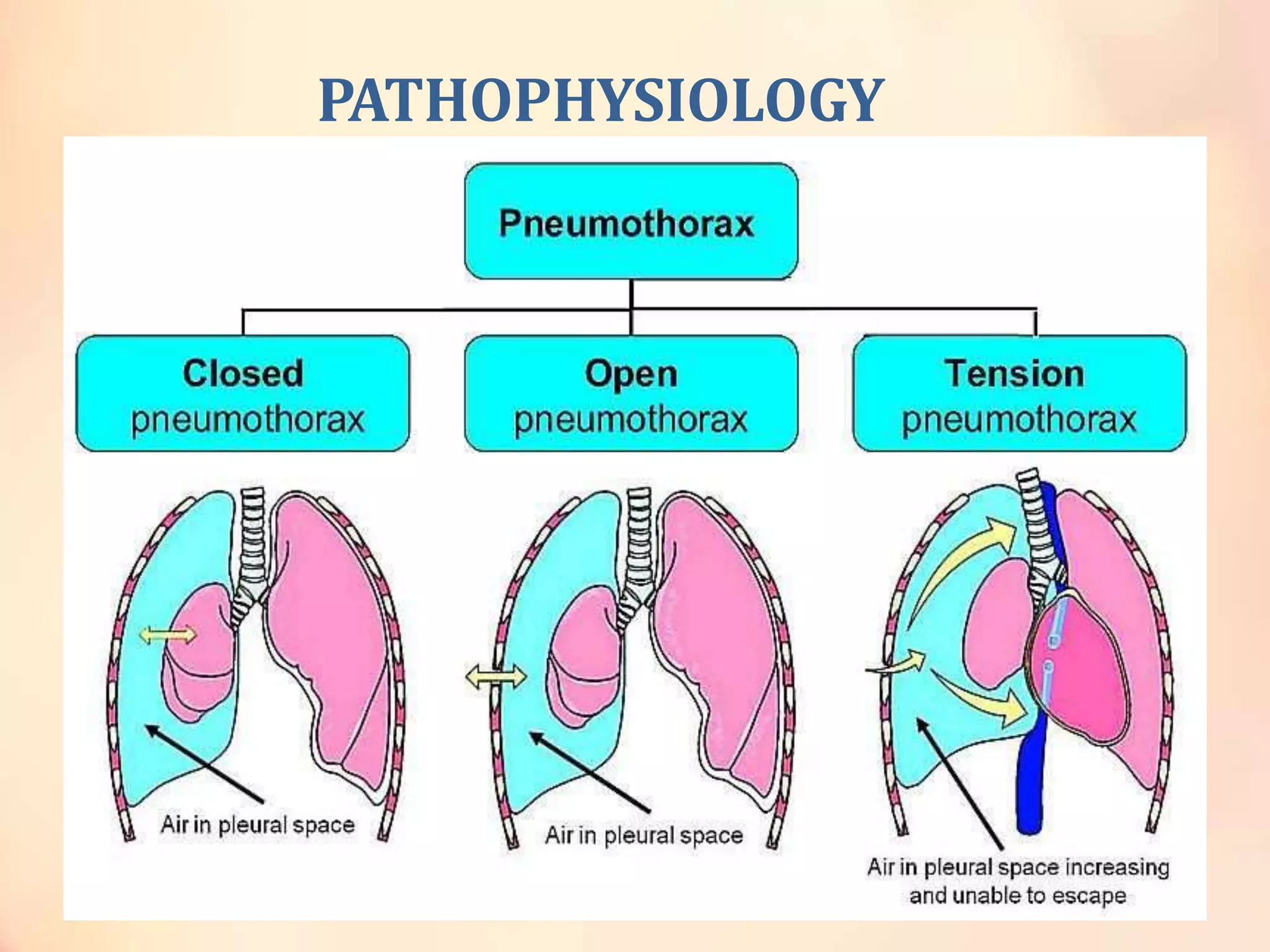
PNEUMOTHORAX
By
DR.P.MADHU HARSHA
2yr pg
RESPIRATORY MEDICINE
ASRAM MEDICAL COLLEGE
Madhu.harsha888@gmail.com
Definition
• collection of air or in the pleural space separating
the lung from the chest wall which may interfere
with normal breathing, causing the lungs to
collapse.”
“Intra” pleural
Space
There are 2 pleural membranes
• visceral pleura
• parietal pleura
The Parietal pleura lines the inside of the thoracic cavity.
The visceral Pleura adheres to the outside of the lung.
CLASSIFICATION
Pneumothorax
Spontaneous
Primary Secondary
Traumatic
Iatrogenic
Interventional
procedures.
Positive pressure
ventilation
Non iatrogenic
Penetrating
trauma
Blunt trauma.
PATHOPHYSIOLOGY
Closed
pneumothorax
Open
pneumothorax
Tension
pneumothorax
The pleural tear
Is sealed
The pleural tear
is open
The pleural tear
act as a ball &
valve mechanism
Ppl < Patm Ppl = Patm Ppl > Patm
PRIMARY SPONTANEOUS
PNEUMOTHORAX
POSSIBLE ETIOLOGY:
• Rupture of apical subpleural blebs.
• Emphysematous changes.
• Leakage of air through some areas rather than rupture
of blebs.
• Broad swings in atm pressure which leads to
distending pressure that leads to blebs rupture.
• Tall & thin individuals, gradient of pleural pressure is
greater from lung base to lung apex in taller
individuals.
• Genetic--- birt hogg dube synd. ( chromosome 17p
mutation in folliculin FLCN gene is responsible).
• Familial – autosomal dominant , individuals with HLA
haplotype A2 B40 were found to be much more to have
pneumothorax.
• Bronchial anamolies –acessory bronchus, missing
bronchus , disproportionate bronchial anatomy.
PATHOPHYSIOLOGY
• Eosinophilic pleuritis is directly related to air in
pleural space.
• Mild pulmonary vascular & perivascular eosinophilia.
• Pul art intimal fibrosis.
• Pul vein intimal fibrosis.
• Presence of abnormalities in pulmonary vessels.
CLINICAL FEATURES(SYMPTOMS)
• Peak age --- early 20s, rare age –40s.
• Chest pain, sob.
• Horners synd (rare)---traction of sympathetic
ganglion produced by shift of mediastenum.
• Pri spontaneous pneumothorax usually develops at
rest.
CLINICAL FEATURES(SIGNS)
• VITALS---normal, slight tachycardia, if >140bpm
tension pneumothorax should be suspected.
• INSPECTION :side of chest with pneumothorax is
larger than normal side.
• PALPATION : tactile fermitus is absent.
• PERCUSSION : HYPERRESONANT NOTE +
• AUSCULITATION : Absent BS , vocal fermitus is absent.
• RT pneumothorax– lower edge of liver is shifted
inferiorly.
ECG CHANGES OF (LEFT SIDED
PNEUMOTHORAX)
• Decresed QRS amplitude.
• RighT sided axis deviation.
• Dimintion of precordial (chest leads v1-v6 ) R voltage.
• Precordial (v1-v6) T wave inverson.
• V2-V6 amplitude decreses
ECG CHANGES OF (RIGHT SIDED
PNEUMOTHORAX)
• Prominent R waves in lead V2.
• Loss of S wave voltage.
• Incresed amplitude in V5-V6.
• PR segments elevation in inferior leads.
• PR segment depression in AVR
DIAGNOSIS
• Demonstration of visceral pleural line on CXR.
• EXPIRATORY film: if there is strong suspition pf
pneumothorax.
• The pleural fluid with pneumothorax- Eosinophilia is
present ( eosinophils exceed 20% after 1 day and 60%
after 7 days)
QUANTIFICATION – 5 methods
• It is done by the following methords:
Lights index
Collins methord.
Rheas methord.
ACCP defenition
BTS defenition
• Collins & rheas methords are recommended.
Lights index
Collins method
• % of pneumothorax = 4.2 + (4.7* ( A + B + C ) ).
• A = distance between partially collapsed lung apex to
the apex of thoracic cavity.
• B = mid points of upper half of collapsed lung.
• C = mid points of lower half of collapsed lung.
RHEA METHOD
• 10% Ppneumothorax for every centimeter of
intrapleural distance.
• This is according to a normogram.
BRITISH THORACIC SOCIETY
defenition for small pneumothorax.
• A pneumothorax is considered small if the rim of air
between the pleura & chestwall at the level of hilum
was less than 2cm.
• Large pneumothorax if its > 2cm
AMERICAN COLLEGE OF CHEST
PHYSICIANS defenition of small
pneumothorax
• A small pneumothorax is the one with the apex to
cupola distance is < 3cm.
• A large pneumothorax if its > 3cm.
Visceral pleural line
Large pneumothorax with
mediastinal shift
Treatment strategies
• There are two basic strategies in treatment of
pneumothorax.
Decrese the likelyhood of recurence.
To get rid of air in pleural space.
Different strategies in management are as
follows
• OBSERVATION.
• SUPLEMENTAL OXYGEN.
• ASPIRATION.
• TUBE THORACOSTOMY.
• INSTILLLING SCLEROSING AGENT (medical
thoracoscopy)
• AUTOLOGUS BLOOD PATCH ( for persistant air leak).
• VATS (video assisted thoracoscopic surgery)
OBSERVATION
• Rate of spontaneous resolution is very low.
• 1.25% of pneumothorax is absorbed per 24hrs.
RECOMENDATION:
 Only patients pneumothorax occupying <15% of
hemithorax should be conducted for this type of
treatment.
SUPPLEMENTAL OXYGEN
• This increases the rate of observation.
• This is done through high flow oxygen through face
mask.
ASPIRATION
• 16 gauze needle is inserted into anterior 2nd ICS at mid
clavicular line after local anesthesia.
• A 3 way stopcock & 60ml syringe are attached to the
catheter.
• Air is manually withdrawn until no more can be aspirated.
• After no more air can be aspirated , the stopcock is closed
and catheter is secured.
• After observation of 4 hrs, a cxr is ordered. If adequate
expansion occurs then pt can be discharged.
• If there is no lung expansion even after 4 liters of air, then
tube thoracostomy should be performed.
• If lung is not expanded in estimated time then VATS can be
performed.
NEEDLE CHEST
DECOMPRESSION
14-gauge or 16-gauge needle with a catheter in the second
intercostal space at the midclavicular line, 1-2 cm from
the sternal edge
TUBE THORACOSTOMY
• Small tubes of 7-14 f should be tried initially because it
creates less trauma.
• It is best inserted using guide-wire technique.
• If lung doesn’t re-expand with smaller tube, then larger tube
can be inserted.
RECOMMENDED:
• no suction should be applied because it creates
reexpansion pul edema.
• Tube should not be removed early because it has likely to
get recurrent.
• If recurrent airleak is present then tube shouldnot be
clamped because it creates tension pneumothorax.
MEDICAL THORACOSCOPY &
INSTILLATION OF SCLEROSING AGENTS
• 50% of primary spontaneous pneumothorax can re-
occur.
• Sclerosing agents used :
Quinacrine.
Olive oil.
Talc slurry.
Tetracycline derivatives.( 500mg doxycycline , 300mg
minocycline)
Talc slurry
• 5-10g of talc is mixed in 250ml NS and given
intrapleurally.
• Recurence rate is < 10%.
• Primary drawback is, a small population develops
ARDS.
TETRACYCLINE DERIVATIVES
• They should be given as soon as lung has been
expanded.
• Pt should be positioned so that tetracyclines come in
contact with apical pleura.
Care to be taken:
• Adminstration of NSAIDS as diclofenac 2mg/kg bw
decreses effectiveness of pleurodesis from mechanical
abrasion.
• Minimise the use of corticosteroids & anti-inflamatory
agents.
• Instillation of sclerosing agents cause severe chronic
debilating pain.
• RECOMMENDED:
All patients with pri/sec spont pneumothorax who are
treated with tube thoracostomy receive an agent unless
they are subjected to thoracoscopy/thoracotomy.
AUTOLOGUS BLOOD PATCH
• 50-100ml of blood is drawn from vein & then promptly
injected without anticoagulation through the chest
tube into pleural space.
• Chest tube shouldn’t be clamped, because with this
airleak, this could lead to tension pneumothorax.
INTRAPLEURAL FIBRIN GLUE FOR
PERSISTANT AIR LEAK
• Diluted regular fibrin glue of 60ml-120ml is injected
into chest tube of pts with persistant BPF.
VATS ( video assisted thoracoscopic
surgery)
• Effective in treatment of spotaneous pneumothorax &
prevention of recurrent pneumothorax.
• OBJECTIVES:
To treat bullous lung disease responsible for
pneumothorax.
To create pleurodesis.
SECONDARY SPONTANEOUS
PNEUMOTHORAX
ETIOLOGY
• COPD (90%)
• AIDS
• LYMPHANGIO LEIOMYOMATOSIS (LAM)
• CYSTIC FIBROSIS
• TB
• SARCOIDOSIS.
• TUMOURS
CLINICAL FEATURES
• Symptoms are more severe than pri spontaneous pneumothorax.
• SOB
• Wheeze.
• Chest pain on side of pneumothorax.
• Cyanosis
• Hypotension
• Type 1 or type2 respiratory failure in some cases.
• Physical examination in spontaneous pneumothorax.(less
helpful)--- hyper expanded lungs with decresed Tactile Fermitus,
hyperresonant note with decresed breath sounds.
DIAGNOSIS
• CXR—
 Copd - loss of elastic recoil of lung , presence of air
trapping.
 normal areas of lung collapse more completely than
diseased areas with bullae & emphysema.
 Deflation of diseased lung- limited by its elastic recoil.
Ultrasound chest
• PLEURAL GLIDING – absent in pneumothorax.
• But in some cases of COPD also this is absent.
• Demonstration of visceral pleural line.
Pleural line with pneumothorax is usually oriented in
convex fashion towards the lateral chest wall.
Pleural line with large bullae is usually concave
towards the lateral chest wall.
CT- CHEST
• To differentiate bullae with pneumothorax.
• If pt with cystic lung disease with increse in sob then a
possibility of pneumothorax should be considered
particularly if hemithoraces are asymetrical in size.
• NOTE: when a person has totally collapsed lung, one
should search for air broncogram in the lung. Air
broncogram is absent when there is an obstructing
endobronchial lesion. If air broncogram is absent , do
broncoscopy before ICD insertion.
TREATMENT
• All patients should be managed by tube thoracostomy.
• Aspiration of air is not recommended because its less sucessful & doesn’t prevent any
recurence.
• Pig-tail catheters can be used for treatment of spont pneumothorax.
• Abg improves within a day after ICD insertion.
• Tube thoracostomy is less efficacioous in treating sec spont pneumothorax. Because
primary spont pnx usually expands within 3 days.
• In copd mean time for lung expansion is 5 days. In 20% cases it takes 7 days.
• Once the lung is expanded, we should attempt to prevent the recurrence of
pneumothorax by VATS (preferred) or medical thoracoscopy with sclerosing agent
instilation.
• Stapling of blebs & pleural abrasion reduces the likelyhood of recurrence to < 5%.
• If patient have diseases such as LAM, CF, IPF, COPD. Thoat might me managed with lung
transplantation.
• If the lung doesn’t expand with in 72 hours if there is a
persistant air leak for >3 days , strong considerations
should be given to performing VATS/ medical thoracoscopy.
• At thoracoscopy, the blebs are excised with stapling
instrument & some other procedure is sone to create a
pleurodesis.
• RECOMMENDED: all sec spont pnx to undergo VATS.
• Blood patch technique is sucessful in treating in aroud 50%
of the pts.
• If persistant air leak is present, endobronchial procedures
can be performed with around 50% cases. The procedures
done are Baloon closure, Fibrin & autologus blood patch,
endobronchial valve.
SPECIAL TYPES OF PNEUMOTHORAX
• Secondary to AIDS
• LAM ( lymphangio leomyomatosis)
• CYSTIC FIBROSIS
• TUBERCULOSIS
• LANGERHAM CELL HISTOCYTOSIS.
• CATAMENIAL PNEUMOTHORAX.
• NEONATAL PNEUMOTHORAX.
• TRAUMATIC PNEUMOTHORAX ( non iatrogenic).
• Secondary to drug abuse.
• PNEUMOTHORAX EX VACUO.
• TENSION PNEUMOTHORAX.
Secondary to AIDS
• Most patients with AIDS who have spontaneous
pneumothorax have a history of pneumocystis
jirovecci infection are on prophylactic pentamidine
and have a recurrence of p.jirovecci infection, ptb, pul
cryptococosis are associated with spont
pneumothorax in pts with AIDS.
• Most pts have CD4 counts < 100 cells/mm3.
RECURRENT PNEUMOTHORAX
• Bullous lung disease.
• Catamenial pneumothorax.
• LAM ( lymphangioleomyomatosis)
• Langerham cell histocytosis.
etiology
• Bacterial pneumonia (mc)- pts with CD4 counts >200
• P.jirovecii- pts with CD4 < 200
• In p.jirovecii the high incidence of pneumothorax are due to
multiple subpleural cavities which are associated with subpleural
necrosis.
• The bullous changes & pulmonary cysts occur due to repeated
episodes of inflamation & cytotoxic effects of HIV on pulmonary
macrophages.
• Most pts have radiological evidence of fibrocystic parenchymal
disease .if these pts are subjected to surgery, there is diffuse
involvement of upper lobes than lower lobes. Areas of necrosis
are usually present in consolidated areas of lung and these areas
are extremely friable on touch.
• Pts with less diffusion capacity are more likely to
develop spontaneous pneumothorax.
• Aerosoled pentamidine doesn’t reach the periphery of
upper lobes.accordingly a lowgrade infection persists
and destroys the lung leading to the development of
cysts & broncopleural fistula.
• Once the pt with AIDS & p.jirovecci infection has
spontaneous pneumothorax, he / she is likely to have
recurrent pneumothorax or a contralateral
pneumothorax.
• .
Treatment
• The necrotic lung surrounding the ruptured cavity, the
spontaneous pneumothorax associated with AIDS & p.jirivecci is
difficult to treat.
• In view of poor results with only ICD tube, alternate therapies are
to be considered. The simplest alternative is placement of
heimelich valve to the chest tube & dischage the pt.
• If the pt doesn’t get managed through hemilich valve, then VATS
should be considered.
• Alternate therapies are : sclerotherapy, thoracotomy, pleurodesis.
• Pleural sclerosis has limited role. It depends on the size of BPF.
• If pleurodesis is planned, DOXYCYCLINE is recommended.
CYSTIC FIBROSIS
• 3.5% Pts have spontaneous pnx.
• 75% cystic fibrosis will have an FEV1<40%
• Occurs more frequently with pts of severe respiratory
impairment.
treatment
• Recurence is 50%
• Primary line of management is tube thoracostomy unless
pneumothorax is small.
• If air leak ceases & lung expands then do thoracoscopy or
instillation of sclerosing agent.
• But CF pulmonary guidelines doesn’t approve anyof the
above because CF pts are candidates for lung
transplantation.
• VATS with stapling of blebs & pleural abrasion.
• Thoracoscopy should also be performed if air leak persists
or lung doesn’t expand after 3 days also.
SECONDARY TO TUBERCULOSIS
• Prevelance 1- 3 %
• All pts with pneumothorax should be treted with
thoracostomy.
• If persistant air leak is present, then SURGERY.
• VATS & thoracotomy ---if recurrent pneumothorax &
persisted airleak > 10 days.
• An atypical segmentectomy is another option but with
high recurrence.
LYMPHANGIOLEIOMYOMATOSIS
• It is a rare condition charecterised by peribronchial,
perivascular & perilymphatic proliferation of
abnormal smooth muscle cells.
• Women of child bearing age.
• Slow progressive sob, chylothorax, recurrent
spontaneous pneumothorax or hemoptysis.
• As the recurence rates are very high chemical
pleurodesis or surgery is recommended after first
pneumothorax.
LANGERHAM CELLS HISTOCYTOSIS
• It is a rare form of ILD that results from specific types
of histocytic cells known as langerham cells in the
lung.
• Treatment
i. Icd tube
ii. Sclerosant
iii. VATS
iv. Pleural abalation.
CATAMENIAL PNEUMOTHORAX
• Occurs in conjunction with menses. Before & within
72 hrs of menses.
• Usually recurrent. (30%
• Mean age 34yrs.
• Classically develop chest pain & sob after 24-48 hrs
after onset of menses.
• Recurrent thoracic/ scapular pain.
• Mental or physical stress usually right sided but left
sided & even bilateral pneumothoraces have been
reported.
pathogenesis
• DIAPHRAGMATIC DEFECT: air gained acess from
peritoneal cavity into pleural cavity through the defect.
• ENDOMETRIOSIS: pleural or diaphragmatic.
• Leakage of air from the lung owing to subpleural
endometrial implants.
DIAGNOSIS & TREATMENT
• Diagnosis is considered if a female of 20 yrs age
develops sypmptoms at menses.
• MEDICAL TREATMENT : Gonadotropin releasing
hormone antagonists that supresses ectopic
endometrium.
• SURGICAL TREATMENT: closure of any diaphragmatic
defects , stapling of blebs & pleural abrasions.
• Coverage of diaphragmatic surface by polyglactin
mesh even when the diaphragm appears normal.
• Bilateral tubal ligations done in some cases prevented
recurrences
NEONATAL PNEUMOTHORAX
• Newborn period
• Present shortly after breath in 1-2% individuals
• Male > females
• Baby has a h/o fetal distress requiring resussitation or
a difficult delivery with evidence of aspiration of
meconieum, blood or mucus.
• Inidence of pneumothorax in infants with resp distress
syndrome is high.
pathogenesis
• Mechanical problems of first expanding the lung.
• Transpulmonary pressure averaging 40cm of h2o
during the first few breaths of life, with occasional
pressures of 100cm of h2o.
• At birth, alveoli open in a rapid sequence, but if
bronchial obstruction occurs from aspiration of blood
or meconeum or mucus, high transpulmonary
pressures may lead to rupture of the lung.
• NOTE : Transpulmonary pressure of 60 cm h2o
ruptures adult lungs.
Clinical features
• None to severe respiratory distress
• Small pnx----no symptoms or mild apnoec spells or
restlessness.
• Large pnx : varying degree of resp distress,
tachypnoea(upto120/min) , grunting, retraction,
cyanosis.
Detection of pneumothorax
• Difficult because of abnormal physical signs.
• Breath sounds are widely transmitted in small
neonatal thorax so its difficult to localize.
• Shift of apical heart impulse away from side of
pneumothorax.
• Infant who develops hypotension as a result of
pneumothorax is at a high risk of having intra
ventricular hemorrhage, because the hypotension
results in cerebral infarction with the intraventricular
hemorrhage occuring after the systemic bp has raised
to normal levels
diagnosis
• Transillumination of chest wall with high intensity
transilluminating light is rapid, accurate & easy way to
make a diagnosis.
• Cxr is important to differentiate between
pneumomediastenum, hyaline membrane disease
congenital cyst, aspiration pneumonia, lobar
emphysema, diaphragmatic hernia.
treatment
• Observation if no symptoms.
• Close observation is necessary because it may lead to tension
pneumothorax.
• Supplemental oxygen speeds up the absorbtion.
• Thoracentesis: icd at anterior axillary line 4-5cm inferior to
nipple in 5th or 6th ics, because other sites placement may caise
breast abnormalities in adult ages.
• Tube thoracostomy should always be done in infants with rds &
pneumothorax because of already poor ventilatory status.
• Usually the air leak is small. Intermittent positive pressure
ventilation can maintain adequate gaseous exchange.
• If air leak is large , then high frequency ventilation maybe the only
methord by which adequate gas exchange can be maintained
IATROGENIC PNEUMOTHORAX
• These are due to any invasive procedures.
• LEADING CAUSES :
i. Trans thoracic needle aspiration (22%)
ii. Subclavian needle stick (22%)
iii. Thoracentesis (19%)
iv. Transbronchial biopsy (10%)
v. Pleural biopsy (9%)
vi. Positive pressure ventilation (7%)
vii. Supra clavicular needle stick (5%)
viii. Nerve block (3%)
ix. miscellaneous (1%)
Clinical features
• It depends on the condition of the patient & on the
initiating procedure.
• If the pt is on mechanicall ventilation:
Vc mode ---peak & plateau pressure increses. Pressure
support----tidal volume decreses
• If pt is on cardiopulmonary resuscitation, its more of
harder side to treat.
• If pt develops pneumothorax while during or after
thoracentesis, pleural biopsy, percutaneous lung
aspiration, there may be no symptoms pertaining to
pneumothorax also.
diagnosis
• The presence of mediastinal emphysema serves as an indicator to
look closely for a pneuomothorax.
i. Anteromedial 38%
ii. Subpulmonic 26%
iii. Apicolateral 22%
iv. Posteromedial 11%
• The above are the frequency of most common site to least
common site in supine radiographs.
• The occurrence of iatrogenic pneumothorax should be
suspected in pts whose sob incresed after the procedure.
• Diagnosis is confirmed by CXR & USG CHEST.
treatment
• When pneumothorax occurs during positive pressure
ventilation, tube thoracostomy should be performed
immediatley or else it causes tension pneumothorax.
• Chest tube should be left in place for at least 48 hours
after the air leak stops, if the patient continues to
receive mech vent.
• <40% hemithorax pneumo-------OBSERVATION.
• >40% Hemithorax & incresing---- aspiration of air.
• Patients are more likely to require a chest tube if thy
have COPD.
TRAUMATIC PNEUMOTHORAX (non-
iatrogenic)
• It can result either from penetrating (any penetrating
object) or non penetrating trauma( rib #).
• Mechanism:
• With sudden chest compression, the alveolar pressure increses
& this may cause alveolar rupture.
diagnosis
• Chest xray : not that sensitive in diagnosis.
• CT scan : pneumothoraces seen only on CT are called
occult pneumothoraces.
• USG CHEST : absence of pleural lung sliding, absence
of comet tail artifact.
treatment
• Most are treated by tube thoracostomy.
• Size of tubes : 10-14f .
• If hemopneumothorax is present, one tube should be placed
superiorly to evacuate the air and another is placed
inferiorly to evacuate blood.
• Usually lung expands within 72hrs, if not VATS should be
planned.
• Some reports dis on pts with persistant air leak or un
expaned lung post 72hrs trauma, they were posted for
thoracoscopy for inspecting the persistant air leak, if there
is no obvious air leak, then 250ml saline is instilled with
slight lung ventilation to see the air leak.once the
identification of leak is done, surgical sclerosant is applied.
• Tube thoracostomy maynot be necessary for small
pneumothoraces.
• Wolfman classified occult pneumothorax as
i. Minuscle (<1cm in greatest anteropost thickness &
seen not more than 4 consiguos ct.)
ii. Anterior (>1cm but not extending beyond
midcoronal line.)
iii. Anterolateral ( extending posteriorly beyond mid
coronal line)
• Minuscle & anterior receive tube thoracostomy.
• These two uncommon diagnostic possibilities lead to
emergency operations.
1. Fracture of trachea & or a main bronchus.
2. Traumatic rupture of esophagus.
• Bronchial rupture should be suspected in pts having
persistant air leak with subcutaneous emphysema,
pneumomediastinum, deep cervical emphysema,
hemoptysis, or rib or clavicular #.
• The possibility of bronchial rupture should be asses by
FOB.
• Rx is surgical repair.
• Traumatic rupture of esophagus usually produces
hydropneumothorax, therefore if a patient is having
traumatic pneumothorax with pleural effusion, the
serum amylase levels should also be considered.
• If amylase levels are elevated then contrast studies of
esophagus should be considered.
Traumatic pneumothorax secondary
to drug abuse.
• Attempt to inject drugs through internal jugular vein
or subclavian vein.
• Treatment is by insertion of chest drain.
PNEUMOTHORAX EX VACUO
• It is secondary to acute bronchial obstruction.
• Recently its said that it’s the development of pneumothorax after
a thoracentesis because the lung is unable to reexpand & fill the
pleural space.
• Acute collapse of the lung results in negative intrapleural pressure
which leads to the accumulation of gas that originated in the
ambient tissues and blood in pleural spaces.
• Inorder to come out of the tissues the pleural pressure would
have to be -60cm/h2o.
• The pneumothoraces developed from transient parenchymal-
pleural fistulae caused by non uniform stress distribution over
visceral pleura that develop during large volume drainage if lung
cannot conform to the shape of thoracic cavity.
TENSION PNEUMOTHORAX
• IT OCCURS WHEN THE INTRA PLEURAL PRESSURE
EXCEEDS THE ATMOSPHERIC PRESSURE.
ETIOLOGY
• Pts receiving positive pressure ventilation by
mechanical ventilation or during resusitation (most).
• It develops due to a one way valve mechanism in
which the valve is open during inspiration & closed
during expiration.
pathophysiology
• The development of tension pneumothorax is followed
by sudden deterioration of cardiopulmonary status of
the patient.
• The main disasterous effect of tension pneumothorax
in patients appears to be decrease of cardiac output &
decrease in Pao2
Clinical manifestations (symptoms)
• It is much more frequent in patients who develop
pneumothorax while receiving mechanical ventilation
or during cardiopulmonary resuscitation.
• Pt appears distressed.
• Increased resp rate(tachypnoea)
• Cyanosis
• Profuse sweating
• Hypotension
• tachycardia
signs
• ABG shows marked hypoxemia & sometimes
respiratory acidosis.
• Involved hemithorax is larger than unaffected side.
• Rib spaces are widened.
• Trachea is shifted to opposite side.
Tension Pneumothorax
diagnosis
• It should be suspected in patients whose condition
suddenly deterirates , who are receiving mechanical
ventilation & who have undergone a procedure that
may cause pneumothorax.
• It occurred during hyperbaric o2 therapy as treatment
for CO poisoning.
• Even a malpositioned chest tube can also cause
tension pneumothorax.
Treatment
• It is a medical emergency.
• High concentration of supplemental o2 should be
given to combat hypoxia.
• 16gauze needle can be placed at 2nd ICS anteriorly at
mid clavicular line.
• ICD insertion is a must to treat the tension
pneumothorax.
NEEDLE CHEST
DECOMPRESSION
14-gauge or 16-gauge needle with a catheter in the second
intercostal space at the midclavicular line, 1-2 cm from
the sternal edge
Needle is
inserted
through lower
part of 2nd
intercostal
space
Bronco-pleural fistulas & alveo-pleural
fistulas.
• Bronco –Pleural fistula : communication between a
mainstem lobar or segmental bronchi & pleural space.
• Theese occur almost exclusively after pneumonectomy,
lobectomy or segmentectomy & always require
surgical management.
• Alveo-pleueal fistula : communication between
pulmonary parenchyma distal to segmental bronchus
& pleural space.
• These rarely require surgery.
AIR LEAKS classification
• Four types are there.
i. Continuous type : the air leak is persistant throughout
the entire respiratory cycle. There is continuous
bubbling. Present in pts with mechanical ventilation &
having large bpf
ii. Inspiratory type : present only during inspiration. With
big apf and small bpf.
iii. Expiratory type : present only during expiration.
Commonly seen after pulmonary surgery, its presence
denotes APF.
iv. Forced expiratory type : present only when patient
coughs or forced expiration.
AIRFIX DEVICE
• It gives the digital readouts of the leakage per breath
per minute.
• They reported that if the leakage volume was less than
1ml/ breath or 20ml/minute, the chesttube can be
removed with no recurence of pneumothorax.
Air leaks & mechanical ventilation
• Difficult to treat.
• Hypoxia rather than hypercapnoea is the main threat to the
patient because the air that exists through the chest tube
has a co2 level comparable to mixed expired air.
• The air that exists through the chest tube is effective in
removing co2 from the patient.
• One approach of decreasing the flow of bpf is placing the
patient in high frequency jet ventilator. Althogh this type of
ventilation decrease the flow through the fistula & improve
gas exchange in experimental animals therfore NOT
RECOMMENDED.
• Multiple agents & devices like silver nitrate, gelfoam,
cyanoacrylate based agents & fibrin agents have been
passed through broncoscope to stop the air leaks.
• The cyanoacrylate agents have been recently improved
with an additive that slows down the drying time to
permit greater tme for modeling of the agent into
fistula site.
• Tetracycline derivatives, talc & diluted fibrin glue have
also been injected into pleural space in an attempt to
treat the air leak.
Post operative BPF & APF
• After pulmonary resections of lesser magnitude than a
pneumonectomy, there is frequently an air leak from a
residual raw parenchymal surface.
• If the lung has expanded & pleural space is obliterated,
then the leak usually stops in 2 / 3 days.
• The persistance of air leak > 7 days is considered as a
prolonged air leak.
• The management of chest tubes post operatively also
influences the duration of air leaks. ( some studies
demonstrated that when chest tubes are managed with
water seal, the air leak sealed sooner than when thy
managed with suction.
• Patients who has large air leaks on POD1 were more
likely to have prolonged air leaks.
• Flutter valves are more effective in management of BPF
than water seal systems for the management of post
operative air leaks.
• BPF is observed in approx 4% of pts after a
pneumonectomy > lobectomy > segmentectomy.
• BPF is more common after resections for inflamatory
diseases of the lung especially in pts with active tb &
positive sputum cultures.
• After pulmonary surgery, bpf may develop immediately or
weeks or months later.
• 1-6 days after surgery : poor closure of bronchial stump.
After surgery, the early fistula is massive & persistant, the
pt frequently develops massive subcutaneous emphysema&
exibit various degrees of respiratory insufficiency.
• 7-10 days after surgery : failure of healing because of
inadequate viable tissue coverage of the stump or as a
result of infection of the fluid within the space & rupture of
empyema through the suture line of the bronchial stump. At
this stage the pt coughs up variable quantities of
serosanguinous frothy fluid from resp tract. The pt should
be placed with affected side down to reduce flooding of
remaining lung
• > 2 weeks after surgery : rupture of frank empyema through
the bronchial stump or sometimes due to the failure of healing
of the stump.
• If bpf occurs in early post op period, it can be managed by
reoperation & repair of bronchial stump.
• Bronchial stump is covered with transposed muscle flap,
pericardial pad of fat, omental pedicle flap.
• If primary repair is unsucessful, the pt should be treated with a
chest tube.
• When the pt had undergone lessthan pneumonectomy, a
fogarty baloon catheter is passed throught the working channel
of broncoscope & occlusion of segments on the side of air leak
is undertaken.
• Materials placed in appropriate bronchus to close the fistula
are, gelfoam, doxycyclin ,blood, fibrin glue, vascular occlusion
coils, self expandable stents, endobronchial one way valves.
• BPF that occur late after surgery are almost always associated
with empyema.
SPECIAL DEVICES
• Heimlich valve. ( to prevent tension pneumothorax in
a patient with icd tube)
• ATRIUM ( dry suction device )
Heimlich valve
• It is a mechanical one way valve.
• It helps the air to escape from the pleura.
• Prevents the air from atmosphere to re enter the
pleural cavity.
• Advantages:
I. Easy ambulation of patient.
II. Doesn’t require water to operate.
ATRIUM ( dry suction kit )
AUTOLOGUS BLOOD PATCH vs TALC
vs TETRACYCLINE PLEURODESIS
• Reference NCBI
• LINK :
https://www.ncbi.nlm.nih.gov/pmc/articles/PMC280
1042/
• TETRACYCLINE : produces inflamatory reaction,
scarring, no patch effect.
• TALC : interleukin mediated neutrophils migration &
monocyte infiltration with inflamation. No patch effect.
• BLOOD : fibronogenic activity, inflamation & irritation
of pleura. Patch effect is present.
leaks closure TIME
• TETRACYCLINES : 3- 5 days.
• BLOOD : 1ST 12 hours there is 72% closure.
• next 48 hours , All leaks are closed.
SUCCESS RATES
• TALC SLURRY : 87.2 %
• AUTOLOGUS BLOOD : 75 %
• TETRACYCLINES : 63.6%
FAILURE RATES
• TETRACYCLINES : 36.4%
• AUTOLOGUS BLOOD : 25%
• TALC SLURRY : 15.8%
COMPLICATIONS
• TALC SLURRY :
• TETRACYCLINES :
Both cause pleural thickening, diffuse fibrosis, decreased
lung function(Restrictive lung diseases).
They may also cause chest pain, fever, SVT.
Tetracycline derivatives cause severe pain so they are
usually instilled with lidocaine.
Rate of recurrence at ipsilateral side was reported as 50%
higher in tetracyclines.
EMPYEMA : tetracyclines may cause it (usually on day
15)
AUTOLOGUS BLOOD
• PROCEDURE 1 : 50-250ml autologus blood is injected
repeated until success.
• PROCEDURE 2 : A single 50cc autologus blood is
injected via ICD tube.
• Not > 50ml should be injected , as blood if injected not
under asceptic conditions it can be a good culture
medium for bacterial growth, later cause EPMPYEMA
COMPLICATIONS OF AUTOLOGUS
BLOOD PLEURODESIS
• Fever
• Effusion
• Empyema (9th day )
Analysis of empyema revealed staphylococus
epidermidis.
Obstruction of catheter also may occur , therfore
flushing with sterile NS may be necessary.
CONCLUSION
• AUTOLOGUS BLOOD IS :
Painless.
Inexpensive.
Simple methord.
Faster cessation of air leaks.
Fewer side effects.
 WHY NEGLECTED THEN ??? Not many case reports published on
this mode of pleurodesis.
 pneumothorax BY DR.P.MADHU HARSHA

pneumothorax BY DR.P.MADHU HARSHA

  • 1.
    PNEUMOTHORAX By DR.P.MADHU HARSHA 2yr pg RESPIRATORYMEDICINE ASRAM MEDICAL COLLEGE Madhu.harsha888@gmail.com
  • 2.
    Definition • collection ofair or in the pleural space separating the lung from the chest wall which may interfere with normal breathing, causing the lungs to collapse.”
  • 3.
    “Intra” pleural Space There are2 pleural membranes • visceral pleura • parietal pleura The Parietal pleura lines the inside of the thoracic cavity. The visceral Pleura adheres to the outside of the lung.
  • 4.
  • 5.
  • 6.
    Closed pneumothorax Open pneumothorax Tension pneumothorax The pleural tear Issealed The pleural tear is open The pleural tear act as a ball & valve mechanism Ppl < Patm Ppl = Patm Ppl > Patm
  • 7.
  • 8.
    POSSIBLE ETIOLOGY: • Ruptureof apical subpleural blebs. • Emphysematous changes. • Leakage of air through some areas rather than rupture of blebs. • Broad swings in atm pressure which leads to distending pressure that leads to blebs rupture. • Tall & thin individuals, gradient of pleural pressure is greater from lung base to lung apex in taller individuals.
  • 9.
    • Genetic--- birthogg dube synd. ( chromosome 17p mutation in folliculin FLCN gene is responsible). • Familial – autosomal dominant , individuals with HLA haplotype A2 B40 were found to be much more to have pneumothorax. • Bronchial anamolies –acessory bronchus, missing bronchus , disproportionate bronchial anatomy.
  • 10.
    PATHOPHYSIOLOGY • Eosinophilic pleuritisis directly related to air in pleural space. • Mild pulmonary vascular & perivascular eosinophilia. • Pul art intimal fibrosis. • Pul vein intimal fibrosis. • Presence of abnormalities in pulmonary vessels.
  • 11.
    CLINICAL FEATURES(SYMPTOMS) • Peakage --- early 20s, rare age –40s. • Chest pain, sob. • Horners synd (rare)---traction of sympathetic ganglion produced by shift of mediastenum. • Pri spontaneous pneumothorax usually develops at rest.
  • 12.
    CLINICAL FEATURES(SIGNS) • VITALS---normal,slight tachycardia, if >140bpm tension pneumothorax should be suspected. • INSPECTION :side of chest with pneumothorax is larger than normal side. • PALPATION : tactile fermitus is absent. • PERCUSSION : HYPERRESONANT NOTE + • AUSCULITATION : Absent BS , vocal fermitus is absent. • RT pneumothorax– lower edge of liver is shifted inferiorly.
  • 13.
    ECG CHANGES OF(LEFT SIDED PNEUMOTHORAX) • Decresed QRS amplitude. • RighT sided axis deviation. • Dimintion of precordial (chest leads v1-v6 ) R voltage. • Precordial (v1-v6) T wave inverson. • V2-V6 amplitude decreses
  • 14.
    ECG CHANGES OF(RIGHT SIDED PNEUMOTHORAX) • Prominent R waves in lead V2. • Loss of S wave voltage. • Incresed amplitude in V5-V6. • PR segments elevation in inferior leads. • PR segment depression in AVR
  • 15.
    DIAGNOSIS • Demonstration ofvisceral pleural line on CXR. • EXPIRATORY film: if there is strong suspition pf pneumothorax. • The pleural fluid with pneumothorax- Eosinophilia is present ( eosinophils exceed 20% after 1 day and 60% after 7 days)
  • 16.
    QUANTIFICATION – 5methods • It is done by the following methords: Lights index Collins methord. Rheas methord. ACCP defenition BTS defenition • Collins & rheas methords are recommended.
  • 17.
  • 19.
    Collins method • %of pneumothorax = 4.2 + (4.7* ( A + B + C ) ). • A = distance between partially collapsed lung apex to the apex of thoracic cavity. • B = mid points of upper half of collapsed lung. • C = mid points of lower half of collapsed lung.
  • 21.
    RHEA METHOD • 10%Ppneumothorax for every centimeter of intrapleural distance. • This is according to a normogram.
  • 23.
    BRITISH THORACIC SOCIETY defenitionfor small pneumothorax. • A pneumothorax is considered small if the rim of air between the pleura & chestwall at the level of hilum was less than 2cm. • Large pneumothorax if its > 2cm
  • 24.
    AMERICAN COLLEGE OFCHEST PHYSICIANS defenition of small pneumothorax • A small pneumothorax is the one with the apex to cupola distance is < 3cm. • A large pneumothorax if its > 3cm.
  • 25.
  • 26.
  • 27.
    Treatment strategies • Thereare two basic strategies in treatment of pneumothorax. Decrese the likelyhood of recurence. To get rid of air in pleural space.
  • 28.
    Different strategies inmanagement are as follows • OBSERVATION. • SUPLEMENTAL OXYGEN. • ASPIRATION. • TUBE THORACOSTOMY. • INSTILLLING SCLEROSING AGENT (medical thoracoscopy) • AUTOLOGUS BLOOD PATCH ( for persistant air leak). • VATS (video assisted thoracoscopic surgery)
  • 29.
    OBSERVATION • Rate ofspontaneous resolution is very low. • 1.25% of pneumothorax is absorbed per 24hrs. RECOMENDATION:  Only patients pneumothorax occupying <15% of hemithorax should be conducted for this type of treatment.
  • 30.
    SUPPLEMENTAL OXYGEN • Thisincreases the rate of observation. • This is done through high flow oxygen through face mask.
  • 31.
    ASPIRATION • 16 gauzeneedle is inserted into anterior 2nd ICS at mid clavicular line after local anesthesia. • A 3 way stopcock & 60ml syringe are attached to the catheter. • Air is manually withdrawn until no more can be aspirated. • After no more air can be aspirated , the stopcock is closed and catheter is secured. • After observation of 4 hrs, a cxr is ordered. If adequate expansion occurs then pt can be discharged. • If there is no lung expansion even after 4 liters of air, then tube thoracostomy should be performed. • If lung is not expanded in estimated time then VATS can be performed.
  • 32.
    NEEDLE CHEST DECOMPRESSION 14-gauge or16-gauge needle with a catheter in the second intercostal space at the midclavicular line, 1-2 cm from the sternal edge
  • 33.
    TUBE THORACOSTOMY • Smalltubes of 7-14 f should be tried initially because it creates less trauma. • It is best inserted using guide-wire technique. • If lung doesn’t re-expand with smaller tube, then larger tube can be inserted. RECOMMENDED: • no suction should be applied because it creates reexpansion pul edema. • Tube should not be removed early because it has likely to get recurrent. • If recurrent airleak is present then tube shouldnot be clamped because it creates tension pneumothorax.
  • 34.
    MEDICAL THORACOSCOPY & INSTILLATIONOF SCLEROSING AGENTS • 50% of primary spontaneous pneumothorax can re- occur. • Sclerosing agents used : Quinacrine. Olive oil. Talc slurry. Tetracycline derivatives.( 500mg doxycycline , 300mg minocycline)
  • 35.
    Talc slurry • 5-10gof talc is mixed in 250ml NS and given intrapleurally. • Recurence rate is < 10%. • Primary drawback is, a small population develops ARDS.
  • 36.
    TETRACYCLINE DERIVATIVES • Theyshould be given as soon as lung has been expanded. • Pt should be positioned so that tetracyclines come in contact with apical pleura.
  • 37.
    Care to betaken: • Adminstration of NSAIDS as diclofenac 2mg/kg bw decreses effectiveness of pleurodesis from mechanical abrasion. • Minimise the use of corticosteroids & anti-inflamatory agents. • Instillation of sclerosing agents cause severe chronic debilating pain. • RECOMMENDED: All patients with pri/sec spont pneumothorax who are treated with tube thoracostomy receive an agent unless they are subjected to thoracoscopy/thoracotomy.
  • 38.
    AUTOLOGUS BLOOD PATCH •50-100ml of blood is drawn from vein & then promptly injected without anticoagulation through the chest tube into pleural space. • Chest tube shouldn’t be clamped, because with this airleak, this could lead to tension pneumothorax.
  • 39.
    INTRAPLEURAL FIBRIN GLUEFOR PERSISTANT AIR LEAK • Diluted regular fibrin glue of 60ml-120ml is injected into chest tube of pts with persistant BPF.
  • 40.
    VATS ( videoassisted thoracoscopic surgery) • Effective in treatment of spotaneous pneumothorax & prevention of recurrent pneumothorax. • OBJECTIVES: To treat bullous lung disease responsible for pneumothorax. To create pleurodesis.
  • 41.
  • 42.
    ETIOLOGY • COPD (90%) •AIDS • LYMPHANGIO LEIOMYOMATOSIS (LAM) • CYSTIC FIBROSIS • TB • SARCOIDOSIS. • TUMOURS
  • 43.
    CLINICAL FEATURES • Symptomsare more severe than pri spontaneous pneumothorax. • SOB • Wheeze. • Chest pain on side of pneumothorax. • Cyanosis • Hypotension • Type 1 or type2 respiratory failure in some cases. • Physical examination in spontaneous pneumothorax.(less helpful)--- hyper expanded lungs with decresed Tactile Fermitus, hyperresonant note with decresed breath sounds.
  • 44.
    DIAGNOSIS • CXR—  Copd- loss of elastic recoil of lung , presence of air trapping.  normal areas of lung collapse more completely than diseased areas with bullae & emphysema.  Deflation of diseased lung- limited by its elastic recoil.
  • 45.
    Ultrasound chest • PLEURALGLIDING – absent in pneumothorax. • But in some cases of COPD also this is absent. • Demonstration of visceral pleural line. Pleural line with pneumothorax is usually oriented in convex fashion towards the lateral chest wall. Pleural line with large bullae is usually concave towards the lateral chest wall.
  • 46.
    CT- CHEST • Todifferentiate bullae with pneumothorax. • If pt with cystic lung disease with increse in sob then a possibility of pneumothorax should be considered particularly if hemithoraces are asymetrical in size. • NOTE: when a person has totally collapsed lung, one should search for air broncogram in the lung. Air broncogram is absent when there is an obstructing endobronchial lesion. If air broncogram is absent , do broncoscopy before ICD insertion.
  • 47.
    TREATMENT • All patientsshould be managed by tube thoracostomy. • Aspiration of air is not recommended because its less sucessful & doesn’t prevent any recurence. • Pig-tail catheters can be used for treatment of spont pneumothorax. • Abg improves within a day after ICD insertion. • Tube thoracostomy is less efficacioous in treating sec spont pneumothorax. Because primary spont pnx usually expands within 3 days. • In copd mean time for lung expansion is 5 days. In 20% cases it takes 7 days. • Once the lung is expanded, we should attempt to prevent the recurrence of pneumothorax by VATS (preferred) or medical thoracoscopy with sclerosing agent instilation. • Stapling of blebs & pleural abrasion reduces the likelyhood of recurrence to < 5%. • If patient have diseases such as LAM, CF, IPF, COPD. Thoat might me managed with lung transplantation.
  • 48.
    • If thelung doesn’t expand with in 72 hours if there is a persistant air leak for >3 days , strong considerations should be given to performing VATS/ medical thoracoscopy. • At thoracoscopy, the blebs are excised with stapling instrument & some other procedure is sone to create a pleurodesis. • RECOMMENDED: all sec spont pnx to undergo VATS. • Blood patch technique is sucessful in treating in aroud 50% of the pts. • If persistant air leak is present, endobronchial procedures can be performed with around 50% cases. The procedures done are Baloon closure, Fibrin & autologus blood patch, endobronchial valve.
  • 49.
    SPECIAL TYPES OFPNEUMOTHORAX • Secondary to AIDS • LAM ( lymphangio leomyomatosis) • CYSTIC FIBROSIS • TUBERCULOSIS • LANGERHAM CELL HISTOCYTOSIS. • CATAMENIAL PNEUMOTHORAX. • NEONATAL PNEUMOTHORAX. • TRAUMATIC PNEUMOTHORAX ( non iatrogenic). • Secondary to drug abuse. • PNEUMOTHORAX EX VACUO. • TENSION PNEUMOTHORAX.
  • 50.
    Secondary to AIDS •Most patients with AIDS who have spontaneous pneumothorax have a history of pneumocystis jirovecci infection are on prophylactic pentamidine and have a recurrence of p.jirovecci infection, ptb, pul cryptococosis are associated with spont pneumothorax in pts with AIDS. • Most pts have CD4 counts < 100 cells/mm3.
  • 51.
    RECURRENT PNEUMOTHORAX • Bullouslung disease. • Catamenial pneumothorax. • LAM ( lymphangioleomyomatosis) • Langerham cell histocytosis.
  • 52.
    etiology • Bacterial pneumonia(mc)- pts with CD4 counts >200 • P.jirovecii- pts with CD4 < 200 • In p.jirovecii the high incidence of pneumothorax are due to multiple subpleural cavities which are associated with subpleural necrosis. • The bullous changes & pulmonary cysts occur due to repeated episodes of inflamation & cytotoxic effects of HIV on pulmonary macrophages. • Most pts have radiological evidence of fibrocystic parenchymal disease .if these pts are subjected to surgery, there is diffuse involvement of upper lobes than lower lobes. Areas of necrosis are usually present in consolidated areas of lung and these areas are extremely friable on touch.
  • 53.
    • Pts withless diffusion capacity are more likely to develop spontaneous pneumothorax. • Aerosoled pentamidine doesn’t reach the periphery of upper lobes.accordingly a lowgrade infection persists and destroys the lung leading to the development of cysts & broncopleural fistula. • Once the pt with AIDS & p.jirovecci infection has spontaneous pneumothorax, he / she is likely to have recurrent pneumothorax or a contralateral pneumothorax. • .
  • 54.
    Treatment • The necroticlung surrounding the ruptured cavity, the spontaneous pneumothorax associated with AIDS & p.jirivecci is difficult to treat. • In view of poor results with only ICD tube, alternate therapies are to be considered. The simplest alternative is placement of heimelich valve to the chest tube & dischage the pt. • If the pt doesn’t get managed through hemilich valve, then VATS should be considered. • Alternate therapies are : sclerotherapy, thoracotomy, pleurodesis. • Pleural sclerosis has limited role. It depends on the size of BPF. • If pleurodesis is planned, DOXYCYCLINE is recommended.
  • 55.
    CYSTIC FIBROSIS • 3.5%Pts have spontaneous pnx. • 75% cystic fibrosis will have an FEV1<40% • Occurs more frequently with pts of severe respiratory impairment.
  • 56.
    treatment • Recurence is50% • Primary line of management is tube thoracostomy unless pneumothorax is small. • If air leak ceases & lung expands then do thoracoscopy or instillation of sclerosing agent. • But CF pulmonary guidelines doesn’t approve anyof the above because CF pts are candidates for lung transplantation. • VATS with stapling of blebs & pleural abrasion. • Thoracoscopy should also be performed if air leak persists or lung doesn’t expand after 3 days also.
  • 57.
    SECONDARY TO TUBERCULOSIS •Prevelance 1- 3 % • All pts with pneumothorax should be treted with thoracostomy. • If persistant air leak is present, then SURGERY. • VATS & thoracotomy ---if recurrent pneumothorax & persisted airleak > 10 days. • An atypical segmentectomy is another option but with high recurrence.
  • 58.
    LYMPHANGIOLEIOMYOMATOSIS • It isa rare condition charecterised by peribronchial, perivascular & perilymphatic proliferation of abnormal smooth muscle cells. • Women of child bearing age. • Slow progressive sob, chylothorax, recurrent spontaneous pneumothorax or hemoptysis. • As the recurence rates are very high chemical pleurodesis or surgery is recommended after first pneumothorax.
  • 59.
    LANGERHAM CELLS HISTOCYTOSIS •It is a rare form of ILD that results from specific types of histocytic cells known as langerham cells in the lung. • Treatment i. Icd tube ii. Sclerosant iii. VATS iv. Pleural abalation.
  • 60.
    CATAMENIAL PNEUMOTHORAX • Occursin conjunction with menses. Before & within 72 hrs of menses. • Usually recurrent. (30% • Mean age 34yrs. • Classically develop chest pain & sob after 24-48 hrs after onset of menses. • Recurrent thoracic/ scapular pain. • Mental or physical stress usually right sided but left sided & even bilateral pneumothoraces have been reported.
  • 61.
    pathogenesis • DIAPHRAGMATIC DEFECT:air gained acess from peritoneal cavity into pleural cavity through the defect. • ENDOMETRIOSIS: pleural or diaphragmatic. • Leakage of air from the lung owing to subpleural endometrial implants.
  • 62.
    DIAGNOSIS & TREATMENT •Diagnosis is considered if a female of 20 yrs age develops sypmptoms at menses. • MEDICAL TREATMENT : Gonadotropin releasing hormone antagonists that supresses ectopic endometrium. • SURGICAL TREATMENT: closure of any diaphragmatic defects , stapling of blebs & pleural abrasions. • Coverage of diaphragmatic surface by polyglactin mesh even when the diaphragm appears normal. • Bilateral tubal ligations done in some cases prevented recurrences
  • 63.
    NEONATAL PNEUMOTHORAX • Newbornperiod • Present shortly after breath in 1-2% individuals • Male > females • Baby has a h/o fetal distress requiring resussitation or a difficult delivery with evidence of aspiration of meconieum, blood or mucus. • Inidence of pneumothorax in infants with resp distress syndrome is high.
  • 64.
    pathogenesis • Mechanical problemsof first expanding the lung. • Transpulmonary pressure averaging 40cm of h2o during the first few breaths of life, with occasional pressures of 100cm of h2o. • At birth, alveoli open in a rapid sequence, but if bronchial obstruction occurs from aspiration of blood or meconeum or mucus, high transpulmonary pressures may lead to rupture of the lung. • NOTE : Transpulmonary pressure of 60 cm h2o ruptures adult lungs.
  • 65.
    Clinical features • Noneto severe respiratory distress • Small pnx----no symptoms or mild apnoec spells or restlessness. • Large pnx : varying degree of resp distress, tachypnoea(upto120/min) , grunting, retraction, cyanosis.
  • 66.
    Detection of pneumothorax •Difficult because of abnormal physical signs. • Breath sounds are widely transmitted in small neonatal thorax so its difficult to localize. • Shift of apical heart impulse away from side of pneumothorax. • Infant who develops hypotension as a result of pneumothorax is at a high risk of having intra ventricular hemorrhage, because the hypotension results in cerebral infarction with the intraventricular hemorrhage occuring after the systemic bp has raised to normal levels
  • 67.
    diagnosis • Transillumination ofchest wall with high intensity transilluminating light is rapid, accurate & easy way to make a diagnosis. • Cxr is important to differentiate between pneumomediastenum, hyaline membrane disease congenital cyst, aspiration pneumonia, lobar emphysema, diaphragmatic hernia.
  • 68.
    treatment • Observation ifno symptoms. • Close observation is necessary because it may lead to tension pneumothorax. • Supplemental oxygen speeds up the absorbtion. • Thoracentesis: icd at anterior axillary line 4-5cm inferior to nipple in 5th or 6th ics, because other sites placement may caise breast abnormalities in adult ages. • Tube thoracostomy should always be done in infants with rds & pneumothorax because of already poor ventilatory status. • Usually the air leak is small. Intermittent positive pressure ventilation can maintain adequate gaseous exchange. • If air leak is large , then high frequency ventilation maybe the only methord by which adequate gas exchange can be maintained
  • 69.
    IATROGENIC PNEUMOTHORAX • Theseare due to any invasive procedures. • LEADING CAUSES : i. Trans thoracic needle aspiration (22%) ii. Subclavian needle stick (22%) iii. Thoracentesis (19%) iv. Transbronchial biopsy (10%) v. Pleural biopsy (9%) vi. Positive pressure ventilation (7%) vii. Supra clavicular needle stick (5%) viii. Nerve block (3%) ix. miscellaneous (1%)
  • 70.
    Clinical features • Itdepends on the condition of the patient & on the initiating procedure. • If the pt is on mechanicall ventilation: Vc mode ---peak & plateau pressure increses. Pressure support----tidal volume decreses • If pt is on cardiopulmonary resuscitation, its more of harder side to treat. • If pt develops pneumothorax while during or after thoracentesis, pleural biopsy, percutaneous lung aspiration, there may be no symptoms pertaining to pneumothorax also.
  • 71.
    diagnosis • The presenceof mediastinal emphysema serves as an indicator to look closely for a pneuomothorax. i. Anteromedial 38% ii. Subpulmonic 26% iii. Apicolateral 22% iv. Posteromedial 11% • The above are the frequency of most common site to least common site in supine radiographs. • The occurrence of iatrogenic pneumothorax should be suspected in pts whose sob incresed after the procedure. • Diagnosis is confirmed by CXR & USG CHEST.
  • 72.
    treatment • When pneumothoraxoccurs during positive pressure ventilation, tube thoracostomy should be performed immediatley or else it causes tension pneumothorax. • Chest tube should be left in place for at least 48 hours after the air leak stops, if the patient continues to receive mech vent. • <40% hemithorax pneumo-------OBSERVATION. • >40% Hemithorax & incresing---- aspiration of air. • Patients are more likely to require a chest tube if thy have COPD.
  • 73.
    TRAUMATIC PNEUMOTHORAX (non- iatrogenic) •It can result either from penetrating (any penetrating object) or non penetrating trauma( rib #). • Mechanism: • With sudden chest compression, the alveolar pressure increses & this may cause alveolar rupture.
  • 74.
    diagnosis • Chest xray: not that sensitive in diagnosis. • CT scan : pneumothoraces seen only on CT are called occult pneumothoraces. • USG CHEST : absence of pleural lung sliding, absence of comet tail artifact.
  • 75.
    treatment • Most aretreated by tube thoracostomy. • Size of tubes : 10-14f . • If hemopneumothorax is present, one tube should be placed superiorly to evacuate the air and another is placed inferiorly to evacuate blood. • Usually lung expands within 72hrs, if not VATS should be planned. • Some reports dis on pts with persistant air leak or un expaned lung post 72hrs trauma, they were posted for thoracoscopy for inspecting the persistant air leak, if there is no obvious air leak, then 250ml saline is instilled with slight lung ventilation to see the air leak.once the identification of leak is done, surgical sclerosant is applied.
  • 76.
    • Tube thoracostomymaynot be necessary for small pneumothoraces. • Wolfman classified occult pneumothorax as i. Minuscle (<1cm in greatest anteropost thickness & seen not more than 4 consiguos ct.) ii. Anterior (>1cm but not extending beyond midcoronal line.) iii. Anterolateral ( extending posteriorly beyond mid coronal line) • Minuscle & anterior receive tube thoracostomy.
  • 77.
    • These twouncommon diagnostic possibilities lead to emergency operations. 1. Fracture of trachea & or a main bronchus. 2. Traumatic rupture of esophagus. • Bronchial rupture should be suspected in pts having persistant air leak with subcutaneous emphysema, pneumomediastinum, deep cervical emphysema, hemoptysis, or rib or clavicular #. • The possibility of bronchial rupture should be asses by FOB. • Rx is surgical repair.
  • 78.
    • Traumatic ruptureof esophagus usually produces hydropneumothorax, therefore if a patient is having traumatic pneumothorax with pleural effusion, the serum amylase levels should also be considered. • If amylase levels are elevated then contrast studies of esophagus should be considered.
  • 79.
    Traumatic pneumothorax secondary todrug abuse. • Attempt to inject drugs through internal jugular vein or subclavian vein. • Treatment is by insertion of chest drain.
  • 80.
    PNEUMOTHORAX EX VACUO •It is secondary to acute bronchial obstruction. • Recently its said that it’s the development of pneumothorax after a thoracentesis because the lung is unable to reexpand & fill the pleural space. • Acute collapse of the lung results in negative intrapleural pressure which leads to the accumulation of gas that originated in the ambient tissues and blood in pleural spaces. • Inorder to come out of the tissues the pleural pressure would have to be -60cm/h2o. • The pneumothoraces developed from transient parenchymal- pleural fistulae caused by non uniform stress distribution over visceral pleura that develop during large volume drainage if lung cannot conform to the shape of thoracic cavity.
  • 81.
    TENSION PNEUMOTHORAX • ITOCCURS WHEN THE INTRA PLEURAL PRESSURE EXCEEDS THE ATMOSPHERIC PRESSURE.
  • 82.
    ETIOLOGY • Pts receivingpositive pressure ventilation by mechanical ventilation or during resusitation (most). • It develops due to a one way valve mechanism in which the valve is open during inspiration & closed during expiration.
  • 83.
    pathophysiology • The developmentof tension pneumothorax is followed by sudden deterioration of cardiopulmonary status of the patient. • The main disasterous effect of tension pneumothorax in patients appears to be decrease of cardiac output & decrease in Pao2
  • 84.
    Clinical manifestations (symptoms) •It is much more frequent in patients who develop pneumothorax while receiving mechanical ventilation or during cardiopulmonary resuscitation. • Pt appears distressed. • Increased resp rate(tachypnoea) • Cyanosis • Profuse sweating • Hypotension • tachycardia
  • 85.
    signs • ABG showsmarked hypoxemia & sometimes respiratory acidosis. • Involved hemithorax is larger than unaffected side. • Rib spaces are widened. • Trachea is shifted to opposite side.
  • 86.
  • 87.
    diagnosis • It shouldbe suspected in patients whose condition suddenly deterirates , who are receiving mechanical ventilation & who have undergone a procedure that may cause pneumothorax. • It occurred during hyperbaric o2 therapy as treatment for CO poisoning. • Even a malpositioned chest tube can also cause tension pneumothorax.
  • 88.
    Treatment • It isa medical emergency. • High concentration of supplemental o2 should be given to combat hypoxia. • 16gauze needle can be placed at 2nd ICS anteriorly at mid clavicular line. • ICD insertion is a must to treat the tension pneumothorax.
  • 89.
    NEEDLE CHEST DECOMPRESSION 14-gauge or16-gauge needle with a catheter in the second intercostal space at the midclavicular line, 1-2 cm from the sternal edge
  • 90.
    Needle is inserted through lower partof 2nd intercostal space
  • 91.
    Bronco-pleural fistulas &alveo-pleural fistulas. • Bronco –Pleural fistula : communication between a mainstem lobar or segmental bronchi & pleural space. • Theese occur almost exclusively after pneumonectomy, lobectomy or segmentectomy & always require surgical management. • Alveo-pleueal fistula : communication between pulmonary parenchyma distal to segmental bronchus & pleural space. • These rarely require surgery.
  • 92.
    AIR LEAKS classification •Four types are there. i. Continuous type : the air leak is persistant throughout the entire respiratory cycle. There is continuous bubbling. Present in pts with mechanical ventilation & having large bpf ii. Inspiratory type : present only during inspiration. With big apf and small bpf. iii. Expiratory type : present only during expiration. Commonly seen after pulmonary surgery, its presence denotes APF. iv. Forced expiratory type : present only when patient coughs or forced expiration.
  • 93.
    AIRFIX DEVICE • Itgives the digital readouts of the leakage per breath per minute. • They reported that if the leakage volume was less than 1ml/ breath or 20ml/minute, the chesttube can be removed with no recurence of pneumothorax.
  • 94.
    Air leaks &mechanical ventilation • Difficult to treat. • Hypoxia rather than hypercapnoea is the main threat to the patient because the air that exists through the chest tube has a co2 level comparable to mixed expired air. • The air that exists through the chest tube is effective in removing co2 from the patient. • One approach of decreasing the flow of bpf is placing the patient in high frequency jet ventilator. Althogh this type of ventilation decrease the flow through the fistula & improve gas exchange in experimental animals therfore NOT RECOMMENDED.
  • 95.
    • Multiple agents& devices like silver nitrate, gelfoam, cyanoacrylate based agents & fibrin agents have been passed through broncoscope to stop the air leaks. • The cyanoacrylate agents have been recently improved with an additive that slows down the drying time to permit greater tme for modeling of the agent into fistula site. • Tetracycline derivatives, talc & diluted fibrin glue have also been injected into pleural space in an attempt to treat the air leak.
  • 96.
    Post operative BPF& APF • After pulmonary resections of lesser magnitude than a pneumonectomy, there is frequently an air leak from a residual raw parenchymal surface. • If the lung has expanded & pleural space is obliterated, then the leak usually stops in 2 / 3 days. • The persistance of air leak > 7 days is considered as a prolonged air leak.
  • 97.
    • The managementof chest tubes post operatively also influences the duration of air leaks. ( some studies demonstrated that when chest tubes are managed with water seal, the air leak sealed sooner than when thy managed with suction. • Patients who has large air leaks on POD1 were more likely to have prolonged air leaks. • Flutter valves are more effective in management of BPF than water seal systems for the management of post operative air leaks. • BPF is observed in approx 4% of pts after a pneumonectomy > lobectomy > segmentectomy. • BPF is more common after resections for inflamatory diseases of the lung especially in pts with active tb & positive sputum cultures.
  • 98.
    • After pulmonarysurgery, bpf may develop immediately or weeks or months later. • 1-6 days after surgery : poor closure of bronchial stump. After surgery, the early fistula is massive & persistant, the pt frequently develops massive subcutaneous emphysema& exibit various degrees of respiratory insufficiency. • 7-10 days after surgery : failure of healing because of inadequate viable tissue coverage of the stump or as a result of infection of the fluid within the space & rupture of empyema through the suture line of the bronchial stump. At this stage the pt coughs up variable quantities of serosanguinous frothy fluid from resp tract. The pt should be placed with affected side down to reduce flooding of remaining lung
  • 99.
    • > 2weeks after surgery : rupture of frank empyema through the bronchial stump or sometimes due to the failure of healing of the stump. • If bpf occurs in early post op period, it can be managed by reoperation & repair of bronchial stump. • Bronchial stump is covered with transposed muscle flap, pericardial pad of fat, omental pedicle flap. • If primary repair is unsucessful, the pt should be treated with a chest tube. • When the pt had undergone lessthan pneumonectomy, a fogarty baloon catheter is passed throught the working channel of broncoscope & occlusion of segments on the side of air leak is undertaken. • Materials placed in appropriate bronchus to close the fistula are, gelfoam, doxycyclin ,blood, fibrin glue, vascular occlusion coils, self expandable stents, endobronchial one way valves. • BPF that occur late after surgery are almost always associated with empyema.
  • 100.
    SPECIAL DEVICES • Heimlichvalve. ( to prevent tension pneumothorax in a patient with icd tube) • ATRIUM ( dry suction device )
  • 101.
  • 105.
    • It isa mechanical one way valve. • It helps the air to escape from the pleura. • Prevents the air from atmosphere to re enter the pleural cavity. • Advantages: I. Easy ambulation of patient. II. Doesn’t require water to operate.
  • 106.
    ATRIUM ( drysuction kit )
  • 108.
    AUTOLOGUS BLOOD PATCHvs TALC vs TETRACYCLINE PLEURODESIS • Reference NCBI • LINK : https://www.ncbi.nlm.nih.gov/pmc/articles/PMC280 1042/
  • 109.
    • TETRACYCLINE :produces inflamatory reaction, scarring, no patch effect. • TALC : interleukin mediated neutrophils migration & monocyte infiltration with inflamation. No patch effect. • BLOOD : fibronogenic activity, inflamation & irritation of pleura. Patch effect is present.
  • 110.
    leaks closure TIME •TETRACYCLINES : 3- 5 days. • BLOOD : 1ST 12 hours there is 72% closure. • next 48 hours , All leaks are closed.
  • 111.
    SUCCESS RATES • TALCSLURRY : 87.2 % • AUTOLOGUS BLOOD : 75 % • TETRACYCLINES : 63.6%
  • 112.
    FAILURE RATES • TETRACYCLINES: 36.4% • AUTOLOGUS BLOOD : 25% • TALC SLURRY : 15.8%
  • 113.
    COMPLICATIONS • TALC SLURRY: • TETRACYCLINES : Both cause pleural thickening, diffuse fibrosis, decreased lung function(Restrictive lung diseases). They may also cause chest pain, fever, SVT. Tetracycline derivatives cause severe pain so they are usually instilled with lidocaine. Rate of recurrence at ipsilateral side was reported as 50% higher in tetracyclines. EMPYEMA : tetracyclines may cause it (usually on day 15)
  • 114.
    AUTOLOGUS BLOOD • PROCEDURE1 : 50-250ml autologus blood is injected repeated until success. • PROCEDURE 2 : A single 50cc autologus blood is injected via ICD tube. • Not > 50ml should be injected , as blood if injected not under asceptic conditions it can be a good culture medium for bacterial growth, later cause EPMPYEMA
  • 115.
    COMPLICATIONS OF AUTOLOGUS BLOODPLEURODESIS • Fever • Effusion • Empyema (9th day ) Analysis of empyema revealed staphylococus epidermidis. Obstruction of catheter also may occur , therfore flushing with sterile NS may be necessary.
  • 116.
    CONCLUSION • AUTOLOGUS BLOODIS : Painless. Inexpensive. Simple methord. Faster cessation of air leaks. Fewer side effects.  WHY NEGLECTED THEN ??? Not many case reports published on this mode of pleurodesis.