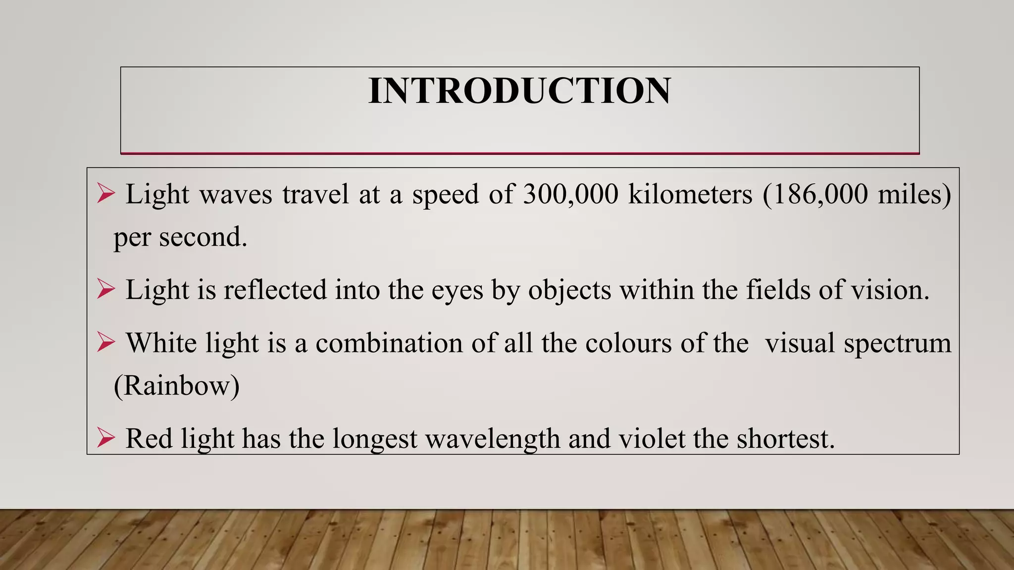
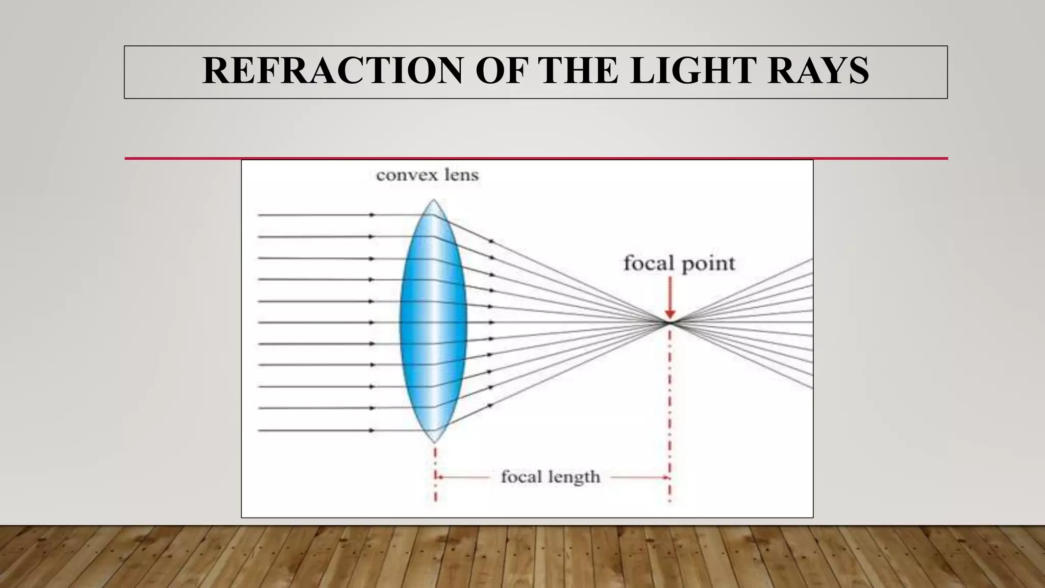
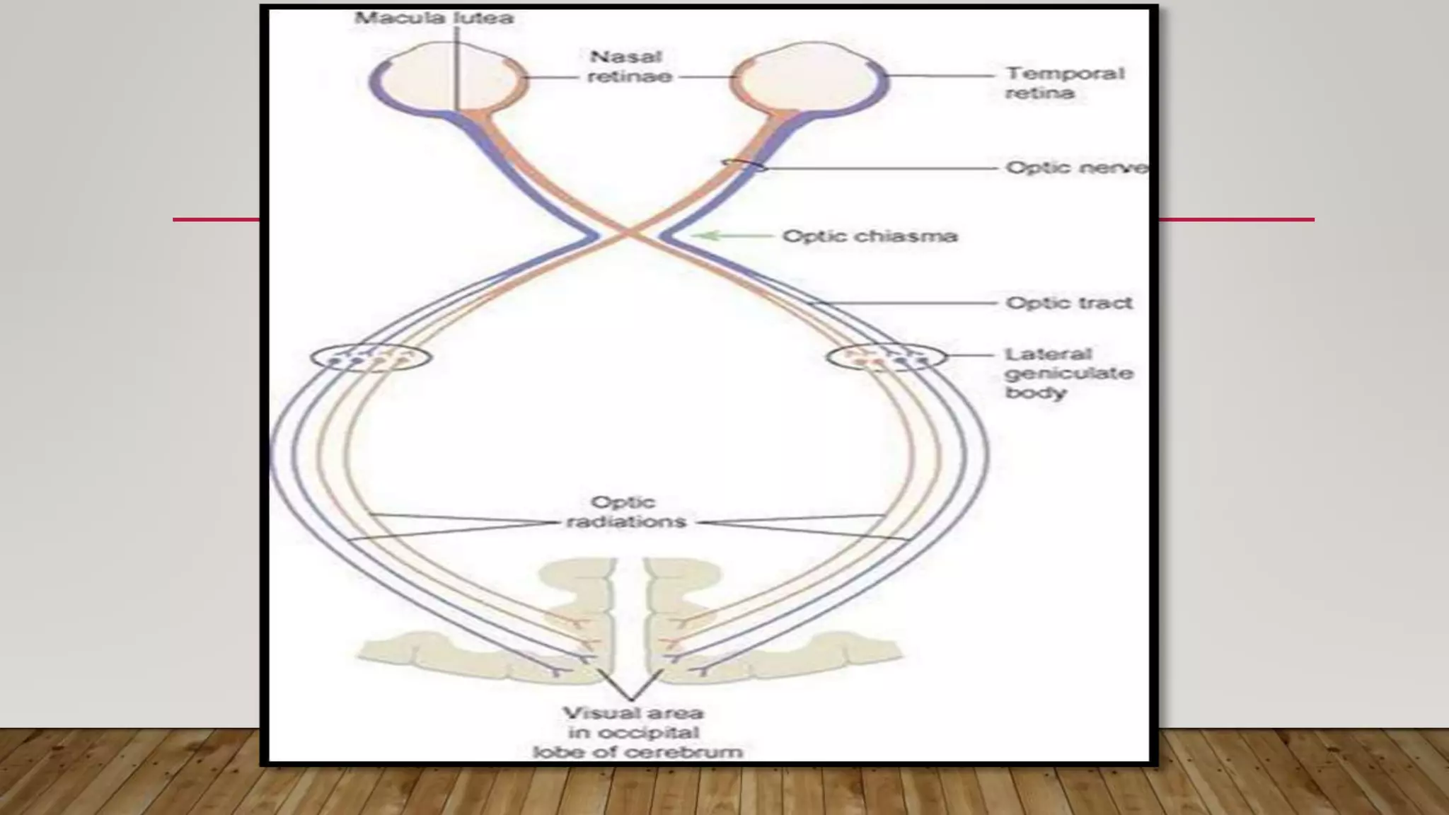
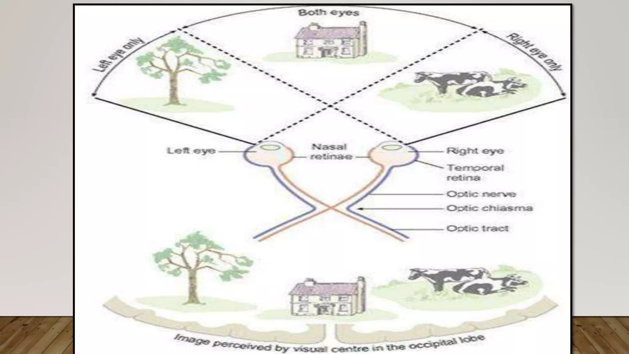
PHYSIOLOGY OF
SIGHT
NIKITA SHARMA
NURSING TUTOR
BECON, JAMMU
INTRODUCTION
 Light waves travel at a speed of 300,000 kilometers (186,000 miles)
per second.
 Light is reflected into the eyes by objects within the fields of vision.
 White light is a combination of all the colours of the visual spectrum
(Rainbow)
 Red light has the longest wavelength and violet the shortest.
THE ELECTROMAGNETIC SPECTRUM
 It is broad, but only a small part is visible to the human eye.
 Beyond the long end are infrared waves (heat), microwaves
and radio waves.
 Beyond the short end are ultraviolet, X-rays and gamma rays.
 UV light is not normally visible because it is absorbed by a
yellow pigment in the lens.
THE ELECTROMAGNETIC SPECTRUM
 Following removal of the (cataract extraction), UV light is visible and
it has been suggested that long-term exposure may damage the retina.
 A specific colour is perceived when only one wavelength is reflected
by the object and all the others are absorbed.
e.g: An object appears red when only the red wavelength is reflected.
Objects appear white when all wavelengths are reflected and black when
they are all absorbed.
THE ELECTROMAGNETIC SPECTRUM
 In order to achieve clear vision, light reflected from objects with in
the visual field is focused on to the retina of each eye.
 The process involved in producing a clear image are:
1. Refraction of the light rays
2. Changing the size of the pupils
3. Accommodation
Although these may be considered as separate processes, effective vison
is dependent upon their coordination.
REFRACTION OF THE LIGHT RAYS
 When the light rays pass from a medium of one density to a medium of a
different density they are bent.
E.g: In the eye, the biconvex lens bends and focuses light rays.
 This principle is used to focus light on the retina.
 Before reaching the retina, light rays pass successively through the
conjunctiva, cornea, aqueous fluid, lens and vitreous body.
They are all denser than air and with the exception of the lens, they have a
constant refractory power, similar to that of water.
FOCUSSING OF AN IMAGE ON THE RETINA
 Light rays reflected from an object are bent by the lens when they enter
the eye. Although the image on the retina is actually upside down.
 The brain adapts to this early in life so that objects are perceived ‘the
right way up’.
 Abnormal refraction with in the eye is corrected using biconvex or
biconcave lenses.
LENS
 The lens is a biconvex elastic transparent body suspended behind the
iris from the ciliary body by the suspensory ligament.
 It is the only structure in the eye that changes its refractory power.
 Light rays entering the eye need to be refracted to focus them on the
retina.
 Light from distant objects needs least refraction and as the object
comes closer, the amount of refraction needed is increased.
LENS
 To increase the refractive power, the ciliary muscle contracts. This moves
the ciliary body inwards towards the lens, relaxing the pull on the
suspensory ligaments and allows the lens to bulge, increasing its convexity.
 This focuses light rays from near objects on the retina. When the ciliary
muscle relaxes it slips backwards, increasing its pull on the suspensory
ligament, making the lens thinner.
 This focuses light rays from distant objects on the retina.
SIZE OF THE PUPILS
 Pupil size influences accommodation by controlling the amount of light
entering the eye.
 In a bright light the pupils are constricted. In a dim light, they are dilated.
 If the pupils were dilated in a bright light, too much light would enter the
eye and damage the sensitive retina.
 In a dim light, if the pupils were constricted, insufficient light would enter
the eye to activate the light-sensitive pigments in the rods and cones which
stimulate the nerve endings in the retina.
SIZE OF THE PUPILS
• The iris consists of one layer of circular and one of radiating smooth
muscle fibres. Contraction of the circular fibres constricts the pupils and
contraction of the radiating fibres dilates it.
• The size of the pupil is controlled by the autonomic nervous system;
sympathetic stimulation dilates the pupils and parasympathetic
stimulation causes constriction.
CONSTRICTION OF THE PUPILS
 This assists accommodating by reducing the width of the beam of light
entering the eye so that it passes through the central cursed part of the
lens.
ACCOMMODATION
CLOSE VISION: In order to focus on near objects, i.e. within about 6
meters, accommodation is required and the eye must make the following
adjustments:
o Constriction of the pupils
o Convergence
o Changing the power of the lens
CONVERGENCE (MOVEMENT OF THE EYEBALLS)
• Light rays from nearby objects enter the two eyes at different angles and
for clear vision they must stimulate corresponding areas of the two retinae.
• Extrinsic muscles move the eyes and to obtain a clear image they rotate
the eyes so that they converge on the object viewed. This coordinated
muscle activity is under autonomic control.
• When there is voluntary movement of the eyes both eyes move and
convergence is maintained.
• The nearer an object is to the eyes the greater the eye rotation needed
to achieve convergence e.g; an individual focusing near the tip of his
nose appears to be ‘cross eyed’ .
• If convergence is not complete the eyes are focused on different
objects or on different points of the same object. There are then two
images sent to the brain and this leads to double vision, diplopia.
CONVERGENCE (MOVEMENT OF THE
EYEBALLS)
• If convergence is not possible, the brain tends to ignore the
impulses received from the divergent eye.
CONVERGENCE (MOVEMENT OF THE
EYEBALLS)
CHANGING THE POWER OF LENS
• Changes in the thickness of the lens are made to focus light on the retina.
• The amount of adjustment depends on the distance of the objects from the
eyes, i.e. the lens is thicker for near vision and its thinnest when focusing on
objects at more than 6 meters distance.
• Looking at nearer objects tires the eyes more quickly, owing to the
continuous use of the ciliary muscle.
• The lens loses its elasticity and stiffens with age, a condition known as
presbyopia.
DISTANT VISION
• Objects more than 6 meters away from the eyes are focused on
the retina without adjustment of the lens or convergence of the
eyes.
FUNCTIONS OF THE RETINA
INTRODUCTION: The retina is the light- sensitive part of the eye. The
light-sensitive nerve cells are the rods and cones and their distribution in
the retina is shown below:
INTRODUCTION
 Light rays cause chemical changes in the light-sensitive pigments in
these cells and they generate nerve impulses which are conducted to the
occipital lobes of the cerebrum via the optic nerves.
 The rods are much more light sensitive than the cones, so they are used
when light levels are low. Stimulation of rods leads to monochromic (black
and white) vision. Rods outnumber cones in the retina by about 16 to 1.
 The cones are sensitive to bright light and colour. The different
wavelengths of visible light, light-sensitive pigments in the cones, resulting
in the perception of different colours.
FUNCTIONS OF THE RETINA
 In bright light, the light rays are focused on the macula lutea.
 Colour blindness is a condition which is transmitted by sex-linked
recessive gene (cone cells)
 The rods are more numerous towards the periphery of the retina. Visual
purple (rhodopsin) is a light-sensitive pigment present only in the rods. It
is degraded by bright light and is quickly regenerated, provided an
adequate supply of vitamin A is available.
FUNCTIONS OF THE RETINA
DARK ADAPTATION: when exposed to bright light, the rhodopsin within
the sensitive rods is completely degraded. This does not affect vision in good
light, when there is enough light to activate the cones.
 However, if the individual moves into a darkened area where the light
intensity is insufficient to stimulate the cones, temporary visual
impairment results whilst the rhodopsin is being regenerated with in the
rods, ‘dark adaptation’. When regeneration of rhodopsin has occurred,
normal sight returns.
FUNCTIONS OF THE RETINA
 In dim evening light, different colours cannot be distinguished because
the light intensity is insufficient to stimulate colour-sensitive pigments in
cones.
 Breakdown and regeneration of the visual pigments in cones is similar
to that of rods.
FUNCTIONS OF THE RETINA
 Binocular or stereoscopic vision enables three-dimensional views
although each eye ‘sees’ a scene from a slightly different angle.
 The visual fields overlap in the middle but the left eye sees more on the
left than can be seen by the other eye and vice-versa.
 The images from the two eyes are fused in the cerebrum so that only one
image is perceived.
BINOCULAR VISION
 Binocular vision provides a much more accurate assessment of one
object relative to another.
E.g; its distance, depth, height and width.
 People with monocular vision may find it difficult, for example, to
judge the speed and distance of an approaching vehicle.
BINOCULAR VISION
Physiology of sight

Physiology of sight

  • 1.
  • 2.
    INTRODUCTION  Light wavestravel at a speed of 300,000 kilometers (186,000 miles) per second.  Light is reflected into the eyes by objects within the fields of vision.  White light is a combination of all the colours of the visual spectrum (Rainbow)  Red light has the longest wavelength and violet the shortest.
  • 4.
    THE ELECTROMAGNETIC SPECTRUM It is broad, but only a small part is visible to the human eye.  Beyond the long end are infrared waves (heat), microwaves and radio waves.  Beyond the short end are ultraviolet, X-rays and gamma rays.  UV light is not normally visible because it is absorbed by a yellow pigment in the lens.
  • 6.
    THE ELECTROMAGNETIC SPECTRUM Following removal of the (cataract extraction), UV light is visible and it has been suggested that long-term exposure may damage the retina.  A specific colour is perceived when only one wavelength is reflected by the object and all the others are absorbed. e.g: An object appears red when only the red wavelength is reflected. Objects appear white when all wavelengths are reflected and black when they are all absorbed.
  • 7.
    THE ELECTROMAGNETIC SPECTRUM In order to achieve clear vision, light reflected from objects with in the visual field is focused on to the retina of each eye.  The process involved in producing a clear image are: 1. Refraction of the light rays 2. Changing the size of the pupils 3. Accommodation Although these may be considered as separate processes, effective vison is dependent upon their coordination.
  • 8.
  • 9.
    REFRACTION OF THELIGHT RAYS  When the light rays pass from a medium of one density to a medium of a different density they are bent. E.g: In the eye, the biconvex lens bends and focuses light rays.  This principle is used to focus light on the retina.  Before reaching the retina, light rays pass successively through the conjunctiva, cornea, aqueous fluid, lens and vitreous body. They are all denser than air and with the exception of the lens, they have a constant refractory power, similar to that of water.
  • 10.
    FOCUSSING OF ANIMAGE ON THE RETINA  Light rays reflected from an object are bent by the lens when they enter the eye. Although the image on the retina is actually upside down.  The brain adapts to this early in life so that objects are perceived ‘the right way up’.  Abnormal refraction with in the eye is corrected using biconvex or biconcave lenses.
  • 12.
    LENS  The lensis a biconvex elastic transparent body suspended behind the iris from the ciliary body by the suspensory ligament.  It is the only structure in the eye that changes its refractory power.  Light rays entering the eye need to be refracted to focus them on the retina.  Light from distant objects needs least refraction and as the object comes closer, the amount of refraction needed is increased.
  • 13.
    LENS  To increasethe refractive power, the ciliary muscle contracts. This moves the ciliary body inwards towards the lens, relaxing the pull on the suspensory ligaments and allows the lens to bulge, increasing its convexity.  This focuses light rays from near objects on the retina. When the ciliary muscle relaxes it slips backwards, increasing its pull on the suspensory ligament, making the lens thinner.  This focuses light rays from distant objects on the retina.
  • 15.
    SIZE OF THEPUPILS  Pupil size influences accommodation by controlling the amount of light entering the eye.  In a bright light the pupils are constricted. In a dim light, they are dilated.  If the pupils were dilated in a bright light, too much light would enter the eye and damage the sensitive retina.  In a dim light, if the pupils were constricted, insufficient light would enter the eye to activate the light-sensitive pigments in the rods and cones which stimulate the nerve endings in the retina.
  • 16.
    SIZE OF THEPUPILS • The iris consists of one layer of circular and one of radiating smooth muscle fibres. Contraction of the circular fibres constricts the pupils and contraction of the radiating fibres dilates it. • The size of the pupil is controlled by the autonomic nervous system; sympathetic stimulation dilates the pupils and parasympathetic stimulation causes constriction.
  • 17.
    CONSTRICTION OF THEPUPILS  This assists accommodating by reducing the width of the beam of light entering the eye so that it passes through the central cursed part of the lens.
  • 18.
    ACCOMMODATION CLOSE VISION: Inorder to focus on near objects, i.e. within about 6 meters, accommodation is required and the eye must make the following adjustments: o Constriction of the pupils o Convergence o Changing the power of the lens
  • 19.
    CONVERGENCE (MOVEMENT OFTHE EYEBALLS) • Light rays from nearby objects enter the two eyes at different angles and for clear vision they must stimulate corresponding areas of the two retinae. • Extrinsic muscles move the eyes and to obtain a clear image they rotate the eyes so that they converge on the object viewed. This coordinated muscle activity is under autonomic control. • When there is voluntary movement of the eyes both eyes move and convergence is maintained.
  • 20.
    • The neareran object is to the eyes the greater the eye rotation needed to achieve convergence e.g; an individual focusing near the tip of his nose appears to be ‘cross eyed’ . • If convergence is not complete the eyes are focused on different objects or on different points of the same object. There are then two images sent to the brain and this leads to double vision, diplopia. CONVERGENCE (MOVEMENT OF THE EYEBALLS)
  • 21.
    • If convergenceis not possible, the brain tends to ignore the impulses received from the divergent eye. CONVERGENCE (MOVEMENT OF THE EYEBALLS)
  • 22.
    CHANGING THE POWEROF LENS • Changes in the thickness of the lens are made to focus light on the retina. • The amount of adjustment depends on the distance of the objects from the eyes, i.e. the lens is thicker for near vision and its thinnest when focusing on objects at more than 6 meters distance. • Looking at nearer objects tires the eyes more quickly, owing to the continuous use of the ciliary muscle. • The lens loses its elasticity and stiffens with age, a condition known as presbyopia.
  • 23.
    DISTANT VISION • Objectsmore than 6 meters away from the eyes are focused on the retina without adjustment of the lens or convergence of the eyes.
  • 24.
    FUNCTIONS OF THERETINA INTRODUCTION: The retina is the light- sensitive part of the eye. The light-sensitive nerve cells are the rods and cones and their distribution in the retina is shown below:
  • 25.
    INTRODUCTION  Light rayscause chemical changes in the light-sensitive pigments in these cells and they generate nerve impulses which are conducted to the occipital lobes of the cerebrum via the optic nerves.
  • 27.
     The rodsare much more light sensitive than the cones, so they are used when light levels are low. Stimulation of rods leads to monochromic (black and white) vision. Rods outnumber cones in the retina by about 16 to 1.  The cones are sensitive to bright light and colour. The different wavelengths of visible light, light-sensitive pigments in the cones, resulting in the perception of different colours. FUNCTIONS OF THE RETINA
  • 28.
     In brightlight, the light rays are focused on the macula lutea.  Colour blindness is a condition which is transmitted by sex-linked recessive gene (cone cells)  The rods are more numerous towards the periphery of the retina. Visual purple (rhodopsin) is a light-sensitive pigment present only in the rods. It is degraded by bright light and is quickly regenerated, provided an adequate supply of vitamin A is available. FUNCTIONS OF THE RETINA
  • 29.
    DARK ADAPTATION: whenexposed to bright light, the rhodopsin within the sensitive rods is completely degraded. This does not affect vision in good light, when there is enough light to activate the cones.  However, if the individual moves into a darkened area where the light intensity is insufficient to stimulate the cones, temporary visual impairment results whilst the rhodopsin is being regenerated with in the rods, ‘dark adaptation’. When regeneration of rhodopsin has occurred, normal sight returns. FUNCTIONS OF THE RETINA
  • 30.
     In dimevening light, different colours cannot be distinguished because the light intensity is insufficient to stimulate colour-sensitive pigments in cones.  Breakdown and regeneration of the visual pigments in cones is similar to that of rods. FUNCTIONS OF THE RETINA
  • 31.
     Binocular orstereoscopic vision enables three-dimensional views although each eye ‘sees’ a scene from a slightly different angle.  The visual fields overlap in the middle but the left eye sees more on the left than can be seen by the other eye and vice-versa.  The images from the two eyes are fused in the cerebrum so that only one image is perceived. BINOCULAR VISION
  • 33.
     Binocular visionprovides a much more accurate assessment of one object relative to another. E.g; its distance, depth, height and width.  People with monocular vision may find it difficult, for example, to judge the speed and distance of an approaching vehicle. BINOCULAR VISION