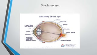
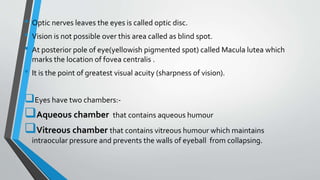
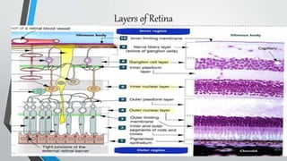
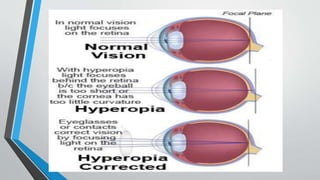
- The eye has three principal layers: an outer fibrous layer, middle vascular layer, and inner nervous layer.
- The middle layer contains the choroid, ciliary body, and iris. The iris regulates the intensity of light entering the eye.
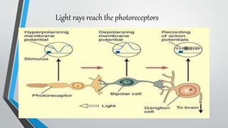
- The inner layer contains the retina, which has photoreceptors (rods and cones) that detect light and transmit signals to the brain.


![[L-3]- Eye- Nov 13, 2019.pdfnbnccncbcncbcnc](https://cdn.slidesharecdn.com/ss_thumbnails/l-3-eye-nov132019-240222105749-6613d5d2-thumbnail.jpg?width=640&height=640&fit=bounds)


![Transport of gases carbon dioxide and oxygen [Autosaved].pptx](https://cdn.slidesharecdn.com/ss_thumbnails/transportofgasesautosaved-250324055730-f2aabc7a-thumbnail.jpg?width=640&height=640&fit=bounds)


