Downloaded 1,423 times


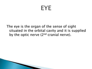
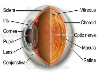
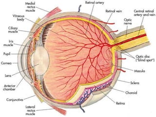

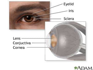
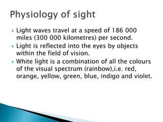
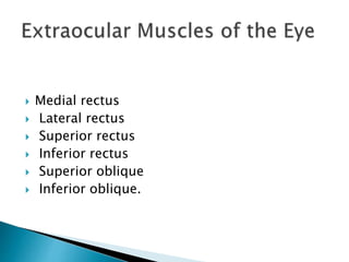
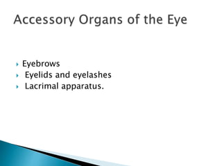

The eye is composed of three layers: the outer fibrous layer, middle vascular layer, and inner nervous tissue layer. It contains structures like the lens, aqueous fluid, and vitreous body. Light enters through the cornea and is refracted by the lens to focus on the retina. The iris and pupil regulate the amount of light entering, and accommodation and refraction allow for vision at varying distances. Dark and light adaptation involve changes in the retina and visual pigments in response to light levels. Accessory structures include extraocular muscles, eyebrows, eyelids, and the lacrimal apparatus.


![concept of safety[1].ppt BSN YEAR 1 SEM1](https://cdn.slidesharecdn.com/ss_thumbnails/conceptofsafety1-240509093204-8977567e-thumbnail.jpg?width=640&height=640&fit=bounds)


