The document provides a detailed overview of the anatomy of the eye, describing the dimensions, structure, and functions of various components including the cornea, sclera, iris, ciliary body, choroid, crystalline lens, vitreous humor, and retina. It highlights the roles these structures play in light refraction, protection, nourishment, and signal conversion. Overall, it emphasizes the complexity and interconnectivity of the eye's anatomy.
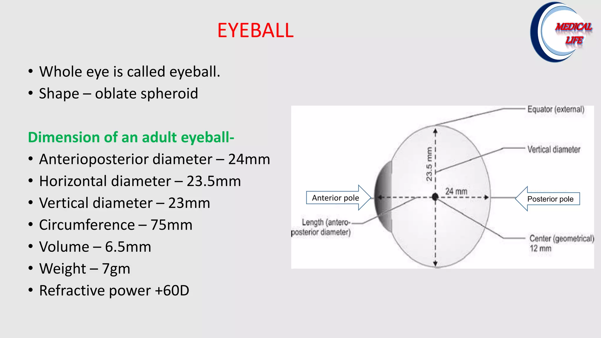
• Whole eyeis called eyeball.
• Shape – oblate spheroid
Dimension of an adult eyeball-
• Anterioposterior diameter – 24mm
• Horizontal diameter – 23.5mm
• Vertical diameter – 23mm
• Circumference – 75mm
• Volume – 6.5mm
• Weight – 7gm
• Refractive power +60D
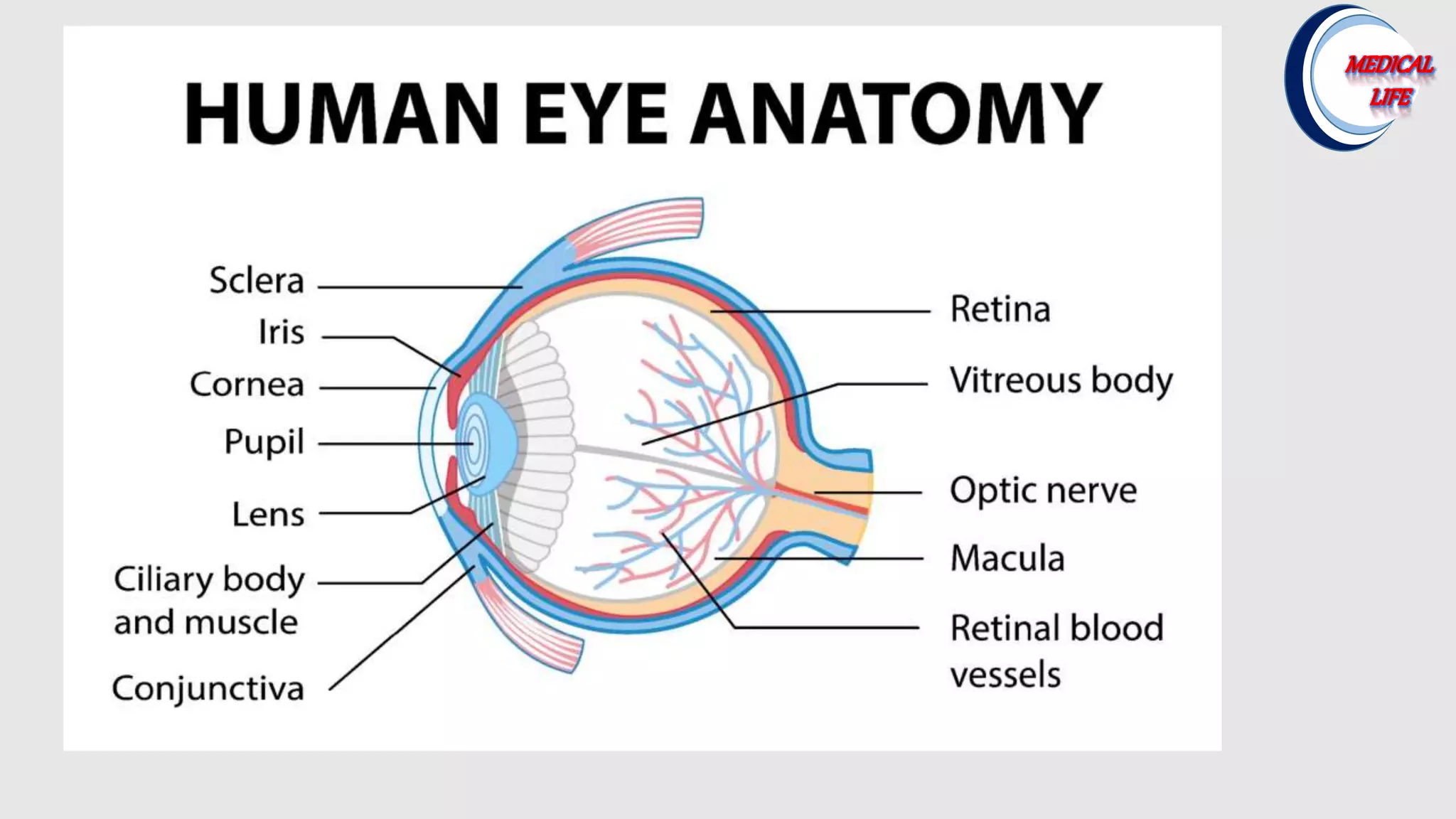
EYEBALL
Anterior pole Posterior pole
3.
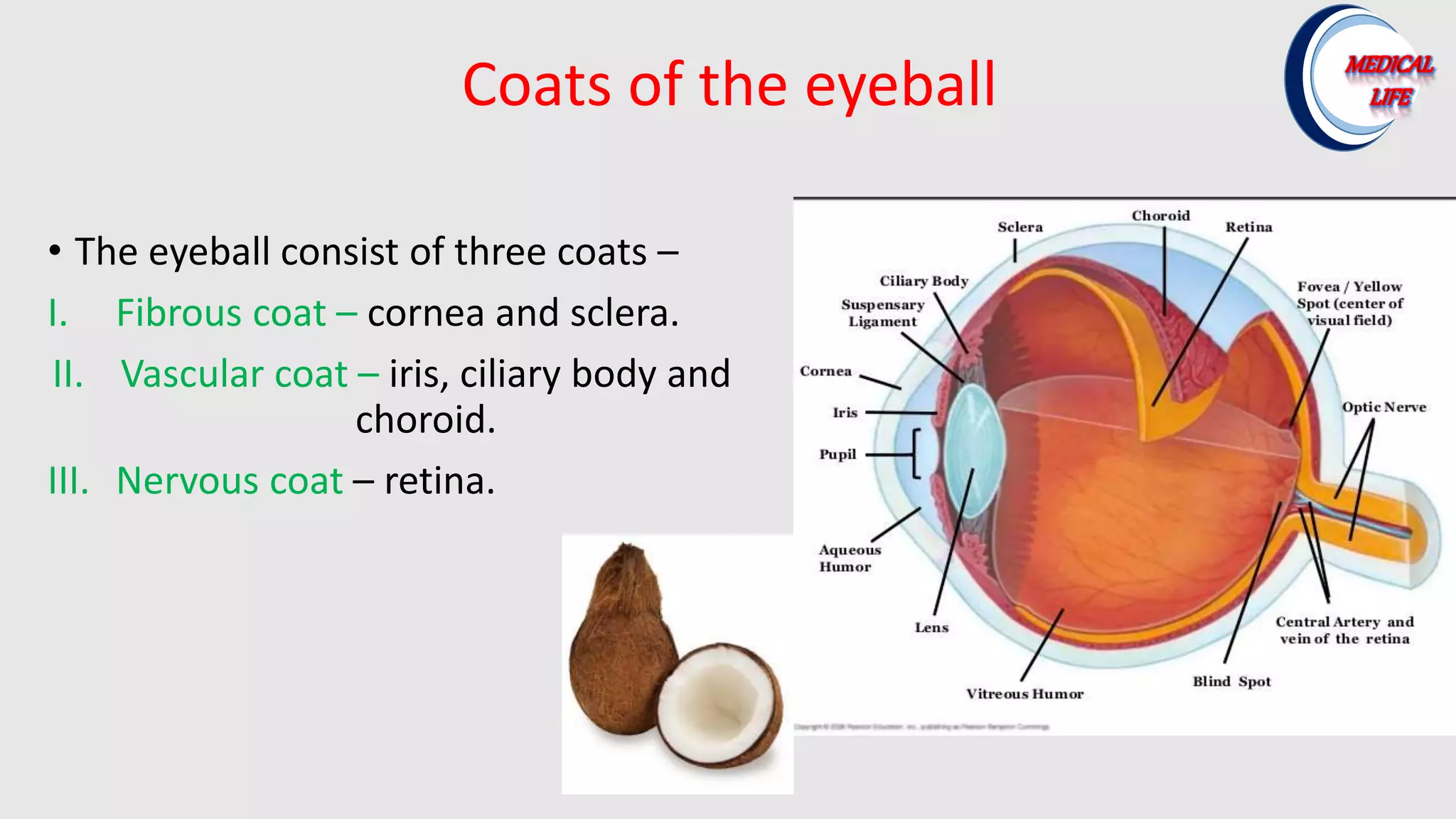
Coats of theeyeball
• The eyeball consist of three coats –
I. Fibrous coat – cornea and sclera.
II. Vascular coat – iris, ciliary body and
choroid.
III. Nervous coat – retina.
4.
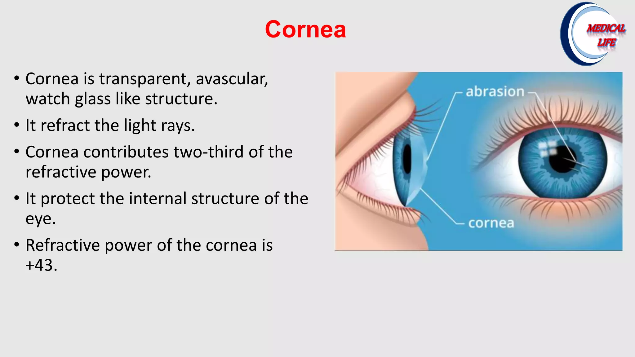
Cornea
• Cornea istransparent, avascular,
watch glass like structure.
• It refract the light rays.
• Cornea contributes two-third of the
refractive power.
• It protect the internal structure of the
eye.
• Refractive power of the cornea is
+43.
5.
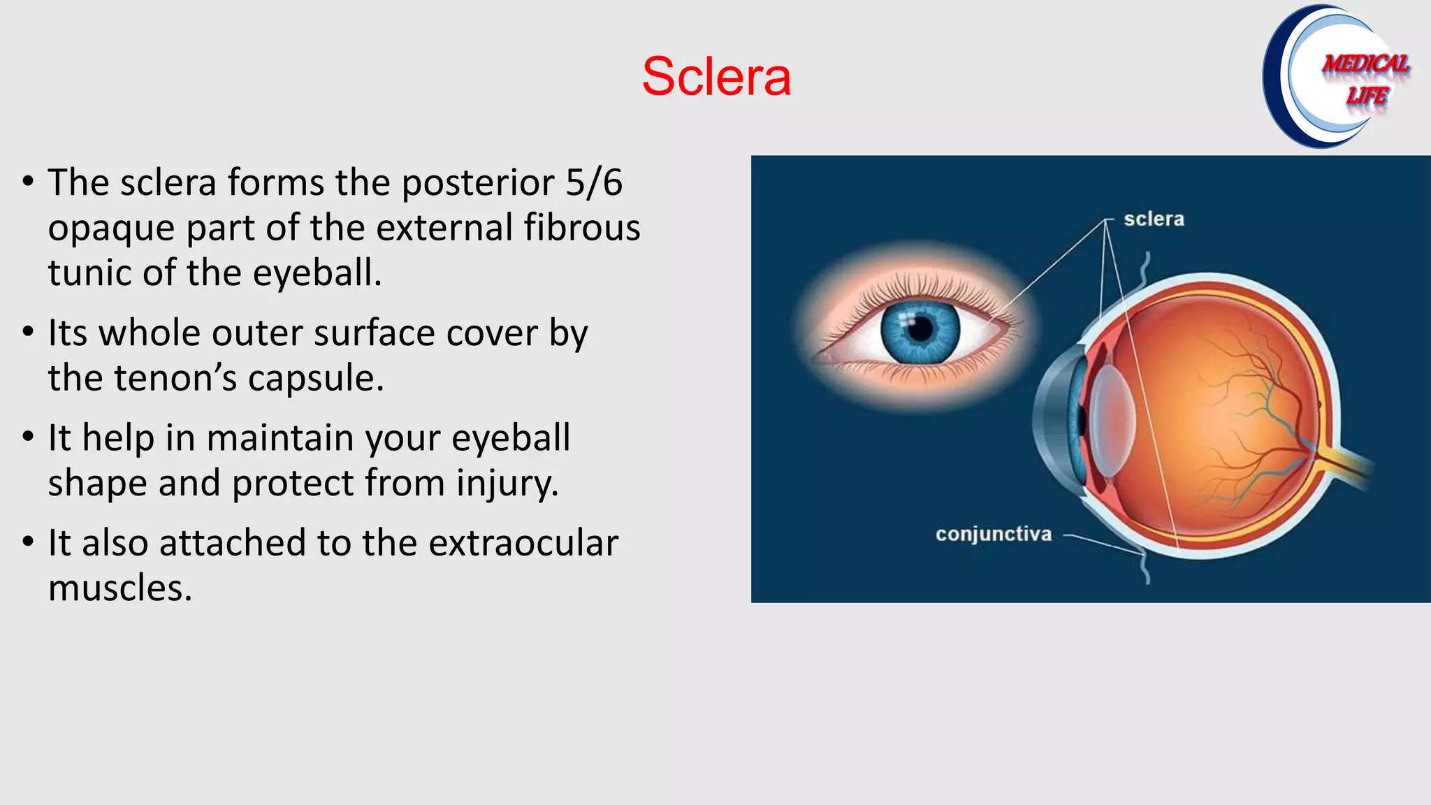
Sclera
• The scleraforms the posterior 5/6
opaque part of the external fibrous
tunic of the eyeball.
• Its whole outer surface cover by
the tenon’s capsule.
• It help in maintain your eyeball
shape and protect from injury.
• It also attached to the extraocular
muscles.
6.
Iris
• Iris isthe colored part of the eye.
• Iris is the anterior most part of the
uveal tract.
• It is circular disk corresponding to the
diaphragm of a camera.
• In its center is an aperture of about
4mm called pupil which regulates the
amount of light reaching the retina.
7.
Ciliary body
• Forwordcontinuation of choroid at ora
serrata.
• It contains ciliary muscle and ciliary
processes.
• The ciliary process produce aqueous
humor .
• The ciliary muscle take part
accommodation.
8.
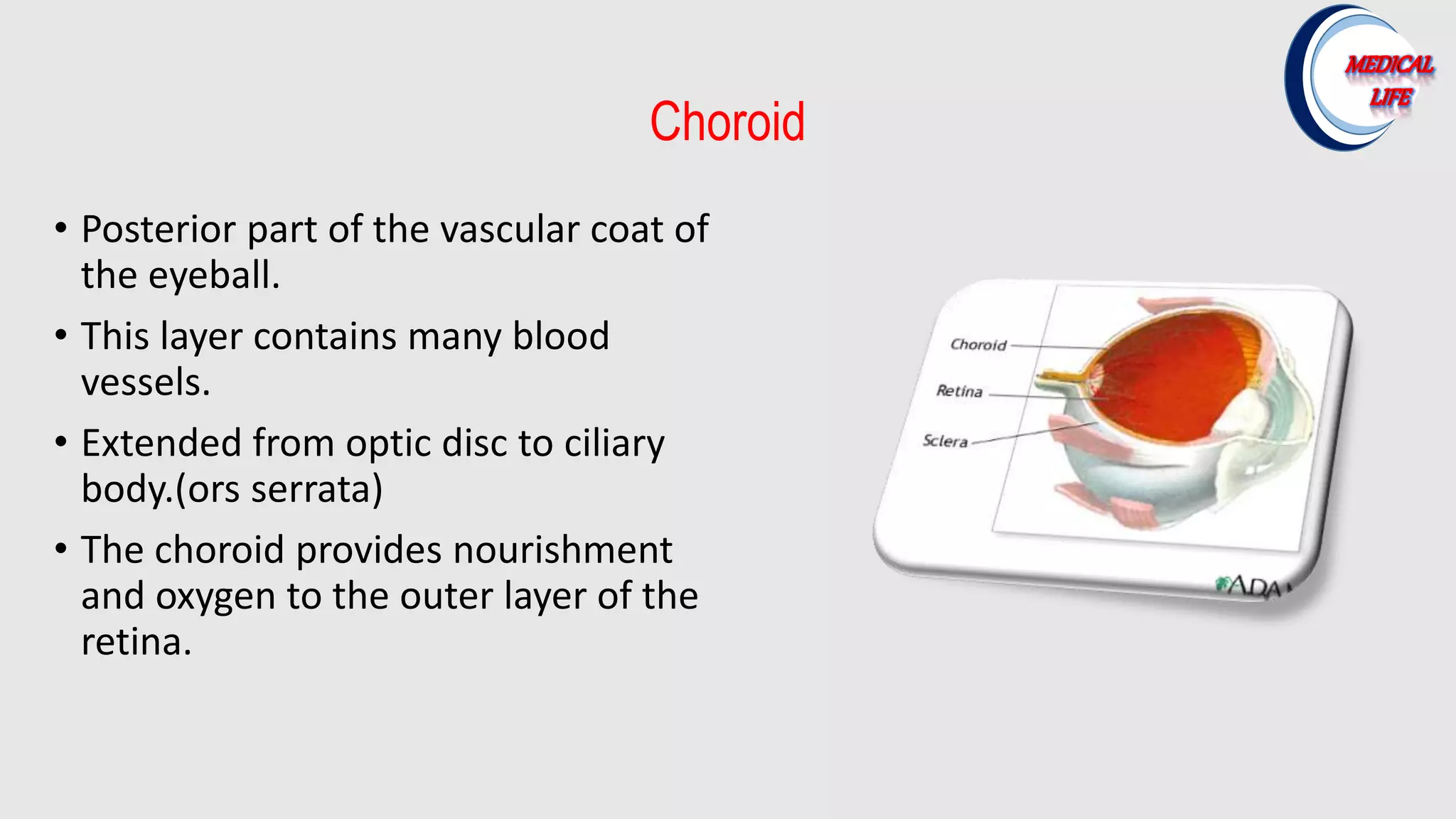
Choroid
• Posterior partof the vascular coat of
the eyeball.
• This layer contains many blood
vessels.
• Extended from optic disc to ciliary
body.(ors serrata)
• The choroid provides nourishment
and oxygen to the outer layer of the
retina.
9.
Crystalline Lens
• Lensis a transparent, biconvex,
crystalline structure.
• Lens lies between posterior surface
of the iris and and anterior surface
of the vitreous.
• It converge the light rays and focus
on retina.
• Refractive power of the crystalline
lens is +17D.
10.
Vitreous Humour
• Vitreoushumours is an inert, transparent,
jelly like structure that fill the posterior 80% of
the cavity of the eyeball.
• It is about 4ml in volume and 4gm in weight.
• It maintain the shape of the eyeball.
• It provide nutrition to the eye.
• Optical function.
11.
Retina
• Retina isthe innermost tunic of the eyeball
• It is transparent, thin, and delicate
membrane.
• It is highly developed tissue of the eye.
• The retina converts light that enters into the
eye into electrical signal and send to the
brain.